外显子组测序ppt课件
- 格式:ppt
- 大小:1.63 MB
- 文档页数:31

全外显子组测序的具体方法及步骤
全外显子组测序(Whole Exome Sequencing,WES)是指利用序列捕获或者靶向技术将全基因组外显子区域DNA 富集后再进行高通量测序的基因组分析方法。
与全基因组重测序相比,全外显子组测序只需针对外显子区域的基因序列测序,覆盖度更深、数据准确性更高,更加简便、经济、高效。
技术优势
高性价,强分析,快速交付
外显子组测序主要用于识别和研究与疾病相关的编码区的基因组变异。
结合大量的公共数据库提供的外显子数据和正常人群数据库, 有利于更好地排除无害突变及解释变异信息之间的关联和致病机理。
技术路线
技术参数
样本要求。


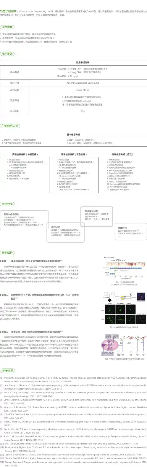
b r i e fc o m m u n i c at i o n sHyperphosphatasia mental retardation (HPMR) syndrome is an autosomal recessive form of mental retardation with distinct facial features and elevated serum alkaline phosphatase. We performed whole-exome sequencing in three siblings of a nonconsanguineous union with HPMR and performed computational inference of regions identical by descent in all siblings to establish PIGV , encoding a member of the GPI-anchor biosynthesis pathway, as the gene mutated in HPMR. We identified homozygous or compound heterozygous mutations in PIGV in three additional families.Recessive mutations are relatively common in the human genome, but their identification remains challenging. Initial efforts at using exome sequencing for disease gene discovery 1 analyzed small num-bers of unrelated individuals, removed variants that are common or not predicted to be deleterious and then searched for genes with such variants in all affected individuals. The analysis of the exome sequences of two siblings and two further unrelated individuals affected by the autosomal recessive Miller syndrome led to the iden-tification of DHODH as the disease gene 2. Subsequently, researchers analyzed whole genome sequences of the same two siblings and their parents to identify chromosomal regions in which both siblings had inherited identical haplotypes from both parents, which allowed thenumber of gene candidates for Miller syndrome to be reduced from 34 to 4, showing that linkage information represents a useful filter for genome sequence data 3. These studies illustrate the utility of sophisti-cated algorithmic analysis in reducing the candidate gene set beyond what can be achieved by a simple intersection filter.HPMR, also known as Mabry syndrome (MIM%239300), was ini-tially described as an autosomal recessive syndrome characterized by mental retardation and greatly elevated alkaline phosphatase levels 4,5. Within a group of individuals with this rare syndrome, a previous study 6 delineated a specific clinical entity characterized by a distinct facial gestalt including hypertelorism, long palpebral fissures, a broad nasal bridge and tip, and a mouth with downturned corners and a thin upper lip, as well as brachytelephalangy. More variable neurological features included seizures and muscular hypotonia 6.Here, DNA from three siblings of nonconsanguineous parents with this subtype of HPMR was analyzed by exome sequenc-ing (Supplementary Figs. 1 and 2 and Supplementary Table 1). Whole-exome sequencing using the ABI SOLiD platform was per-formed following enrichment of exonic sequences using Agilent’s SureSelect whole-exome enrichment. Called variants were filtered to exclude variants not found in all affected persons as well as common variants identified in the dbSNP130 or HapMap databases, which left 14 candidate genes on multiple chromosomes (Table 1 and Supplementary Tables 2–4).In this work, we developed a statistical model that allowed us to infer regions that are identical by descent (IBD) from the exome sequences of only the affected children of a family in which an auto-somal recessive disorder segregates. In consanguineous families, affected siblings share two haplotypes that are inherited from a single common ancestor at the disease locus and are thus homozygous by descent. In nonconsanguineous families, the affected children inherit identical maternal and paternal haplotypes in a region surrounding the disease gene, meaning that both haplotypes originated from the same maternal and paternal haplotype but are not necessarily from an identical ancestor (IBD = 2).We developed an algorithm based on a Hidden Markov Model (HMM), a type of Bayesian network that is used to infer a sequence of hidden (that is, unobservable) states. We used the HMM algorithm to identify chromosomal regions with IBD = 2 in the presence of noisy (that is, potentially erroneous) sequence data. It is not possible to measure the IBD = 2 state directly; it is only possible to determine whether the genotypes of the siblings are compatible with identity-by-state status, that is, whether each sibling has the same homozygousIdentity-by-descent filtering of exome sequence dataidentifies PIGV mutations in hyperphosphatasia mental retardation syndromePeter M Krawitz 1–3,11, Michal R Schweiger 1,2,11,Christian Rödelsperger 1–3, Carlo Marcelis 4, Uwe Kölsch 5, Christian Meisel 5, Friederike Stephani 4, Taroh Kinoshita 6, Yoshiko Murakami 6, Sebastian Bauer 2, Melanie Isau 1,Axel Fischer 1, Andreas Dahl 1, Martin Kerick 1, Jochen Hecht 1,3, Sebastian Köhler 2, Marten Jäger 2, Johannes Grünhagen 2, Birgit Jonske de Condor 2, Sandra Doelken 2, Han G Brunner 4, Peter Meinecke 7, Eberhard Passarge 8, Miles D Thompson 9,David E Cole 9, Denise Horn 2, Tony Roscioli 4,10, Stefan Mundlos 1–3 & Peter N Robinson 1–31Max Planck Institute for Molecular Genetics, Berlin, Germany. 2Institut für Medizinische Genetik, Charité Universitätsmedizin Berlin, Berlin, Germany.3Berlin-Brandenburg Center for Regenerative Therapies (BCRT), Charité -Universitätsmedizin Berlin, Berlin, Germany. 4Department of Human Genetics, UniversityMedical Centre St. Radboud, Nijmegen, The Netherlands. 5Institut für Medizinische Immunologie, Charité Universitätsmedizin Berlin, Berlin, Germany. 6Department of Immunoregulation, Research Institute for Microbial Diseases, Osaka University, Osaka, Japan. 7Medizinische Genetik, Altonaer Kinderkrankenhaus, Hamburg, Germany. 8Institut für Humangenetik, Universitätsklinikum Essen, Essen, Germany. 9Department of Laboratory Medicine and Pathobiology, University of Toronto, Toronto, Ontario, Canada. 10Department of Molecular and Clinical Genetics, University of Sydney, Sydney, Australia. 11These authors contributed equally to this work. Correspondence should be addressed to S.M. (stefan.mundlos@charite.de) or P .N.R. (peter.robinson@charite.de).Received 29 March; accepted 3 August; published online 29 August 2010; doi:10.1038/ng.653© 2010 N a t u r e A m e r i c a , I n c . A l l r i g h t s r e s e r v e d.b r i e fc o m m u n i c at i o n sor heterozygous genotype, a situation which we refer to as IBS*. In our model, every genetic locus was either IBD = 2 or IBD ≠ 2. The HMM was then used to predict the most likely sequence of IBD = 2 or IBD ≠ 2 chromosomal segments on the basis of the observed exome sequences of two or more affected siblings (Supplementary Fig. 1 and Supplementary Methods ).HMM analysis decreased the search space to about 20% of the tran-scribed genome, reducing the number of candidate genes with muta-tions present in all three siblings from 14 to 2 (Table 1, Supplementary Table 5 and Supplementary Figs. 3–5). The two mutations, c.[859G>A]+[859G>A] in SLC9A1 and c.[1022C>A]+[1022C>A] in PIGV , were located within a 13-Mb homozygous block that was part of a larger 35-Mb IBD = 2 block. Runs of homozygosity of up to 4 Mb can occur in the European population even in individuals with no shared ancestors in the previous five to ten generations 7. Both variants were confirmed with ABI Sanger sequencing and were not detected in 200 healthy, unrelated central European individuals. Further homozygous and compound heterozygous mutations were detected in PIGV in individuals from the families designated B 8, C 9 and D 10 (Supplementary Note and Supplementary Tables 6 and 7). All of these missense mutations affect evolutionarily highly conserved residues of PIGV (Fig. 1a ).PIGV, the second mannosyltransferase in the GPI anchor bio-synthesis pathway 11, appeared to be of particular interest because alkaline phosphatase is a GPI-anchored protein. Over 100m ammalian proteins are modified by a glycosylphosphatidylinosi tol (GPI) anchor at their C terminus. The highly conserved back-bone structure of the GPI anchor is synthesized in the endoplasmicr eticulum through at least nine sequential reaction steps mediated by at least 18 proteins. GPI-anchored proteins comprise functionally divergent classes including hydrolytic enzymes, receptors, adhesion molecules and proteins with roles in the immune system 12. Little is known to date about the phenotypic consequences of mutationsof the GPI pathway in mammals. Abrogation of GPI biosynthesis in mice by knockdown of Piga , which encodes a protein that is involved in the first step of GPI-anchor biosynthesis, results in embryonic lethality 13. Somatic loss-of-function mutations in PIGA in hematopoietic stem cells are associated with paroxysmaln octurnal hemoglobinuria 14, primarily because the progeny of affected stem cells are deficient in the GPI-anchored complement regulatory proteins CD55 and CD59, leading to the intravascular hemolysis characteristic of the disease. A promoter mutation in PIGM , encoding a subunit of the complex transferring the first mannose, reduces PIGM expression by over 90% and leads to an autosomal recessive syndrome characterized by hepatic venous thrombosis and absence seizures 15.Defects in the GPI biosynthesis pathway can result in down-regulation of GPI-anchored proteins but not necessarily in a uniform reduction of all such proteins 12. We therefore examined the surface expression of the GPI anchor itself on leukocytes of three indivi-duals with HPMR using Alexa488-conjugated inactivated aerolysin (FLAER). All three subjects showed a substantial reduction of GPI-anchor expression. Correspondingly, expression of the GPI-anchored protein CD16 was markedly reduced (Supplementary Fig. 6). Wild-type PIGV cDNA and PIGV cDNA containing the p.Ala341Glu alteration were transiently transfected into PIGV-deficient Chinese hamster ovary (CHO) cells 11 to assess their effect on protein expression. Cells transfected with themutant constructdid not restore surface expression of GPI-anchored marker proteins (Fig. 1b ), possibly because expressed PIGV protein levels were substantially reduced (Fig. 1c ).Human Mouse Rat Dog Cow Horse FrogZebrafishLumenPIGVCytosolNH 2p.Gln256Lysp.Ala341Glu p.Ala341ValCD59CD55GAPDH12337 kD37 kDPIGV0.0011.50.4p.His385Pro QAHCOOHab cfigure 1 Identification of PIGV mutations in individuals with HPMRsyndrome. (a ) The homozygous PIGV mutation c.[1022C>A]+[1022C>A]; p.[Ala341Glu]+[Ala341Glu] was detected via whole exome sequencing in family A. Further homozygous and compound heterozygous mutations affecting evolutionarily highly conserved residues were found in threeunrelated families: c.[1022C>A]+[1154A>C]; p.[Ala341Glu]+[His385Pro] in family B, c.[766C>A]+[766C>A]; p.[Gln256Lys]+[Gln256Lys] in family C, and c.[1022C>A]+[1022C>T]; p.[Ala341Glu]+[Ala341Val] in family D. (b ) PIGV-deficient CHO cells were transiently transfected with wild-type (dashed lines) or p.Ala341Glu mutant (solid lines) PIGV cDNA in a weak expression vector or the empty vector (gray shadow). Wild-type PIGV efficiently restored the surface expression of CD59 (left) and CD55 (right), whereas p.Ala341Glu mutant PIGV induced only very low levels of CD59 and CD55. (c ) PIGV protein levels were assessed 2 d after transfection of a control vector (lane 1), wild-type PIGV (lane 2) and PIGV with p.Ala341Glu (lane 3). The numbers beneath the gel indicate the relative intensity of PIGV to GAPDH expression.table 1 number of genes with nonsynonymous variants and acceptor or donor splice site mutationsA1A2A3A1 & A2 & A3FilterHomozygous heterozygous All Homozygous heterozygous All Homozygous heterozygous All Homozygous heterozygous All NS/SS2,7529343,3852,9001,0903,6402,8061,0703,6251,7282731,928Not in dbSNP13018235216218472622003823512214IBD = 2165212052517623202Sanger validated22Reducing the search space to the identical by descent (IBD = 2) regions and filtering out all common variants decreased the number of genes with nonsynonymous variants and acceptor or donor splice site mutation to two candidate genes. NS, nonsynonymous; SS, acceptor or donor splice site mutations.© 2010 N a t u r e A m e r i c a , I n c . A l l r i g h t s r e s e r v e d .b r i e fc o m m u n i c at i o n sIn summary, we have identified PIGV mutations in HPMR using whole-exome capture and SOLiD sequencing in combination with an HMM algorithm to identify regions with IBD = 2 in siblings affected with autosomal recessive disorders. Our algorithm can be used in combination with other bioinformatic filters to streamline gene dis-covery in future exome sequencing projects.Accession codes. The mutations in this work were numbered according to transcripts available in GenBank under the codes NM_003047.3 (SLC9A1) and NM_017837.2 (PIGV ).Note: Supplementary information is available on the Nature Genetics website.ACKNowlEDGMENTSThis work was supported by a grant from the Deutsche Forschungsgemeinschaft (SFB 665) to S.M., by a grant from Bundesministerium für Bildung und Forschung (BMBF , project number 0313911) and an Australian National Health and Medical Research Council international research training fellowship to T.R., and by a grant of the Canadian Institutes of Health Research and Epilepsy Canada to M.D.T. We thank B. Fischer, U. Kornak, M. Ralser, E. van Beusekom, U. Marchfelder and D. Lefeber for their assistance in this project.AUTHoR CoNTRIBUTIoNSM.R.S., M.I. and A.D. performed targeted exome resequencing. P .M.K., C.R., A.F., M.K., S.B., S.K., M.J. and P .N.R. performed bioinformatic analysis. P .M.K., C. Marcelis, J.G., B.J.d.C., F.S. and T.R. performed mutation analysis andgenotyping. D.H., C. Marcelis, M.D.T., D.E.C., S.D., P .M., E.P ., T.R. and H.G.B.contributed to clinical evaluation of the affected individuals and delineation of the phenotype. P .M.K., U.K. and C. Meisel performed flow cytometric analysis. Y.M. and T.K. performed analysis of wild-type and A341E PIGV clones. P .M.K., M.R.S., D.H., J.H., H.G.B., P .N.R. and S.M. carried out the project planning and preparation of the manuscript.CoMPETING FINANCIAl INTERESTSThe authors declare no competing financial interests.Published online at /naturegenetics/.Reprints and permissions information is available online at /reprintsandpermissions/.1. Ng, S.B. et al. Nat. Genet. 42, 30–35 (2010).2. Ng, S.B. et al. Nature 461, 272–276 (2009).3. Roach, J.C. et al. Science 328, 636–639 (2010).4. Mabry, C.C. et al. J. Pediatr. 77, 74–85 (1970).5.Kruse, K., Hanefeld, F ., Kohlschutter, A., Rosskamp, R. & Gross-Selbeck, G. J. Pediatr. 112, 436–439 (1988).6. Horn, D., Schottmann, G. & Meinecke, P . Eur. J. Med. Genet. 53, 85–88 (2010).7. McQuillan, R. et al. Am. J. Hum. Genet. 83, 359–372 (2008).8. Rabe, P . et al. Am. J. Med. Genet. 41, 350–354 (1991).9. Marcelis, C.L., Rieu, P ., Beemer, F . & Brunner, H.G. Clin. Dysmorphol. 16, 73–76(2007).10. Thompson, M.D. et al. Am. J. Med. Genet. 152a , 1661–1669 (2010).11. Kang, J.Y. et al. J. Biol. Chem. 280, 9489–9497 (2005).12. Kinoshita, T., Fujita, M. & Maeda, Y. J. Biochem. 144, 287–294 (2008).13. Nozaki, M. et al. Lab. Invest. 79, 293–299 (1999).14. Takeda, J. et al. Cell 73, 703–711 (1993).15. Almeida, A.M. et al. Nat. Med. 12, 846–851 (2006).© 2010 N a t u r e A m e r i c a , I n c . A l l r i g h t s r e s e r v e d.。

外显子测序生物学重复-概述说明以及解释1.引言1.1 概述外显子测序(exome sequencing)是一种基于高通量测序技术的生物学研究方法,其目的是对生物体中的外显子区域进行快速、准确地测序和分析。
外显子是基因组中编码蛋白质的片段,它们占据了整个基因组的仅0.5至1.5的区域,但却承载着80以上的已知致病突变。
因此,外显子测序被广泛应用于寻找蛋白质编码基因的突变,以及与遗传性疾病、肿瘤和其他复杂疾病相关的致病突变的鉴定和研究。
外显子测序的基本原理是使用高通量测序技术对DNA样本进行测序,然后利用生物信息学方法将测序结果与参考基因组进行比对和分析,从而确定样本中外显子的序列和存在突变的位置。
与全基因组测序相比,外显子测序具有较低的成本和更高的效率,因为外显子相对较小且具有较高的功能重要性,可以更准确地筛选和鉴定潜在致病突变。
外显子测序在生物学研究中的应用广泛而重要。
它不仅可以用于研究人类遗传性疾病和肿瘤突变,还可应用于农业、畜牧业和其他生物领域的基因组学研究。
通过对不同个体的外显子进行测序,我们可以了解个体间的遗传差异、突变积累和遗传进化规律,为人类进化和适应性研究提供重要依据。
然而,外显子测序也面临一些挑战。
首先,由于外显子区域相对较小,它只能提供关于外显子的信息,对非编码区域的突变鉴定有限。
其次,外显子测序在处理复杂疾病和疾病相关基因组变异时可能会遇到困难,因为这些变异可能位于基因的调控区域或与功能相关的非编码RNA中。
此外,外显子测序对测序深度和准确性要求较高,因此需要高质量的测序平台和数据分析方法的支持。
总之,外显子测序作为一种高效、准确的测序技术,在生物学研究和临床诊断中发挥着重要作用。
随着技术的不断发展和应用的不断扩大,外显子测序将为我们揭示生物体的基因组变异与功能之间的关系,为疾病的早期诊断和个性化治疗提供更多可能性。
同时,对于生物学重复的研究也为我们提供了全新的视角和理解,有助于揭示生命的奥秘和进化的规律。

人外显子测序药明康德基因中心,陆桂1. 什么是外显子测序(whole exon sequencing)?外显子组测序是指利用序列捕获技术将全基因组外显子区域DNA捕捉并富集后进行高通量测序的基因组分析方法。
外显子测序相对于基因组重测序成本较低,对研究基因的SNP、Indel 等具有较大的优势,但无法研究基因组结构变异如染色体断裂重组等。
2. 外显子捕获试剂盒有哪些?目前主要有Roche、Illumina和Agilent三家的外显子捕获试剂。
Nimblegen和Illumina的捕获试剂盒中的探针是DNA探针,化学性质稳;Agilent的捕获试剂盒是RNA探针,有可能RNA 不是很稳定。
3. 外显子捕获效率是什么?外显子测序过程中要用到杂交过程。
在人的染色体上有许多与外显子有同源性的部分,这些有同源性的部分很可能在杂交过程中也被捕获下来。
所以,测到的序列中,有一部分不是外显子序列。
我们把测序得是外显子的部分占全部测序序列的比列称为捕获效率。
Nimblegen大约是70%Agilent大约是60%Illumina大约是50%4. 外显子测序一般建议做多少倍的覆盖?一般做100X或者150X。
较高的覆盖倍数,对于测异质性的遗传变质,可以发现小比例的突变。
另外,外显子测序的覆盖不是很均匀,这样较高的平均覆盖率有利于保证大部分的区域有足够的覆盖倍数。
5. 外显子测序能够测出多大的片段缺失?大致能测出50bp的片段缺失。
目前的测序主要还是用Hiseq 2000,单侧的测长就是100bp。
由于外显子测序的覆盖很不平均,所以如果有大段的缺失,无法判断是因为杂交没有捕获到,还是因为缺失。
目前能够测到的,就是在一个read中发现的缺失。
一个read的长度也就是100bp,所以大到50bp以下的片段缺失可以从外显子测序中测出来。
6. 外显子捕获可以做CNV吗?外显子测序因为有一个杂交捕获的过程,这样就会有一个杂交捕获效率的问题。