炎症不同种类(英文)different inflammation types
- 格式:docx
- 大小:15.12 KB
- 文档页数:2


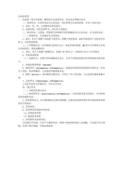
炎症的类型一、炎症的一般分类原则:概括的分为变质性炎、渗出性炎和增生性炎。
二、变质性炎:以变质变化为主的炎症,渗出和增生改变较轻微,多见于急性炎症。
1、部位:肝、肾、心和脑等实质性器官。
2、疾病举例:急性重型肝炎,流行性乙型脑炎。
三、渗出性炎:以浆液、纤维蛋白原和嗜中性粒细胞渗出为主的炎症,多为急性炎症。
(一)浆液性炎:以浆液渗出为其特征。
1、部位:发生于粘膜--浆液性卡他性炎;浆膜--体腔积液;疏松结缔组织--局部炎性水肿2、对机体的影响(二)纤维素性炎:以纤维蛋白原渗出为主,继而形成纤维素。
HE切片中纤维素呈红染交织的网状、条状或颗粒状。
1、部位:发生于粘膜--假膜性炎;浆膜--如"绒毛心";肺组织--见于大叶性肺炎2、对机体的影响(三)化脓性炎:以嗜中性粒细胞渗出为主,并有不同程度的组织坏死和脓液形成为特点。
1、表面化脓和积脓(empyema)2、蜂窝织炎(phlegmonous inflammation):指疏松结缔组织的弥漫性化脓性炎,常发生于皮肤、肌肉和阑尾。
主由溶血性链球菌引起。
3、脓肿(abscess):指局限性化脓性炎,可发生于皮下和内脏。
主由金黄色葡萄球菌引起。
4、出血性炎(hemorrhagic inflammation)上述各型炎症可单独发生,亦可合并存在。
四、增生性炎(一)非特异性增生性炎(二)肉芽肿性炎(granulomatous inflammation):以肉芽肿形成为其特点,多为特殊类型的慢性炎症。
1、肉芽肿的定义:由巨噬细胞及其演化的细胞,呈限局性浸润和增生所形成的境界清楚的结节状病灶。
2、常见病因3、肉芽肿的形成条件和组成(1)异物性肉芽肿(2)感染性肉芽肿4、肉芽肿性炎病理变化:以结核结节为例:中央--干酪样坏死;周围--放射状排列的上皮细胞;可见郎罕氏巨细胞;外围--淋巴细胞、纤维结缔组织。

细胞和组织的适应、损伤与修复萎缩:atrophy化生:metaplasia变性:degeneration坏死:necrosis坏疽:gangrene机化:organization肉芽组织:granulation tissue炎症炎症:inflammation变质:alteration渗出:exudation蜂窝织炎:phlegmonous inflammation 脓肿:abscess窦道:sinus瘘管:fistula肉芽肿:granuloma局部血液循环障碍淤血:congestion血栓形成:thrombosis血栓:thrombus栓塞:embolism栓子:embolus梗死:infarct肿瘤肿瘤:tumor异型性:atypia转移:metastasis癌:carcinoma肉瘤:sarcoma恶病质:cachexia心血管系统疾病动脉粥样硬化:atherosclerosis高血压:hypertension风湿病:rheumatism呼系统疾病大叶性肺炎:lobar pneumonia小叶性肺炎:lobular pneumonia原发综合症:primary complex消化系统疾病慢性消化性溃疡:chronic peptic ulcer病毒性肝炎:viral hepatitis肝硬化:liver cirrhosis假小叶:pseudolobule泌尿系统疾病肾小球肾炎:glomerulonephritis肾病综合症:nephritic syndrome病生健康:health疾病:disease脑死亡:brain death水肿:edema缺氧:hypoxia血氧饱和度:oxygen saturation心力衰竭:heaert failure呼吸衰竭:respiratory failure肺性脑病:pulmonary encephalopathy 肝性脑病:hepatic encepholopathy。


Histological types
1 Alterative inflammation
(1) Alterative changes are the most obvious, exudative and proliferative changes are slighter.
(2) Commonly seen in parenchyma organs
(3) Causes: Virus, toxin, chemical poison, etc.
(4) e.g: fulminant hepatitis, type B epidemic encephalitis, poliomyelitis, caseous pneumonia, etc.
2 Exudative inflammation
Excess of a particular component of the inflammatory exudates imparts distinctive features.
(1) Serous inflammation
①Watery, low protein content, derived from blood or serosal lining cells.
②Commonly seen in:
Mucous membrane, serosa, lung, loose connective tissue, skin.
③Examples: Blister formation following burning, the pleural effusion associated with tuberculosis, common cold, etc.
(2) Fibrinous inflammation
①Much fibrin due to coagulation of large fibrinogen outpouring.
②Causes: Shigella
Streptococcus peneumoniae
Corynebacterium diphtheriae
Hg poison
Uremia
③Commonly seen in
a. Serosa
b. Mucous membrane: Pseudo-membrane
c. Lung
④Examples: Rheumatic, pericarditis, dysentery, diphtheriae, lobular peneumonir, etc.
(3) Suppurative inflammation
①Definition: Much exudate with lots of neutrophils and liquefied nerotic tissue (pus) occurs. Pus composed of: dead and dying neutrophils, liquefied tissue, pyogenic organisms.
②Causes: staphylolcocci, pneumococci, gonococci, gram-negative rods, and some nonhemolytic streptococci.
③Types:
a. Abscess: a localised collection of pus in an organ or tissue.
Abscess could formation:
Ulcer: localized defect in an epithelial surface due to necrosis.
Sinus: an abnormal tract leading from a cavity to the surface.
Fistula: a tract open at both ends, through which abnormal communicaton between two surface is established.
b. Phlegmonous inflammation
Definition: wide-spread purulent inflammation in loose tissue, and appendix
Causes: hemolytic streptococci
c. Surface purulent inflammation and Empyema
Surface purulent inflammation: purulent inflammation of mucosal or serosa surface. Empyema: a collection of pus in a hollow viscus, e. g. in the gall-bladder or appendix.
(4) Haemorrhagic inflammation:
Inflammation associated with conspicuous haemorrhage as a result of vascular damage, e. g. meningococcaemia.
(5) Catarrhal inflammation:
Inflammation of mucosal surfaces with hypersecretion of mucus, e.g. common cold
3 Proliferative inflammation
(1) General proliferative inflammation
Proliferative constitutions:
Fibroblasts, endothelium, macrophage, and sometimes coated-epithelium, adenocytes, parenchymal cells, etc. commonly seen in chronic inflammation
(2) Special types
①Inflammatory granuloma (granulomatous inflammation)
Definition: Nodular area of histocytes (macrophages)
Proliferation that has been transformed into epithelioid cells, surrounded by a collar of lymphocytes, occasionally fibroblasts and plasma cells.
(3)Types:
①Infective granuloma:tuberculosis, syphilis, sarcoidosis, cat-scratch fever, leprosy, brucellosis, some of the mycotic infections, etc.
Example: granuloma of tuberculosis focal aggregation of monocyter that have undergone alteration to epithelioid cells. Or Caseous necrosis is in the center. The enclosing wall contains several large mutinucleate giant cells, the Langhans type, with peripheral orientation of the nuclel, and many lymphcytes and fibroblasts.
②Foreign body’s granuloma
Features: foreign bodies
Foreign dody giant cell: seen in association with particulate insoluble material. Nuclei scattered throughout cytoplasm. No caseous necrosis
③Inflammatory polyp
④Inflammatory pseudo-tumor。