医学影像学专业英语8Test Yourself On the ABCsOf Heart Disese
- 格式:ppt
- 大小:1.38 MB
- 文档页数:64
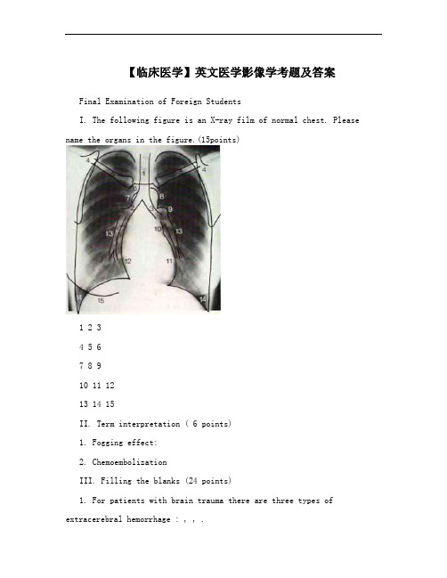
【临床医学】英文医学影像学考题及答案Final Examination of Foreign StudentsI. The following figure is an X-ray film of normal chest. Please name the organs in the figure.(15points)1 2 34 5 67 8 910 11 1213 14 15II. Term interpretation ( 6 points)1. Fogging effect:2. ChemoembolizationIII. Filling the blanks (24 points)1. For patients with brain trauma there are three types of extracerebral hemorrhage : , , .2. On X-ray film, there are four fundamental radiographic densities. Bones appear . Muscles appear . Fat appears .Air and gases appear .3. The indications of embolization conclude____________,______________, ______________, ________ ____, _______ ____, .4. The aim of inferior vena cava filter placement was to____________________. 5. Endovascular stent has two kinds of unfold types: _________ _and_______________.6. Angiography can be performed by ____________, _____________,_______________ technique.7. Give some example of non-vascular intervention:__________________,__________________, ______________________, ______________________.8. The main limitation of angioplasty was__________________________.IV. Answer the questions. (35 points)1. Please write down the common site of hypertensive cerebral hemorrhage and its CT appearance (10 points)2. Please write down CT and MRI appearance of meningioma. (14 points)3. What is the principle of transcatheter arterial infusion? (6 points)4. Please give an introduction of angioplasty and its main technique. (5 points)V. According to the clinical materials and radiologic examination , please write down the radiologic appearance and your impression. (20 points) 1.Case 1A 38-year-old man with a history of fever, cough, rusted sputum and left chest 9pain for one week. The white blood cell count was 12.0×10/L. Theposteroanterior and lateral view films as follows:2.Case 2A 34-year-old man, He suffered from low grade fever in the afternoon, cough, weakness for half a month. The findings of CT scan as follows:3.Case 3A-70-year-old femal with a history of colon carcinoma.4.Case 4A 38-year-old man with myasthenia gravis for half a year.5.Case5A 68-year-old female with a history of smoking for 30 years. The clinical symptom was cough, hemoptysis for two months.2002-2003 Final Examination of Foreign Students (Answers)I.1 trachea2 right main bronchus3 left main bronchus4 scapula5 clavicle6 sternum7 azygos vein8 aortic arch9 left pulmonary artery 10 upper left cardiac border 11 lower left cardiac border 12 right atrium 13 lower lobe arteries 14 lateral costophrenic angle 15 breast II.1. Fogging effect: During 2~3 weeks after cerebral infarction, the density of the infarcted lesion increased and became isodense. This is due to reduction in edema ,leakage of proteins from cell lysis and invasion of macrophage.2. Chemoembolization: A technique that combines transcatheterarterial infusion of chemotherapy drugs and embolization.III.1. epidural hemorrhage, subdural hemorrhage, subarachnoid hemorrhage2. black, gray-black, gray, white3. control of hemorrage, devascularization of tumors,. ablation of organ or tissue, varicocele,modification of blood flow, perigraft leak and back bleeding, following stent-graft,localization of small bowel angiodysplasia (choose 5)4. prevent the pummonary embolism5. balloon-expandble stent, self-expandable stent6.DSA, MRA, CTA7. percutaneous biopsy, percutaneous drainage & aspiration, transcatheter dilate nonvasclular lumen, percutaneous interstitial therapy8. restenosisIV.1.Very common reason is hypertension and the common site are basal ganglion and thalamus.Acute hemorrhage: hyperdense, 60-80Hu ( hemoglobin )Subacute hemorrhage: attenuation decreased with time (1.5 Hu per day);start periphery progress centrally; isodense between 1 to 6 weeks; peripheral enhancementChronic hemorrhage: hypodenseResidua of hemorrage: hypodense foci (37%); slitlike lesion (25%); calcification (10%); no abnormal (27%)2.CT: Sharp circumscribed round or smoothly lobulated mass;Connected with dura or inner table with a broad bas.Homogenous hyperdense (75%), isodense.Calcification 20~25%,diffuse, focal, sandlike, sunburst, globular, rimlike. Bone destructure or hyperosteogenySoft tissue mass in scalpEdema: non except for venous or sinus compressedNecrosis or cyst formation : seldom 8~25%Hemorrhage:uncommonEnhancement :obvious and uniform enhancementMRI: Isointense with gray matter on T1WI ,T2WI;CSF cleft and pial vessels around tumor; Flow void vessels within tumor;Enhanced rapidly and intensely,Dural tail sign. Dura adjacent to tumor show liner enhancemet. suggestive not specific for meningioma, also can be seen in schwannoma, gliblastoma multiforme, metastases3. Drugs administrated through veins were diluted, combined with protein, the level of drugs in the target organ is low, untarget organs was also affected, side effects was seriousTAI increase the level of drugs in the target organ, prolong the contact time with lesion, enhance the effectiveness, lower the side effects4. Ballon angioplasty. Endovascular stent. Atherectomy/thrombous removel. Laserangioplasty. Ultrasound angioplasty.V.1.case1: left lower lobe; confluent patchy opacities; obliterate the posterior left hemidiaphragm on the lateral view; impression: left lower lobe pneumonia2.case2: multiple(diffuse); small(military) nodules throughout both lungs; homogeneous density; impression: military tuberculosis3.case3: multiple nodules; bilateral lung fields; well-defined margins; homogeneous density; impression: pulmonary metastases4.case4: anterior mediastinum(anterior to the great vessels); mass; well-defined margins; soft tissue density; homogeneous density; impresson: thymoma5.case5: right hilum enlarged; the wall of the bronchus thickened; soft tissue density mass; mediastinal and right pulmonary artery involvement; obviously and heterogeneously enhancement; paratracheal and subcarinal lymph nodes enlarged; impression: right central bronchial carcinoma involved mediastinal and right pulmonary artery, mediastinal lymph nodes metastases。
医学影像学简答题1(Medical imaging simplified answer 1)3. Short answergeneral1. Brief description of medical X-ray characteristicsThe X ray is an electromagnetic wave with penetration; Fluorescence effect; Photographic effects and biological effects. Its penetration is related to material density, thickness and wavelength of X ray, and fluorescence effect is the basis of fluoroscopy. Photographic effect is the basis of X-ray photography; The ionization effect, which involves changes in human biology, is the basis of radiological protection and radiotherapy.2. The basic principle of X-ray imaging is describedOn the one hand, it is based on the penetration of the X ray, the fluorescence effect and the photographic effect, and on the other hand, the difference between the density and thickness of the body tissue. When the X rays penetrate various tissues of the human body, it is absorbed in different degrees so that there is a difference in the amount of X on the screen or in the X-ray. This allows for contrast between the black and white contrast on the screen or in the X-ray.Bones, joint systems1. The X-ray performance of acute and chronic suppurative osteomyelitisThe suppurative osteomyelitis is caused by staphylococcus aureus in the bone marrow, good hair in children and juvenile, long backbone epiphysis good hair. The early (2 weeks) may have the following soft tissue changes: 1. Two, subcutaneous tissue and muscle intersections blurred, bone may not have obvious change. Bone changes were seen after 2 weeks. The localized osteoporosis was started in the epiphyseal cancellous bone. Subsequently, most dispersed irregular form of bone destruction. Edge blur. In the future, the bone destruction area may merge into a large area of destruction. And gradually extend to the backbone. Can be accompanied by pathological fracture. Osteocortical destruction can form the subperiosteum abscess and stimulate the periosteum to cause periosteal hyperplasia. The new bone with low density is parallel to the backbone. Later, as the course of illness extended. The new osteogenesis is obvious and can form the shell. Osteonecrosis is caused by the emergence of periosteum and thrombotic arteritis. The X-ray shows the dead bone formed along the axis of the bone, which is very dense. If the lesion is close to the joint, the abscess can destroy the bone cortex of the dry epiphysis and enter the joint synovial card. Cause suppurative arthritis. The X line is the swelling of the joint capsule. The gap in the joint is widened early and even dislocated. Late narrowing. Osteopenia. When acute suppurative osteomyelitis is not treated promptly and adequately. It can be transformed into chronic suppurative osteomyelitis. The X-ray showed a large number of osteogenic hyperplasia, thickening of periosteum and fusion with cortex, which was stratified or lacy, thickening of the bony cortex, and narrowing of the medullary cavity. The backbone thickened. Irregular appearance, if not recovered,can still be seen bone destruction and dead bone.2. Describe the X - ray performance of the spine tuberculosisIt is the most common person of bone and joint tuberculosis. Good for children and young people. With lumbar multiple hair. The X - line performance is mainly osteoporosis and cancellous bone fracture. The attachment is less cumulative. Vertebral bodies often collapse due to bone damage, flattening or wedges. When the lesion is involved in the vertebral body, the lower margin of the bone. The rupture of the intervertebral cartilage plate is caused by the use of the broken cortex. When the intervertebral disc is invaded, the intervertebral space is narrowed. Even disappearing, the adjacent vertebral bodies are embedded and fused. At the same time, the lesion can produce a large number of caseous necrosis material in the destruction of bone, and the cold abscess is formed in the soft tissues around the spinal column.The X line is characterized by the presence of a fusiform soft shadow on both sides of the vertebral body, known as a lateral abscess. In addition, due to pathological fracture. The lateral spine of the spine can be seen to change the curvature of the spine. The post-emergence deformity.3. Test the X-ray performance of vitamin D deficiency ricketsBecause vitamin D is not a cause of calcium and phosphorus metabolism, osteoid tissue in bone is deficient in calcium salt deposits. Systemic metabolic bone disease. The X-ray showed a decrease in bone density in the general bone. Bone trabeculaeare rare, fuzzy, margin roughness, bony cortex thinned, stratified change. In the areas where bone metabolism is more active, such as the occurrence of bone epiphysis, the low density edge is blurred, and the epiphyseal calcification zone is irregular, blurred, thin and disappeared. In the middle of the epiphyses, there is a tortuous deformation in the middle of the epiphysis, with a very irregular margin and a hairbrush shape. The gap between the epiphysis and the epiphysis is widened. The corner of the epiphysis was altered by bone spur. The front of the thoracic ribs is a wide mouth. At the same time, the weight-bearing long bone is often bent and deformed. (O leg, X leg, etc.), a small number of patients can have a blue branch fracture healing X-ray performance: the temporary calcification belt reappears, the cup mouth shape depression and brush change are relieved and disappeared. The epiphyseal space is normal. Bone density increases and bone cortex thickens. Bone epiphysis increases, the density increases, and the bone deformation is prolonged.4. Test the differential diagnosis of benign and malignant bone tumors from the characteristics of X-ray.Benign:No transfer: no transfer.Growth condition: slow growth, non-invasion and adjacent tissues, but can be oppressive.Local bone changes: swelling bone damage, clear line with normal bone, sharp edge, thinning of the bony cortex, andexpansion can maintain continuity.Periosteum hyperplasia: generally no periosteal proliferation, can have a small amount of periosteum hyperplasia after pathological fracture, and periosteal new bone is not damage the surrounding soft tissue changes: no swelling or lump shadow more, if there are any lump, the edge is clear.Angiography: vascular differentiation is normal, and the tumor can be used to compress blood vessels.Malignant:Transfer: transfer.Growth: rapid growth, transsexual and adjacent tissue organs.Local bone changes: invasive bone fracture, blurred boundary and normal bone boundary, uneven edges, irregular fracture and defect, and bone formation of tumor.Periosteal hyperplasia: multiple forms of periosteal hyperplasia, and can be destroyed by tumor.The surrounding soft tissue changes: the growth of the soft tissue is not clear from the surrounding tissue.Angiography: it can be seen that tumor blood vessels are more and more disordered, the tumor staining and arteriovenous fistula, and the blood supply artery thickening and the blood vessel erosion become rigid, and the edge damage and so on.5. Take the femoral neck fracture as an example.1. Delayed healing or non-healing of fracture; X - ray showed delayed bone scab, with few or no presence, delayed or prolonged fracture line.Two, false joint formation: the X ray shows the bone bushy with the end of the bone, and there is a clear line between the two sides of the broken end.3. Fracture deformity healing: X - ray shows bone formation Angle, rotation, shortening deformity.4. After trauma, osteoporosis.5. Bone and joint infection; For acute chronic bone, arthritis X ray performance.6. Bone ischemic necrosis: increased femoral bone density and deformation.7. Joint rigidity: it is caused by adhesion to the joint, often with osteoporosis and soft tissue atrophy.Viii. Degeneration of joints: change after chronic bone injury.9. Ossified myositis: calcification in different degree of soft tissue after fracture.The respiratory system1. What methods are used in chest imaging examination?1. Chest perspective 2, (positive and lateral) 3, high - kilovol-meter 4, body layer photography 5, bronchography 6, CT 7, MRI2. What are the basic X-ray manifestations of lung lesions?A, exudative lesions: show the edge blur, density uniform shape shadow, range from flocculus to big leaf, when lesions involving the big leaf, its shape is in line with lung and sharp edges, air-bronchogram and visible.2. Fibrosis change: the expression is high density, the boundary is clear, walking rigid, irregular shape of the line shape.Iii. Proliferative lesion: localized nodules or petals, with high density, relatively clear edges, and generally no fusion trend.4. Calcified venereal changes: the appearance is sharp, the density is extremely high, the shape is different, the size of the speckle shape or plaque shape.Voids: 1. Wormwood vacuous cavity: manifested in a large number of pulmonary real changes with multiple small permeable areas. The form is irregular, it is wormlike. 2. Thin wall cavity: hollow wall thickness < 3mm, boundary clear, smooth circular light zone. 3. Thick wall hole: wall thickness > 3mm, the holeis round or irregular, peripheral or unreal change area, the inner wall is smooth and neat or concave, the hole can have or airless plane.Six, mass lesions, benign tumor characterized by round or oval, smooth boundary, density uniform spherical density shadow, malignant tumors are lobulated, the boundary is not sharp, can have a short nap or umbilical concave), central necrosis.3. With a solid shadow on one side of the chest, which diseases should be considered? What aspects should be analyzed in the identification?One, a large number of pleural effusion, one side of the lung, the one side of the lung, the one side of the pleural hypertrophy, the one side of the pleural hypertrophy, the one side of the lung, the one side of the lung and the one side of the lungShould note: when identifying a, mediastinal position 2, diaphragmatic level three, five, four, thoracic rib gap width size on a flat piece of six, observe whether air-bronchogram, observe whether the main bronchus is unobstructed in layer 7, combined with clinical data4. What are the direct and indirect X-ray signs of bronchial lung cancer (central type)?1. Lumps, located in the lung area, are rounded or lobule.2. Endobronchial polyps filling defect.3. The bronchial wall thickened and the lumen was narrow or blocked, with rat tail or cup.Ii. Indirect symptoms: 1. Obstructive pulmonary disease,The horizontal and pulmonary masses of the upper lobe of the upper lobe of the upper lobe of the upper lobe.2. Obstructive pneumonia: repeated attacks and slow absorption of exudative lesions.3. Obstructive emphysema: the air volume of the blocked lung is increased, and the brightness is increased.5. Typical X-ray manifestations of large leaf pneumonia?Lobule pneumonia may involve most or all of the lobes. The former is characterized by uniformity of density, and the shadow of the edge is indistinct. The edge is clear, with the interleaf crack as the boundary, its shape with the pulmonary lobe, the contour is consistent, its inside visible bronchi meteorology. Different forms of lobule pneumonia vary.6. Typical X-ray manifestations of acute hemorrhagic disseminated tuberculosis?The early two lung density of the lesion showed a change of hair glass. In about 10 days, the two lungs showed diffuse uniform distribution, the same size, uniform density of miliary nodules. The two lung textures are not clear.The circulatory system1. Simple mitral stenosis X-ray performance?The heart increases, the left atrium and right ventricle are enlarged, and the left heart is often significantly enlarged.The main reason for the reduction of the general aortic ball is the reduction of left ventricle blood elimination, aortic dysplasia or the left rotation of the heart and big blood vessels, and the aortic arch folds.The left ventricle shrank, the apex of the heart moved, the lower part of the heart was straight.4. Mitral membrane calcification, direct sign.5. Pulmonary congestion or interstitial edema, upper pulmonary vein dilation, lower pulmonary veins. Sometimes it can be seen that the diameter of 1 ~ 2mm in diameter can be seen in the lung field, which is composed of hemosiderosis.2. The X line of high blood heart disease is shown as?One, the heart is aortic type, the left ventricle segment increases, becomes round, the heart apex is in the phrenic, the cardiac phrenic horn shows acute Angle, the left ventricle is prominent, overlaps with the spine.Second, the left ventricle is increasing to the left, and the apex of the heart is often under the diaphragm.3. The perspective can be seen that the opposite pulsation.When left heart failure, the left atrium increases, and pulmonary congestion and pulmonary edema appear.5. Severe, the heart is generally enlarged, but the left ventricular enlargement is the main.The aorta has dilation, extension, and circuity.3. X-ray performance of pulmonary heart disease?Changes in pulmonary hypertension and chronic pulmonary diseaseOne, pulmonary hypertension, often occurs before the heart shape changes.Second, the right ventricle enlarges, the heart is in the mitral valve type, the heart rate is more than the normal person not much. Some cases: the heart is smaller than normal, and is related to the low level of the pulmonary emphysema.Three, chronic pulmonary disease, chronic bronchitis, extensive lung tissue fibrosis and emphysema.4. X ray performance of congenital heart disease atrial septal defect?When the defect is small, the size and shape of the heart andshape are normal or change.The heart is of mitral valve type, often moderate increase.Two, right atrium and right ventricular enlargement,The major characteristic changes of atrial septal defect were significantly increased in the right atrium.3. The pulmonary artery protruding, the pulsating enhancement, the pulmonary portal angiectasia. There are often lungmen dancing.In the left atrium, the left ventricle and aorta decreased, while the first left ventricle enlarged.5. Pulmonary hyperemia and later pulmonary hypertension.5. The X ray performance of common Fallot tetralogy?1. The heart is generally not enlarged, the heart is blunt, the upper warped is a sheep's nose, the heart lumbar depression, if there is a third ventricle forming, the heart is flat, or slightly raised.Second, the right ventricle increases.The left ventricle narrowed with decreased blood flow, the left atrium was generally unchanged, and the right atrium was mild to moderate due to increased blood flow and increased right ventricular pressure.4. The lung door shrinks and the lung vessels are slim.The aorta is widened and shifted to the right.The digestive system1. According to what characteristics can the organ of the digestive system be divided into two categories? Where are the organs?According to the characteristic of the digestive organ is the real organ or the hollow viscera, the digestive organ is divided into two categories. The liver and pancreas belong to the substantial organ. Esophagus, stomach, duodenum, large, small intestine and biliary system belong to hollow viscera.2. What kind of inspection methods and imaging methods are used in the two main types of digestive tract and cavity?The liver and pancreas of parenchyma were mainly used for CT, ultrasound and mri. After the general sweep; When necessary, CT iodine contrast agent was enhanced, and magnetic resonance was enhanced with gadolinium contrast agent.The hollow viscera was mainly used for routine X-ray examination, the gastrointestinal tract was radiographed by barium, and the bile was used for the contrast of iodine3. The X-ray signs of benign and malignant ulcers are identified.A benign ulcer protrudes from the gastric cavity. The ulcer is located within the contour of the stomach2. The shape of the shadow: the benign ulcer is relatively small and round, and the malignancy is larger and more shallow.Three, niche mouth: benign ulcer with mucosal edema, width is consistent, sometimes under pressure to change form malignant ulcer niches mouth cancer tissue invasion, forming ring levee involuntary pressure, change or more cancer nodules form refers to the indentation, sharp corners.Iv. Benign ulcer stomach constriction peristaltic direct niches, malignant ulcer is more than 1 cm from the niches, peristalsis disappears.4. Differentiation of esophageal foreign body and trachea foreign bodyTake the coin foreign object as an example, because the diameter of the esophagus is small, the left and right diameters are wider, so the esophageal foreign body is in a circular position, and the lateral position view is striped. The trachea foreign body is opposite, because the trachea half annular cartilage is absent is facing the rear, so the maximum diameter of the foreign body is the front and rear direction. The positive view is long and long, while the lateral position is round.5. Identification of jejunum, ileum and intestinal obstruction? How to diagnose low - level intestinal obstruction based on flatslice?The intestinal mucosa is a fish-bone arrangement perpendicular to the vertical axis of the intestinal tube. The mucosa of ileus is only two intestinal wall lines. The most significant expansion of the obstruction tube diameter is the semilinar fold.The high intestinal obstruction is mainly manifested in the left middle and upper abdominal multiple qi level, the stomach also sees the liquid level, the lower abdomen and the pelvic cavity of the lower abdomen and the lower gas. Low intestinal obstruction, the expression is the whole abdomen several stair - shaped gas levelUrinary system1. Differential diagnosis of urinary calculus. (points)1. Gallstones: the form is polygon, the surrounding density is high, the central density is low, sometimes there is the high density core. Lateral photography is located in front of the spine.Lymph node calcification: form irregular punctate, structure, and has no fixed position, to move a large degree (e.g., mesenteric lymph node calcification) imaging of the renal pelvis can understand outside or in the urinary tract.3. Intestinal contents (coprolites or drugs) : the position is not constant, the repeated photo position can be changed ordisappeared, and the bowel will disappear.Iv. Venous stone (pelvic cavity) : small, round, circular or concentric round dense shadow, the edges are neat, often for both sides and multiple, the position is more than partial, when necessary retrograde contrast imaging is identified.2. X-ray manifestations of renal tuberculosis. (points)Flat slice: the kidney contour area can protrude, terminal form shrinksCalcification: diffuse, cloudy, spottedAngiography: wormhole destruction, renal cortical abscess and vacuous formation, pyelonephrosis, renal pelvis, renal calyx (peripheral imformation, deformed stenosis), renalself-truncation3. The X-ray of typical urinary calculi. (points)Kidney: sliced: mulberry, layered, antlerContrast: density, higher density, filling defect, obstructionUreter: flat slice: the long axis is consistent with the ureterAbdominal segment: side of the lumbar spineThe sacroiliac segment: the sacroiliac jointPelvic segment: roughly parallel to the pelvic rimLower end of ureter: polymorphismContrast: positive, negative and catheter relationship, obstruction of waterBladder calculi: above the symphysis pubis, the midline of the pelvic cavity changes with positionUrethra: the posterior urethra: the symphysis of the pubic bone and the posterior urethra4. Several common radiographic reflux X - ray manifestations.1. Tubule reflux: the radiate dense shadow radiated from the center of the kidney to the cortex.Second, the kidney sinus reflux: it appears as the irregular Angle or band dense shadow around the fornix, and the author appears in an irregular shape.Iii. Circumfluence of the blood vessels: the arch of the arch is shown as the arch of the arch.Iv. Lymphatic reflux: it is shown as a slender, meandering, curved silhouette that walks in the direction of the renal gate.5. Various imaging examinations and USES of urinary system. (points)IVP: the shape of the renal pelvis, renal calices, ureters, and bladder, and the function of the renal excretion2. Retrograde pyelography: used for IVP display (such as renal dysfunction) or not for IVP (such as liver and kidney function, iodine allergy)Bladder angiography: excretory method: the urethral stricture cannot be intubated or at the same time, the upper urinary tract should be examinedRetrograde: observe bladder size, shape, position to diagnose bladder disease4. Urethrography: mostly used for urethral stricture, calculi, congenital malformation, etc5. Retroperitoneal aerated angiography: showing the renal, adrenal profile and retroperitoneal mass and the relationship with the kidney6. Arteriography: diagnosis of vascular lesions and adrenal neoplastic lesionsCentral, five official system1. Evaluation of CT in the treatment of sinus tumors.CT diagnosis of smaller tumors is of great value and can be determined in its origin and scope. Benign tumor margins are clear and orderly, without bone damage. But it is difficult todetermine the pathological nature. The mucous cyst showed an enlarged sinus cavity and increased density. CT is of great value in diagnosis of malignant tumor. In the early stage of osteopenia, there was a shadow of mass in the sinus cavity, and the sinus cavity was seen in the sinus cavity. The sinus wall can be damaged early, and the adjacent structure can be shown as the nasal cavity, the invasion of the orbit and the scope.2. CT manifestations of meningiomas.The CT findings of typical meningiomas are high in density, with clear edges, spherical or subleaf lesions, and cranial bones, which are connected to the cerebellum. There was no edema or slight edema in the oven. The general performance of the enhanced scanning was significantly enhanced.。

医学影像技术专业英语课后题答案1、28.The question is very difficult. ______ can answer it. [单选题] *A.EveryoneB.No one(正确答案)C.SomeoneD.Anyone2、She is a girl, _______ name is Lily. [单选题] *A. whose(正确答案)B. whoC. whichD. that3、She found her wallet()she lost it. [单选题] *A. where(正确答案)B. whenC. in whichD.that4、He is going to _______ a party this evening. [单选题] *A. hold(正确答案)B. makeC. needD. hear5、The rain is very heavy _______ we have to stay at home. [单选题] *A. butB. becauseC. so(正确答案)D. and6、I’m still unable to make myself_____in the discussion, which worries me a lot. [单选题]*A.understandB.understood(正确答案)C.understandingD.to be understood7、I can’t hear you _______. Please speak a little louder. [单选题] *A. clearly(正确答案)B. lovelyC. widelyD. carelessly8、18.Monica wants to be a _______. She is good at sports and she loves teaching others. [单选题] *A.coach(正确答案)B.secretaryC.architectD.waiter9、Neither she nor her friends ______ been to Haikou. [单选题] *A. have(正确答案)B. hasC. hadD. having10、The young man had decided to give up the chance of studying abroad, _____ surprised his parents a lot. [单选题] *A. whenB. whereC. which(正确答案)D. that11、The Chinese team are working hard _______ honors in the Olympic Games. [单选题] *A. to win(正确答案)B. winC. winningD. won12、I don’t like snakes, so I ______ read anything about snakes.()[单选题] *A. alwaysB. usuallyC. oftenD. never(正确答案)13、Can I _______ your order now? [单选题] *A. makeB. likeC. giveD. take(正确答案)14、You have coughed for several days, Bill. Stop smoking, _______ you’ll get better soon. [单选题] *A. butB. afterC. orD. and(正确答案)15、We _______ play basketball after school. [单选题] *A. were used toB. used to(正确答案)C. use toD. are used to16、He is a student of _______. [单选题] *A. Class SecondB. the Class TwoC. Class Two(正确答案)D. Second Two17、Simon does not()his fellow workers because they often argue over trivial matters. [单选题] *A. get on with(正确答案)B. come up withC. do away withD. go on with18、This species has nearly ()because its habitat is being destroyed. [单选题] *A. used upB. died out(正确答案)C. gone upD. got rid of19、We had a(an)_____with him about this problem last night. [单选题] *A.explanationB.impressionC.exhibitionD.discussion(正确答案)20、( ) _____ New York _____ London have traffic problems. [单选题] *A. All…andB. Neither….norC. Both…and(正确答案)D. Either…or21、13.—Will you come to my party?—I am not ________ . [单选题] *A.mindB.sure(正确答案)C.happyD.Sorry22、My brother is _______ actor. He works very hard. [单选题] *A. aB. an(正确答案)C. theD. one23、He went to America last Friday. Alice came to the airport to _______ him _______. [单选题] *A. take; offB. see; off(正确答案)C. send; upD. put; away24、_________ we don't stop climate change, many animals and plants in the world will be gone. [单选题] *A.AlthoughB.WhileC.If(正确答案)D.Until25、--Jimmy, you are supposed to?_______ your toys now.--Yes, mom. [单选题] *A. put upB. put onC. put away(正确答案)D. put down26、In 2019 we moved to Boston,()my grandparents are living. [单选题] *A. whoB. whenC. where(正确答案)D. for which27、78.—Welcome to China. I hope you'll enjoy the ________.—Thank you. [单选题] *A.tour(正确答案)B.sizeC.nameD.colour28、A?pen _______ writing. [单选题] *A. is used toB. used toC. is used for(正确答案)D. used for29、______this story, and you will realize that not everything can be bought with money. [单选题] *A. ReadingB. ReadC. To readD.Being read(正确答案)30、His new appointment takes()from the beginning of next month. [单选题] *A. placeB. effect(正确答案)C. postD. office。
期末考试批次专业:202001-医学影像技术(专升本)课程:大学英语3(专升本)1. (单选题) The room is comfortable ____.( )(本题2.0分)A、to liveB、to live inC、livingD、living in标准答案:B2. (单选题) I command that all spindles in my kingdom ____ ( )(本题2.0分)A、burnB、must burnC、be burnedD、must be burned标准答案:C3. (单选题) We ____ separate and say good-bye to each other sooner orlater.( )(本题2.0分)A、boundB、bound toC、are bound toD、are bound标准答案:C4. (单选题) Such a company collects ____ information about men and women.( )(本题2.0分)A、a great deal ofB、manyC、a great number ofD、 a few标准答案:A5. (单选题) I regretted ____ in my Sunday best.( )(本题2.0分)A 、not dressed B 、having not dressed C 、dressed not D 、 not having dressed标准答案:D6. (单选题) He kept the bag ____ so that his sister couldn ’t see the modest gift.( )(本题2.0分)A 、close B 、closing C 、closed D 、 to close标准答案:C7. (单选题) He was dressed very well, ____ to me.( )(本题2.0分)A 、to compare B 、compared C 、comparing D 、 compare标准答案:B8. (单选题) The bottle of soap bubble liquid ____ 57 cents.( )(本题2.0分)A 、spent B 、cost C 、took D 、 paid标准答案:B9. (单选题) I ____ wrong before.( )(本题2.0分)A 、must count B 、must have counted C 、must be counting D 、 must be counted标准答案:B10. (单选题) The doctor ’s report ____ that her death was due to heart disease.( )(本题2.0分)A、indicatingB、indicatedC、indicatesD、will indicate标准答案:B11. (单选题) Medical researchers reached the ____ long ago that smoking is aserious hazard to health.( )(本题2.0分)A、decisionB、investigationC、conclusionD、explanation标准答案:C12. (单选题) George Washington, the first President of the U.S., is known ____the Father of His Country.( )(本题2.0分)A、forB、likeC、asD、because标准答案:C13. (单选题) They have invented a new ____ washing machine.( )(本题2.0分)A、typesB、kinds ofC、kindD、type of标准答案:D14. (单选题) The movie we are going to see is said to be ______ the life story of an American general.( )(本题2.0分)A 、based on B 、said about C 、talked on D 、 spoken about标准答案:A15. (单选题) The fences along the middle of the road is intended to protect vehicles from crashing ____ each other.( )(本题2.0分)A 、on B 、into C 、with D 、 onto标准答案:B16. (单选题) Supporters of gun control have worked for many years to ban the sale of ____ handguns in America.( )(本题2.0分)A 、dead B 、dying C 、died D 、 deadly标准答案:D17. (单选题) My native town, which was ____ rather small, has now been built into one of the biggest cities in the province.( )(本题2.0分)A 、occasionally B 、originally C 、suddenly D 、 terrible标准答案:B18. (单选题) The speaker said something about the actors first and then _____ to talk about the film.( )(本题2.0分)A 、go on B 、proceededC 、produced D 、 went on with标准答案:B19. (单选题) We had a long debate _____ whether we should spend so much money on space technology.( )(本题2.0分)A 、as to B 、so as C 、as about D 、 so as to标准答案:A20. (单选题) Henry couldn ’t take part in the sports meeting ____ his leg got broken.( )(本题2.0分)A 、so B 、because C 、because of D 、 so that标准答案:B21. (单选题) The earlier the better, as far _____ I am concerned.( )(本题2.0分)A 、as B 、so C 、so that D 、 such that标准答案:A22. (单选题) The meeting was ___ held the following week.( )(本题2.0分)A 、been B 、to C 、to be D 、 being标准答案:C23. (单选题) The girl was _____ into accepting the gift.( )(本题2.0分)A、saidB、toldC、spokenD、talked标准答案:D24. (单选题) By careful examination, the doctors hope to ____ the source ofthe infection.( )(本题2.0分)A、make sureB、set offC、get upD、track down标准答案:D25. (单选题) The coat fits the boy perfectly now, but he will ____ it in a year’s time.( )(本题2.0分)A、outgrowB、up growC、out growD、upright标准答案:A26. (单选题) Johnny’s mouth watered _____ the sight of the big pudding.( )(本题2.0分)A、atB、inC、onD、for标准答案:A27. (单选题) ______ must be done _____ the house before we can live in it.( )(本题2.0分)A、Repairing, toB、Repairs, withC、Repair work, withD、Repairs, to标准答案:D28. (单选题) He knew that ______ him to help her, she could and wouldsucceed.( )(本题2.0分)A、withoutB、withC、inD、for标准答案:B29. (单选题) A fresh idea occurred ______ me at the sight of her coming.( )(本题2.0分)A、inB、fromC、withD、to标准答案:D30. (单选题) The accident resulted ______ the death of two passengers.( )(本题2.0分)A、inB、toC、byD、at标准答案:A31. (阅读理解题)32.After practicing as a surgeon for several years. Dr. Ginoux decided to apply for membership in the American College of Surgeons(美国外科医生学会), a highly selective and distinguished professional organization.As part of the application procedure(手续), Dr. Ginoux was asked to prepare a list of all the operations performed in the precious seven years. Slowly, as she worked on the longlist, she began to feel uncertain. She began to question some of her decisions. Had she used the best technique in that case? Maybe, in this case, she should have run one more test before operation? On the other hand, maybe she should have… Would the doctors on the selection committee understand that, as the only trained surgeon in the area, she usually could not get advice from others and therefore, had to rely completely on her own judgment? For the first time, Dr. Ginoux felt lonely and isolated.The longer Dr. Ginoux worked on the application forms, the more depressed she became. As hope faded, she wondered if a “country doctor” had a realistic chance of being accepted by the American College of Surgeons.(1). (单选题) Dr. Ginoux was working in _______ .( )(本题4.0分)A、a large cityB、the American College of SurgeonsC、an area far from any big cityD、 a selective organization标准答案:C(2). (单选题) The application forms must include _______ .( )(本题4.0分)A、the decision procedureB、a record of all the operationsC、the best techniqueD、 a list of advice and judgments标准答案:B(3). (单选题) It was most probable that Dr. Ginoux was _______ .( )(本题4.0分)A、 a member in that organizationB、 a well-trained surgeonC、 a graduate from the American College of SurgeonsD、 a distinguished surgeon in America标准答案:B(4). (单选题) When she was filling the application forms, Dr. Ginoux began to be ______ .( )(本题4.0分)A、realisticB、distinguishedC、perplexedD、decisive标准答案:C(5). (单选题) When filling the forms, Dr. Ginoux felt depressed because______ .( )(本题4.0分)A、she didn’t perform enough operationsB、some operations were unsuccessfulC、she didn’t get advice from the selection cmmitteeD、she was doubtful about her operations标准答案:D32. (阅读理解题)33.Ask three people to look out the same window at a busy street corner and tell you what they see. Chances are you will receive three different answers. Each person sees the same scene, but each perceives something different about it.Perceiving goes on in our minds. Of the three people who look out of the window, one may say that he sees a policeman giving a motorist a ticket. Another may say that he sees a rush-hour traffic jam at theintersection. The third may tell you that he sees a woman trying to cross the street with four children in tow. For perception is the minds’ interpretation of what the senses ---- in this case our eyes ----- tell us.Many psychologists today are working to try to determine just how a person experiences or perceives the world around him. Using a scientific approach, these psychologists set up experiments in which they can control all of the factors. By measuring and charting the results of many experiments, they are trying to find out what makes different people perceive totally different things about the same scene.(1). (单选题) Seeing and perceiving are _____ .( )(本题4.0分)A、the same actionB、two separate actionsC、two actions carried on entirely by the eyesD、several actions that take place at different times标准答案:B(2). (单选题) Perceiving is an action that takes place( )(本题4.0分)A、in our eyesB、only when we think very hard about somethingC、only under the direction of a psychologistD、in every person’s mind标准答案:D(3). (单选题) Perception involves what ______ .( )(本题4.0分)A、our senses tell usB、our minds interpretC、we see with our eyes onlyD、Both标准答案:D(4). (单选题) People perceive different things about the same scene because ______ .( )(本题4.0分)A、they see different thingsB、they cannot agreeC、some have better eyesightD、None of these标准答案:D(5). (单选题) The best title for this selection is _____ .( )(本题4.0分)A、How We SeeB、Learning About Our Minds Through ScienceC、What Psychologists PerceiveD、How To Become An Experimental Psychologist标准答案:B。
医学影像学专业的英语Medical Imaging Major in EnglishMedical imaging refers to the techniques and processes used to create images of the human body for clinical purposes. It plays a crucial role in the diagnosis and treatment of various medical conditions. The field of medical imaging has developed rapidly in recent years, with new technologies and techniques constantly being introduced to improve the quality and accuracy of diagnostic images.Students majoring in medical imaging are required to have a solid understanding of both medical concepts and technical skills in order to successfully pursue a career in this field. The curriculum of a medical imaging major typically includes courses in anatomy, physiology, radiographic imaging, medical terminology, and patient care. In addition, students also learn how to operate imaging equipment such as X-ray machines, CT scanners, MRI machines, and ultrasound machines.One of the challenges faced by students studying medical imaging is the need to develop proficiency in the English language. English is the international language of medicine, and healthcare professionals around the world use English to communicate with each other and share research findings. As a result, students majoring in medical imaging must be able to read, write, and speak English proficiently in order to succeed in their academic and professional endeavors.To help students improve their English language skills, many universities offer English language courses specifically designed for medicalimaging majors. These courses cover a wide range of topics, including medical vocabulary, professional communication skills, and academic writing. By enrolling in these courses, students can enhance their ability to understand and communicate complex medical concepts in English.In addition to formal English language courses, students majoring in medical imaging can also benefit from self-study methods to improve their English proficiency. Reading medical journals, watching educational videos, and participating in online forums are all effective ways to practice English language skills in a medical context. By incorporating English language learning into their daily routine, students can gradually build their confidence and proficiency in using English for academic and professional purposes.Furthermore, students majoring in medical imaging can also take advantage of international exchange programs and study abroad opportunities to immerse themselves in an English-speaking environment. By studying in countries where English is the primary language, students can gain firsthand experience using English in real-world medical settings and interactions. This can greatly enhance their language skills and cultural competence, making them more competitive in the global healthcare industry.In conclusion, proficiency in the English language is essential for students majoring in medical imaging to succeed in their academic and professional careers. By actively engaging in English language learning activities, taking advantage of specialized courses, and participating in international exchange programs, students can develop the language skillsneeded to excel in the field of medical imaging. With dedication and practice, students can confidently navigate the world of medical imaging, communicate effectively with colleagues and patients, and contribute to the advancement of healthcare worldwide.。
医学英语专业八级Medical English is a specialized field of study that focuses on the language used in the medical profession. It is designed for individuals who are studying or working in healthcare, such as doctors, nurses, and medical researchers.The Medical English Proficiency Test, also known as the Medical English Level 8 Exam, assesses a person's ability to understand and communicate in English within a medical context. The exam covers various topics related to healthcare, including medical terminology, anatomy, physiology, diseases, treatments, and medical procedures.To prepare for the Medical English Level 8 Exam, individuals need to have a strong foundation in English language skills, including reading, writing, listening, and speaking. They should also be familiar with medical terminology and have a good understanding of medical concepts and practices.Some resources that can help individuals prepare for the exam include:1. Medical English textbooks: There are several textbooks available that specifically cover medical English language skills and vocabulary. These books typically include practice exercises and sample scenarios to help individuals improve their medical English proficiency.2. Medical English courses: Many universities and language institutes offer specialized courses in Medical English. Thesecourses provide comprehensive instruction in medical terminology, medical communication skills, and language used in specific healthcare settings.3. Online resources: There are numerous websites and online platforms that offer practice exercises, quizzes, and study materials for Medical English. These resources can help individuals improve their medical vocabulary and comprehension skills.4. Medical journals and articles: Reading medical journals and articles can help individuals familiarize themselves with medical terminology and current research in their field. This can also improve their reading and comprehension skills in English.5. Practice conversations: Practicing conversations with native English speakers or fellow medical professionals can help individuals improve their speaking and listening skills in a medical context. This can be done through language exchange programs, role-playing scenarios, or mock patient consultations.It is important to note that the Medical English Level 8 Exam is typically required for individuals who plan to work or study in an English-speaking country, where a strong command of medical English is necessary. Achieving a high score on the exam demonstrates a person's proficiency in using English within a medical context and can enhance job prospects and career advancement opportunities in the healthcare field.。
Chapter 1Passage 1 Human BodyIn this passage you will learn:1. Classification of organ systems2. Structure and function of each organ system3.Associated medical termsTo understand the human body it is necessary to understand how its parts are puttogether and how they function. The study of the body's structure is called anatomy; thestudy of the body's function is known as physiology. Other studies of human body include biology, cytology, embryology, histology, endocrinology, hematology, immunology, psychology etc.了解人体各部分的组成及其功能,对于认识人体是必需的。
研究人体结构的科学叫解剖学;研究人体功能的科学叫生理学。
其他研究人体的科学包括生物学、细胞学、胚胎学、组织学、内分泌学、血液学、遗传学、免疫学、心理学等等。
Anatomists find it useful to divide the human body into ten systems, that is, the skeletal system, the muscular system, the circulatory system, the respiratory system, the digestive system, the urinary system, the endocrine system, the nervous system, the reproductivesystem and the skin. The principal parts of each of these systems are described in thisarticle.解剖学家发现把整个人体分成骨骼、肌肉、循环、呼吸、消化、泌尿、内分泌、神经、生殖系统以及感觉器官的做法是很有帮助的。
放射影像学复试英文自我介绍英文回答:Good morning/afternoon, esteemed professors. It is with great honor and gratitude that I stand before you today to introduce myself as a candidate for the Master's program in Radiological Imaging at your prestigious university.My name is [Your Name], and I am a recent graduate of [Your University] with a Bachelor's degree in [Your Major]. Throughout my academic journey, I have developed a deep fascination for the field of radiological imaging, particularly its profound impact on medical diagnosis and patient care. The opportunity to pursue an advanced degree in this specialized area at your renowned institution would be a dream come true.During my undergraduate studies, I excelled in courses that laid the foundation for my interest in radiological imaging, such as anatomy, physiology, and physics. I spentcountless hours studying the complexities of the human body and the principles of medical imaging, eagerly absorbing knowledge that I knew would prove invaluable in my future career.My coursework provided me with a solid theoretical understanding of the various modalities used inradiological imaging, including X-ray, computed tomography (CT), magnetic resonance imaging (MRI), and ultrasound. I am particularly intrigued by the potential of advanced imaging techniques, such as positron emission tomography (PET) and functional MRI (fMRI), to provide unparalleled insights into the physiological and metabolic processes of the human body.Beyond academic pursuits, I have actively sought out opportunities to gain practical experience in the field of radiological imaging. As a volunteer at [Hospital Name], I assisted with patient care and observed a wide range of imaging procedures firsthand. This invaluable experience allowed me to witness the transformative power of medical imaging in diagnosing and managing a diverse array ofdiseases and injuries.Furthermore, I have actively participated in research projects related to radiological imaging. Under the guidance of [Professor's Name], I investigated the use of artificial intelligence (AI) in medical image analysis, specifically focusing on the detection and classification of lesions in CT scans. This experience not only deepened my understanding of the technical aspects of radiological imaging but also sparked my interest in the cutting-edge advancements that are shaping the future of this field.I am convinced that the Master's program in Radiological Imaging at your university offers the ideal platform for me to expand my knowledge, refine my skills, and contribute to the advancement of this vital field. The program's emphasis on research, clinical rotations, and interdisciplinary collaboration would provide me with the comprehensive training necessary to excel as a medical imaging professional.My passion for radiological imaging is unwavering, andI am eager to make meaningful contributions to this field through research, innovation, and patient care. I am confident that my academic background, practical experience, and research interests align perfectly with the program's objectives.Thank you for considering my application. I am eager to share my enthusiasm for radiological imaging with the university community and to demonstrate my commitment to becoming a highly skilled and compassionate medical imaging professional.中文回答:各位尊敬的教授,早上/下午好。