Characterization and Identification of Two Opportunistic Human Bacterial Pathogens in Rice
- 格式:pdf
- 大小:218.18 KB
- 文档页数:5

植物水解酶植物水解酶是植物体内产生的一类酶,可以在水解反应中催化底物的降解。
这种酶能够帮助植物在各种重要生理过程中发挥作用,包括种子发芽、叶片老化、植物防御等。
下面将介绍植物水解酶的几个重要类型及其相关参考内容。
1. 淀粉酶/糖化酶:这是一类植物水解酶,能够水解淀粉和其他多糖,使其转化为可溶性糖。
淀粉酶在植物中起到重要的能量储存和供应作用。
相关参考内容包括:- Niittylä, T., Messerli, G., Trevisan, M., Chen, J., Smith, A.M. & Zeeman, S.C. (2004). A previously unknown maltose transporter essential for starch degradation in leaves. Science, 303(5662), 87-89.- Miao, S., Lv, Y., Li, P., Yin, X., Gao, J., & Wang, Y. (2018). Characterization of a glucanase gene from rice and its expression in response to biotic and abiotic stresses. Plant Molecular Biology Reporter, 36(4), etail number: 65.2. 蛋白酶:这些酶能够水解植物细胞中的蛋白质,参与细胞的正常代谢途径。
同时,在植物的生长和发育过程中,蛋白酶也发挥重要作用。
相关参考内容包括:- Chen, Y., Lv, Q., Wang, Y., & Tian, Y. (2020). Classification and potential biotechnological applications of plant proteases. Journal of Biotechnology, 326, 177-189.- Zeng, T., He, X., Tian, Z., Xia, X., & Yin, W. (2019). Identification and characterization of a novel aspartic protease gene in rubber tree (Hevea brasiliensis). Journal of Plant Physiology, 235, 79-87.3. 脂肪酶:这些酶参与植物体内脂质的水解过程,使其转化为辅助生长的营养物质。

组蛋白甲基化阅读蛋白的英文缩写英文回答:The term "chromatin readers" refers to proteins that recognize specific patterns of histone methylation and initiate appropriate biological responses. These readers contain diverse structural domains that allow them to engage with methylated chromatin and mediate downstream signaling events. Chromatin readers play crucial roles in gene regulation, DNA repair, and other nuclear processes.Here is a list of the major classes of histone methyl reader domains along with their abbreviations:Chromodomain (CHD)。
MBD family of methyl-CpG binding domain proteins (MBD)。
Tudor domain (TUD)。
Zinc finger domain (ZnF)。
Plant homeodomain (PHD)。
Double chromodomain (DCHD)。
E3 ubiquitin ligase (E3)。
Among these domains, CHD, Tudor, and MBD are the most commonly found, and their presence usually defines aprotein as a chromatin reader. The DCHD domain is found in a subset of chromatin readers, and it typically engages with methylated CpG islands. PHD domains recognize specific patterns of histone methylation and are often involved in recruiting effector proteins to methylated chromatin. Zinc finger domains can also bind methylated histones, but they have a broader specificity than other reader domains. E3 ubiquitin ligases are chromatin readers that target methylated histones for ubiquitination, leading to their degradation or modification.Chromatin readers have diverse biological functionsthat are dictated by the specific histone methylation marks they recognize. For example, the reader protein CBX3 is involved in gene silencing and heterochromatin formation, while the reader protein 53BP1 is involved in DNA repair and checkpoint signaling. Other chromatin readers, such as the MBD family proteins, are involved in transcriptional regulation and DNA methylation maintenance.Overall, the identification and characterization of chromatin readers have provided valuable insights into the mechanisms of gene regulation and other nuclear processes. These readers are essential components of the epigenetic machinery that governs cellular development and function.中文回答:组蛋白甲基化读取蛋白是指能够识别特定组蛋白甲基化模式并启动适当生物反应的蛋白质。

医学微生物学英语Microbiology is a fascinating field that delves into the intricate world of microscopic organisms, playing a pivotal role in the realm of medicine. From the study of bacteria, viruses, and other microbes, to the understanding of their impact on human health, microbiology has been at the forefront of scientific advancements.One of the primary focuses of medical microbiology is the identification and characterization of pathogenic microorganisms. These are the microbes that can cause various diseases and infections in the human body. By understanding the unique features and behaviors of these microbes, medical professionals can develop effective strategies for diagnosis, treatment, and prevention of infectious diseases.The field of medical microbiology encompasses a wide range of specialized areas. Bacteriology, for instance, involves the study of bacteria, their structure, metabolism, and the ways in which they can either benefit or harm human health. Virology, on the other hand, focuses on the study of viruses, their genetic composition, and their ability to infect and replicate within host cells.Another important aspect of medical microbiology is the study of the human microbiome. The human body is home to a vast and diverse community of microorganisms, collectively known as the microbiome. These microbes play a crucial role in maintaining a healthy immune system, aiding in the digestion of food, and even influencing the development of the brain and nervous system.Understanding the delicate balance of the microbiome and how it can be disrupted by factors such as diet, antibiotic use, and environmental exposures is a critical area of research in medical microbiology. Imbalances in the microbiome have been linked to a variety of health conditions, including inflammatory bowel diseases, obesity, and even certain mental health disorders.In the realm of diagnosis and treatment, medical microbiologists play a vital role. They develop and refine techniques for the rapid and accurate identification of infectious agents, allowing healthcare providers to make informed decisions about the most appropriate course of action. This can include the use of advanced laboratory techniques, such as polymerase chain reaction (PCR) and next-generation sequencing, to detect the presence of specific microbes.Moreover, medical microbiologists contribute to the development of new antimicrobial agents, such as antibiotics and antiviral drugs, to combat the growing threat of drug-resistant microbes. As pathogensevolve and become more resilient, the need for innovative and effective therapies becomes increasingly urgent.Beyond the clinical setting, medical microbiologists also play a crucial role in public health and epidemiology. They investigate outbreaks of infectious diseases, tracing the sources and transmission patterns of microbes, and implementing strategies to control and prevent the spread of these diseases within communities.The field of medical microbiology is constantly evolving, with new discoveries and advancements being made every day. From the development of cutting-edge diagnostic tools to the exploration of the human microbiome, the contributions of medical microbiologists have a profound impact on the health and well-being of individuals and populations worldwide.As we continue to navigate the complex and ever-changing landscape of infectious diseases, the expertise and dedication of medical microbiologists will remain essential in our efforts to maintain and improve global health.。
专利名称:Identification and characterization of a novel alpha-amylase from maize endosperm发明人:Martha G. James,Alan M. Myers,Christophe Colleoni,Kevin D. Stokes申请号:US10952551申请日:20040927公开号:US07270988B2公开日:20070918专利内容由知识产权出版社提供摘要:SHE, a Starch Hydrolytic Enzyme active in maize endosperm (), and the cDNA sequence encoding SHE are disclosed. The specificity of native, purified SHE is similar, in general terms, to previously known alpha-amylases. However, the activity of SHE toward amylopectin results in hydrolysis products that are distinctly different from those of other alpha-amylases. SHE, and its homologous equivalents in other plants such as rice, , apple and potato, can be used in starch processing for generating different, e.g., larger sized, alpha-limit dextrins for industrial use, as compared to those generated by previously known alpha-amylases or other starch hydrolytic enzymes. In addition, modification of the expression of this enzyme in transgenic maize plants or in other transgenic organisms (including bacteria, yeast, and other plant species) can be useful for the generation of novel starch forms or altered starch metabolism.申请人:Martha G. James,Alan M. Myers,Christophe Colleoni,Kevin D. Stokes地址:Des Moines IA US,Ames IA US,Arques FR,Ames IA US国籍:US,US,FR,US代理机构:Weingarten, Schurgin, Gagnebin & Lebovici LLP 更多信息请下载全文后查看。
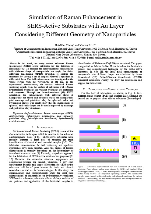
Simulation of Raman Enhancement inSERS-Active Substrates with Au Layer Considering Different Geometry of NanoparticlesHui-Wen Cheng1 and Yiming Li1,2,3,*1Institute of Communications Engineering, National Chiao Tung University, 1001 Ta-Hsueh Road, Hsinchu 300, Taiwan 2Department of Electrical Engineering, National Chiao Tung University, 1001 Ta-Hsueh Road, Hsinchu 300, Taiwan3National Nano Device Laboratories, Hsinchu 300, Taiwan*Tel: +886 3 5712121 ext 52974; Fax: +886 3 5726639; E-mail: ymli@.twAbstract-In this work, we study surface enhanced Raman spectroscopy (SERS) active substrates for the detection of Rhodamine 6G. To examine the electromagnetic enhancement, with different shape of nanoparticle, we apply the finite-difference timedomain (FDTD) algorithm to analyze the structures by solving a set of coupled Maxwell’s equations in differential form. The field enhancements are investigated in the visible regime with the wavelength of 633 nm. In the experimental measurement, the surface enhanced Raman scattering signals from the surface of substrates with 12-hour hydrothermal treatment and without treatment are performed and compared. Through the three-dimensional (3D) FDTD calculation, the enhancements with different shape of nanoparticle are tested and obtained which are nanoparticle, gold nanocage and gold/silver alloy for spherical, cubic and pyramidical shapes. The results show that the enhancement of spherical and cubic shapes can be much improved by nanocage and gold/siliver alloy structures.Keywords- Surface-Enhanced Raman spectroscopy (SERS), electromagnetic enhancement, nanoparticle, gold nanocage, gold/silver alloy, finite-difference time-domain, hydrothermally treated substrate.I.I NTRODUCTIONSurface-enhanced Raman Scattering (SERS) is one of the characterization techniques, which is sensitive to the enhanced electromagnetic fields [1-6]. SERS-active substrates have recently attracted a great deal of attention for rapid identification of chemical and bacterial samples [5-7]. The fabricated nanostructures for both bottom-up and top-down approaches have been reported. And, the degree of Raman enhancement is strongly dependent on the morphology of formulated nanostructures [8]. Recently, a top-down approach for the fabrication of SERS-active substrate was proposed [9-12]. However, the expensive substrate, equipments and complicated process are needed. Therefore, a low cost, environment friendly and simple fabrication for SERS-active substrates will be of great interest for basic and clinical researchers as well as for biotechnologies. In this study, we experimentally and computationally study the local field enhancements of nanoparticles on hydrothermally roughened SERS-active substrates, where the effects of shape and size of Au particles and application of the fabricated samples in identification of Rhdamine 6G (R6G) are examined. This paper is organized as follows. In Sec. II, we introduce the fabrication process and computational technique for the SERS-active substrates. In Sec. III, the local field enhancements of nanoparticle with different shapes are calculated by three-dimensional (3D) finite-difference time-domain (FDTD) numerical simulation. Finally, we draw the conclusions and suggest the future work.II.F ABRICATION AND C OMPUTATIONAL T ECHNIQUEFor the flow of fabrication, as shown in Fig. 1, first, buffered oxide etchant (BOE) and standard RCA cleaning arecarried out to prepare clean silicon substrates (Boron-doped(i)(ii)(iii)(iv)Figure 1. Schematic representation for the fabrication of SERS-active substrate. First, silicon wafers were cleaned by BOE and standard RCA cleaning procedures. Then, Ti films were deposited on the pre-cleaned silicon wafers using reactive DC magnetron sputtering system. The asdeposited samples were cleaved and treated under hydrothermal conditions for various durations. Subsequently, Au was thermal evaporated onto the hydrothermally roughened substrates for sensing. .Figure. 2 (a) The AFM image of titanium thin films treated under hydrothermal condition for 12 hours treatment duration. (b) The plot of simulated substrate which is part of real substrate, where the matrix of nanoparticles is 3 x 5 due to periodical property of the simulated structure.p<100>). Then, 100-nm-thick titanium films are deposited on the pre-cleaned silicon wafers using reactive DC magnetron sputtering system. The as-deposited sample is cleaved into 0.5 cm x 1 cm squares and rinsed with ethanol, and de-ionized water. Subsequently, the sample is put into a 23 mL Teflon-lined stainless steel autoclave filled with 20 mL distilled water, which is sealed, and heated at 200o C for 2, 4, 6, 8, 10, and 12 hours, respectively. Then the treated sample is cooled to room temperature naturally, washed with distilled water for several times, and dried with a stream of cylinder air. For example, the image of Fig. 2(a) shows the AFM images represent titanium thin films treated under hydrothermal conditions for 12 hours treatment duration.The image of Fig. 2(b) shows the plane view of the gold-coated nanoparticular structure, where the matrix of nanoparticles is 3 x 5 due to periodical property of the simulated structure. Numerical simulation using a 3D FDTD method is conducted to investigate the local field enhancement of substrate [13-15]. The Maxwell’s curl equations in linear, isotropic, nondispersive, lossy materials are,E t BK K K×∇−=∂∂ (1) ,1B J t E KK K K ×∇+−=∂∂μεε (2) ,0=⋅∇B K(3)Figure 3. The simulation procedure of solving the Maxwell’s equations.,ερ=⋅∇E K K (4)where E K and B Kare the vectors of electric and magnetic fields, respectively, ϵ and μ are permeability and permittivity and J K and ρ are the current density vector and charge density. For a globally defined curvilinear space, Maxwell’s equations are easily implemented in their differential form, where Faraday’s law is Eq. (1) and Ampere’s law is Eq. (2).The FDTD method solves Maxwell’s equations by first discretizing all equations via central differences in time and space. Then, based upon a 3D Yee’s mesh and components of the electric and magnetic fields at points, the discretized spacing in the x, y, and z directions adopted in our simulation are |x | = 0.01 um, |y | = 0.01 um and |z | = 0.01 um, where the time step Δt is 0.0004 and the time duration T is 3 in units of femtoseconds. The discretized equations are iteratively solved in a leapfrog manner, alternating between computing the E andH fields at subsequent Δt/2 intervals, as shown in Fig. 3. Notably, we employ the perfectly matched layer as the simulation domain boundaries in which both electric and magnetic conductivities are introduced in such a way that wave impedance remains constant, absorbing the energy withoutinducing reflections. III. R ESULTS AND D ISCUSSIONIn order to have less light absorption, the larger scattering of substrate is better to achieve larger field enhancement. For chemical sensing, the hydrothermally roughened substrates are treated with aqueous solutions of 10-4 M R6G. The The chemical structure of R6G is shown in Fig. 4(a). Fig. 4(b) shows that the characteristic Raman vibrational modes of R6G immobilized on the substrate with or without hydrothermal treatment. The substrate with hydrothermal treatment showsFigure4. (a) Chemical structure of Rhodamine 6G (R6G). The molecule is widely used for SERS measurements. (b) The Raman spectra for R6G (10-4 M) immobilized on hydrothermally untreated (blue) and treated (orange)substrates.Au Au Au AgFigure 5. Gold nanoparticle, gold nanocage and gold/silver alloy (from left to right) for spherical, cubic and pyramidical shapes, respectively.larger intensity than that without hydrothermal treatment due to the roughness on the surface [16]. According to the Beckmann-Kirchhoff theory, the roughened surface has larger scattering on the surface of substrate so that the intensity can be enhanced. Through using the FDTD simulation, the evaluation of electric field on the substrates is carried out by the directing light with a wave length of 633 nm.Notably the nanosensor also can be fabricated by other synthesis methods to achieve different shape of nanoparticles.Figure 6. The plot of electric field enhancement factor versus different samples.Figure. 7. The top view of electric field distribution with spherical shape of (a) Au nanoparticle, (b) Au nanocage and (c) Au/Ag alloy, respectively.Here, we consider gold nanoparticle, gold nanocage and gold/silver alloy (from left to right) for spherical, cubic and pyramidical shapes, as shown in Fig 5. The simulation results show that the electric field (E x ) enhancement of nanoparticle with cubic shape is larger than that with spherical and pyramid shapes, as shown in Fig. 6. To improve the enhancement, the structure is considered to fabricate by different synthesized structures for spherical, cubic and pyramidical shapes, respectively. The synthesized structures are illustrated in Fig. 5, which are the gold nanocage (middle one) and gold/silver alloy with empty and silver inside, respectively. From the results of Fig. 6, the Au/Ag alloy and gold nanocage are adopted for spherical and cubic shapes because the enhancement is much improved. For pyramid, the E x enhancement of nanocage or metal alloy is almost the same. These results can be explained by distribution of electric field. The corresponding distributions of electric field are shown in Fig. 7, 8 and 9, respectively. For spherical shape, the enhancement of Au nanoparticle is locallyFigure. 8. Top views of electric field distribution with the cubic shape of (a)Au nanoparticle, (b) Au nanocage, (c)and Au/Ag alloy, respectively.Figure. 9. Top views of electric field distribution with the pyramidical shape of (a) Au nanoparticle, (b) Au nanocage, and (c) Au/Ag alloy, respectively.increased, as shown in Fig. 7(a). Considering the nanocage structure, the enhancement of whole plane is increased, compared with nanopartilce structure, due to empty inside, as shown in Fig. 7(b). With silver inside, the enhancement can be much improved by different materials, as shown in Fig. 7(c). For cubic shape, the distributions of nanoparticle and nanocage are quite similar, as shown in Figs. 8(a) and 8(c). It is obviously that the larger enhancement can be obtained by nanocage due to tips on the corners, as shown in Fig. 8(b). For the pyramidical shape, the electric field can not be improved by synthesized structures so that the distributions are quite similar, as shown in Fig. 9.IV. C ONCLUTIONSIn conclusion, we have successfully prepared SERS-active substrates with low background for the detection of both Rhodamine 6G. The enhancement could be controlled bytuning the surface roughness of the substrates through varying treatment duration. Through FDTD simulation, the field enhancement of spherical and cubic shape nanoparticles can be enhanced by using Au/Ag alloy and naocage samples, where the different shape of nanoparticles also can be fabricated by other synthesis method for local field enhancement in diverse nanosensor applications.A CKNOWLEDGMENTThis work was supported in part by National Science Council (NSC), Taiwan under Contracts No. NSC-97-2221-E-009-154-MY2 and No. NSC-99-2221-E-009-175.R EFERENCES[1] R. P. Van Duyne, J. C. Hulteen, D. A. Treichel, “Atomic forcemicroscopy and surface-enhanced raman spectroscopy,” J. Chem. Phys., vol. 99 , pp. 2101-2115, 1993.[2] J. C. Hulteen, R. P. Van Duyne, “Nanosphere lithography: A materialsgeneral fabrication process for periodic particle array surfaces,” J. Vac. Sci. Technol. A13, pp. 1553-1558, 1995.[3] F. Adrian, “Surface enhanced Raman scattering by surface plasmonenhancement of electromagnetic fields near spheroidal particles on a roughened metal surface,” Chem. Phys. Lett., vol. 78, pp. 45-49, 1981. [4] M. Moskovits, ” Surface-enhanced spectroscopy,” Rev. Mod. Phys. 57,pp. 783-826, 1985.[5] P. L. Stiles, J. A. Dieringer, N. C. Shah, and R. P. Van Duyne, “Surface-Enhanced Raman spectroscopy,” Annu. Rev. Analyt. Chem., vol. 1, pp. 601-626, 2008.[6] R. M. Jarvis and R. Goodacre, “Characterization and identification ofbacteria using SERS,” Chem. Soc. Rev. 37, pp. 931-936, 2008.[7] Y. S. Huh, A. J. Chung and D. Erickson, “Enhanced Ramanspectroscopy and its application to molecular and cellular analysis,” Microfluid. Nanofluid., vol. 6, pp. 285-297, 2009.[8] K. L. Kelly, E. Coronado, L. L. Zhao and G. C. Schatz, “The opticalproperties of metal nanoparticles: the influence of size, shape, and dielectric environment,” J. Phys. Chem. B, vol. 107, pp. 668-677, 2003. [9] P. F. Liao, J. G. Bergman, D. S. Chemla, A.Wokaun, J. Melngailis, A.M. Hawryluk, and N. P. Economou, “Surface-enhanced Raman scattering from microlithographic silver particle surfaces,” Chem. Phys. Lett., vol. 82, pp. 355-359, 1981.[10] N. Felidj, J. Aubard, G. Levi, J. R. Krenn, A. Hohenau, G. Schider, A.Leitner, and F. R. Aussenegg, “Optimized surface-enhanced Raman scattering on gold nanoparticle arrays,” Appl. Phys. Lett., vol. 82, pp. 3095-3097, 2003.[11] J. A. Dieringer, A. D. McFarland, N. C. Shah, D. A. Stuart, A. V.Whitney, C. R. Yonzon, M. A. Young, X. Zhang, and R. P. Van Duyne, “Surface enhanced Raman spectroscopy: new materials, concepts, characterization tools, and applications,” Faraday Discuss., vol. 132, pp. 9-26, 2006.[12] S. B. Chaney, S. Shanmukh, R. A. Dluhy, and Y. -P. Zhao, Appl. Phys.Lett., “Aligned silver nanorod arrays produce high sensitivity surface-enhanced Raman spectroscopy substrates,” vol. 87, 031908, 2005.[13] Y. Li and H.-W. Cheng, “Optimal configuration of hydrogen-embrittlement-fabricated nanogaps for surface-conduction electron-emitter display,” IEEE Trans. Nanotech. vol. 8, pp. 671-677, 2009.[14] A. Taflove and S.C. Hagness, Computational Electrodynamics: TheFinite-Difference Time-Domain Method, 2nd ed., Artech House, Boston, MA, 2000.[15] Y. Li, H.-W. Cheng, “Numerical simulation of field emission efficiencyof anodic aluminum oxide carbon nanotube field emitter in the triode structure,” Comp. Phys. Commun.. vol. 179, pp. 107-111. 2008.[16] R. Li Voti, G. L. Leahu, S. Gaetani, C. Sibilia, V. Violante, E. Castagna,and M. Bertolotti., “Light scattering from a rough metal surface: theory and experiment,” J. Opt. Soc. Am. B, vol. 26, pp. 1585-1593, 2009.。

ftir红外atr校准流程FTIR (Fourier Transform Infrared) and ATR (Attenuated Total Reflection) are common techniques used in the field of spectroscopy for chemical analysis. Both techniques require regular calibration to ensure the accuracy and reliability of the results obtained. 红外光谱(FTIR)和衰减全反射(ATR)是化学分析领域常用的技术。
这些技术需要定期校准,以确保所得结果的准确性和可靠性。
The calibration process for FTIR and ATR instruments involves several key steps to ensure that the instrument is accurately measuring the sample's infrared absorption. 校准FTIR和ATR仪器涉及几个关键步骤,以确保仪器准确测量样品的红外吸收。
Firstly, the instrument must be checked for any potential issues or errors. This includes inspecting the optics, checking for any contamination or damage, and ensuring that all components are functioning properly. 首先,必须检查仪器是否存在潜在问题或错误。
这包括检查光学部件,检查是否有任何污染或损坏,并确保所有组件的正常运行。
傅里叶红外光谱英文缩写The abbreviation for Fourier Transform Infrared Spectroscopy (FTIR).Fourier Transform Infrared Spectroscopy (FTIR) is a widely used analytical technique for the identification and characterization of organic and inorganic compounds. FTIR measures the absorption of infrared radiation by a sample, providing information about its chemical structure and composition. The technique is based on the principle that different chemical bonds absorb infrared radiation at specific frequencies, allowing for the identification of functional groups and molecular structure.FTIR has many applications in various fields, including pharmaceuticals, polymers, food and beverage, environmental analysis, and forensic science. In pharmaceuticals, FTIR is used to identify and quantify active ingredients and excipients in drug formulations, as well as to monitor the stability of pharmaceutical products. In the polymer industry, FTIR is used to analyze the composition and structure of polymer materials, such as plastics and rubbers, to ensure product quality and performance.In environmental analysis, FTIR is used to detect and identify pollutants in air, water, and soil, as well as to monitor the degradation of environmental contaminants. In forensic science, FTIR is used to analyze trace evidence, such as fibers, paints, and drugs, for criminal investigations.FTIR works by passing infrared radiation through a sample and measuring the absorbance of the radiation at different wavelengths. The resulting spectrum, called an infrared spectrum, provides a unique "fingerprint" of the sample's chemical composition. FTIR instruments can analyze samples in various physical forms, including liquids, solids, and gases, making it a versatile tool for different analytical needs.FTIR instruments can be equipped with additional accessories, such as attenuated total reflectance (ATR) or diffuse reflectance, to analyze samples without the needfor sample preparation. In addition, FTIR can be coupled with other analytical techniques, such as gas chromatography (GC-FTIR) or mass spectrometry (MS-FTIR), to provide complementary information about complex samples.FTIR has several advantages, including rapid analysis, high sensitivity, and non-destructive nature, making it an essential tool for quality control, research, and development in various industries. FTIR is also relatively easy to use, with modern instruments featuring user-friendly software for data acquisition and analysis.In conclusion, Fourier Transform Infrared Spectroscopy (FTIR) is a powerful analytical technique with diverse applications in pharmaceuticals, polymers, environmental analysis, and forensic science. Its ability to provide detailed information about chemical structure and composition makes it an indispensable tool for research, development, and quality control in various industries.傅里叶红外光谱英文缩写为FTIR。
兽医微生物学英语Veterinary Microbiology is a branch of microbiology that specifically focuses on the study of microorganisms that cause diseases in animals. This field is crucial for understanding and controlling infectious diseases in animals, which can also have implications for human health.One of the key areas of study in veterinary microbiology is the identification and characterization of pathogenic microorganisms. This involves isolating and culturing microorganisms from infected animals, and then using various techniques such as microscopy, biochemical tests, and molecular methods to identify and characterize the pathogens.Understanding the mechanisms of pathogenesis is another important aspect of veterinary microbiology. This involves studying how microorganisms cause disease in animals, including the role of virulence factors, host-pathogen interactions, and immune responses. By understanding these mechanisms, veterinarians can develop more effective strategies for preventing and treating infectious diseases in animals.Epidemiology is also a critical component of veterinary microbiology. This involves studying the distribution and spread of infectious diseases in animal populations, aswell as identifying risk factors and potential sources of infection. Epidemiological studies are essential for implementing control measures and preventing outbreaks of infectious diseases in animal populations.In addition to studying infectious diseases, veterinary microbiologists also play a key role in monitoring and controlling zoonotic diseases, which are diseases that can be transmitted from animals to humans. By understanding the epidemiology and transmission dynamics of zoonotic diseases, veterinarians can help prevent the spread of these diseases to humans and protect public health.In summary, veterinary microbiology is a diverse and essential field that encompasses the study of pathogenic microorganisms, mechanisms of pathogenesis, epidemiology, and zoonotic diseases. By advancing our understanding of infectious diseases in animals, veterinary microbiologists contribute to the health and well-being of both animals and humans.兽医微生物学是微生物学的一个分支,专门研究导致动物疾病的微生物。
《昆虫学报》投稿须知2014《昆虫学报》是中国昆虫学会和中国科学院动物研究所共同主办的昆虫学学术刊物,1950 年创刊,国内外公开发行,2002 年起由季刊改为双月刊。
本刊为全国核心期刊,曾被SCI 收录,现被《生物学文摘》(BA )、《昆虫学文摘》(EA )、《化学文摘》(CA )、俄罗斯《文摘杂志》(AJ )、英国CAB 文摘数据库和德国“ ISPI Pest Directory Database ”等国内外重要文摘和数据库收录。
1 .办刊宗旨传播报道昆虫学研究最新成果和动态,推动国内外昆虫学界学术交流,发展我国的昆虫学事业,为国家的科技、经济和社会发展服务。
2 .征稿范围本刊面向国内外征稿,登载有关昆虫形态、生理、生化、病理、毒理、生态和系统学等昆虫学各分支学科的中英文原始研究论文、简报和综述等。
本刊鼓励英文投稿。
每期设四个固定栏目(生理与生化,毒理与抗性,生态与害虫管理,进化与系统学),并根据需要增加病理和医学昆虫等不固定栏目。
系统学方面主要接受订正和综述性论文,一般不接受单纯报道新分类单元或新记录的论文。
3 .内容要求内容完整,论点明确,数据可靠,文字简明通畅,引用资料请注明文献出处,注意保守国家机密,文责自负。
中文稿件使用昆虫名称和名词应参照全国科学技术名词审定委员会2000 年公布的《昆虫学名词》;其他名词和名称请参照全国科学技术名词审定委员会公布的相关名词规范;无规范可参考时,宜沿用最广、最久且意义无误的名词和名称。
文稿题目要求准确、简短明了,系统学论文题目要求给出所研究分类单元隶属的纲、目和科名。
摘要应包含研究目的、关键方法、主要结果和结论等。
关键词应能反映文稿的内容范畴,其选择可参考《昆虫学文摘》(EA )所列主题词,应能方便计算机检索,不能是一句短语。
观察标本须指明存放地。
涉及核酸序列时,应提供在国际核酸数据库登记注册的核酸序列号。
4 .格式要求4.1 文字来稿用5 号宋体字、A4 纸隔行打印。
clinical microbiology 检验英文原版书Title: Clinical Microbiology: An Essential Guide for Laboratory TestingIntroduction:Clinical microbiology plays a crucial role in the diagnosis and management of infectious diseases. It involves the laboratory testing of various specimens to identify and characterize microorganisms. In this article, we will explore the fundamental aspects of clinical microbiology as covered in the English original textbook.I. Overview of Clinical Microbiology:1.1 Importance of Clinical Microbiology:- Clinical microbiology aids in the identification of infectious agents, guiding treatment decisions.- It helps in the surveillance and monitoring of antimicrobial resistance.- It plays a vital role in outbreak investigations and infection control measures.1.2 Specimen Collection and Handling:- Proper collection and transportation of specimens ensure accurate and reliable results.- Different types of specimens, such as blood, urine, respiratory secretions, and wound swabs, require specific collection techniques.- Special considerations, such as timing and transport media, are essential to maintain the viability of microorganisms.1.3 Laboratory Techniques:- Microscopy techniques, including gram staining and acid-fast staining, allow for the visualization of microorganisms.- Culture-based methods involve the isolation and identification of microorganisms on various selective and differential media.- Molecular techniques, such as polymerase chain reaction (PCR), enable the detection and characterization of microorganisms at the genetic level.II. Bacterial Infections:2.1 Identification and Characterization:- Bacterial identification relies on phenotypic characteristics, including colony morphology, biochemical tests, and serological methods.- Advanced techniques, such as matrix-assisted laser desorption/ionization time-of-flight mass spectrometry (MALDI-TOF MS), provide rapid and accurate identification.- Antimicrobial susceptibility testing determines the susceptibility or resistance of bacterial isolates to specific antibiotics.2.2 Virulence Factors and Pathogenesis:- Understanding the virulence factors of bacteria helps in assessing their pathogenic potential.- Bacterial toxins, adhesins, and capsules contribute to the ability of bacteria to cause disease.- Molecular techniques, such as gene sequencing, aid in the identification and characterization of virulence factors.2.3 Antibiotic Resistance:- The emergence of antibiotic-resistant bacteria poses a significant challenge in clinical practice.- Mechanisms of antibiotic resistance include enzymatic inactivation, altered target sites, and efflux pumps.- Laboratory testing methods, such as disk diffusion and minimum inhibitory concentration (MIC) determination, help in monitoring and detecting antibiotic resistance.III. Viral Infections:3.1 Diagnostic Techniques:- Serological assays, such as enzyme-linked immunosorbent assay (ELISA), detect antibodies produced in response to viral infections.- Molecular methods, such as PCR and nucleic acid amplification tests (NAATs), directly detect viral genetic material.- Viral culture allows for the isolation and identification of viruses in specialized cell lines.3.2 Antiviral Therapy:- Laboratory testing helps in determining the susceptibility of viruses to antiviral drugs.- Drug resistance testing identifies mutations in viral genes associated with reduced drug effectiveness.- Viral load testing monitors the effectiveness of antiviral therapy by quantifying viral RNA or DNA.3.3 Emerging Viral Infections:- Clinical microbiology plays a crucial role in the detection and surveillance of emerging viral infections.- Rapid diagnostic tests, such as point-of-care assays, aid in the early identification of viral outbreaks.- Molecular techniques, such as next-generation sequencing (NGS), provide insights into the genetic diversity and evolution of emerging viruses.IV. Fungal and Parasitic Infections:4.1 Laboratory Diagnosis:- Direct microscopic examination of clinical specimens allows for the detection of fungal elements and parasites.- Culture-based methods help in the isolation and identification of fungal and parasitic organisms.- Molecular techniques, such as PCR and DNA sequencing, enhance the accuracy and speed of diagnosis.4.2 Antifungal and Antiparasitic Susceptibility Testing:- Laboratory testing determines the susceptibility or resistance of fungal and parasitic isolates to antifungal and antiparasitic drugs.- Methods, such as broth microdilution and agar dilution, provide quantitative susceptibility results.- Interpretive criteria assist in guiding treatment decisions based on the susceptibility profile of the organism.4.3 Epidemiology and Control Measures:- Clinical microbiology contributes to the surveillance and monitoring of fungal and parasitic infections.- Molecular typing techniques, such as multilocus sequence typing (MLST), aid in the investigation of outbreaks.- Infection control measures, including appropriate specimen handling and disinfection protocols, help prevent the spread of fungal and parasitic infections.V. Quality Assurance and Laboratory Management:5.1 Quality Control in Clinical Microbiology:- Quality control measures ensure the accuracy and reliability of laboratory results.- Internal quality control involves the use of known reference materials and regular monitoring of test performance.- External quality assessment programs provide proficiency testing to evaluate laboratory performance.5.2 Laboratory Safety:- Adherence to biosafety guidelines is essential to protect laboratory personnel and prevent laboratory-acquired infections.- Proper use of personal protective equipment, safe handling of infectious materials, and appropriate waste disposal are crucial aspects of laboratory safety.- Risk assessment and implementation of safety protocols minimize the potential for accidents and exposure to hazardous agents.5.3 Emerging Technologies and Future Perspectives:- Advances in technology, such as automation and artificial intelligence, are shaping the future of clinical microbiology.- Rapid diagnostic tests and point-of-care devices enable timely and accurate diagnosis at the patient's bedside.- Integration of big data analytics and genomics holds promise for personalized medicine and precision infectious disease management.Conclusion:Clinical microbiology is a multidisciplinary field that plays a vital role in the diagnosis, management, and prevention of infectious diseases. The English original textbook on clinical microbiology provides comprehensive coverage of the fundamental concepts, laboratory techniques, and practical applications in this field. By understanding the various aspects discussed in the book, healthcare professionals can enhance theirknowledge and skills in clinical microbiology, ultimately improving patient care and public health outcomes.。