Chapter15
- 格式:ppt
- 大小:504.50 KB
- 文档页数:56


Chapter 15 Monopoly 垄断§1. 垄断Monopoly一.对比竞争企业是价格接受者,垄断企业是价格制定者。
a competitive firm is a price taker, a monopoly firm is a price maker二.定义垄断企业:作为一种没有相近替代品的产品的唯一卖者的企业A firm is considered a monopoly if it is the sole seller of its product & its product does not have close substitutes. 如果一个企业是其产品唯一的卖者,而且如果其产品并没有相近的替代品,这个企业就是垄断者。
§2. 为什么会产生垄断Why Monopolies Arise一.(1)垄断的基本原因fundamental cause:进入障碍barriers to entry(2)进入障碍的三个主要来源Barriers to entry have three sources1.垄断资源:生产所需要的关键资源由一家企业拥有Ownership of a key resource.2.政府管制:政府给予一个企业排他性地生产某种产品或服务的权利The government gives a single firm the exclusive right to produce some good.3.生产流程:生产成本使一个生产者比大量生产者更有效率Costs of production make a single producer more efficient than a large number of producers.二.垄断资源Monopoly Resources虽然关键资源的排他性所有权是垄断的一个潜在原因,但垄断很少产生于这种原因Although exclusive ownership of a key resource is a potential source of monopoly, in practice monopolies rarely arise for this reason.三.政府制造的垄断Government-Created Monopolies1.政府给予一个企业排他性地出售某种物品或劳务的权利,限制其他企业进入市场,从而造成垄断。
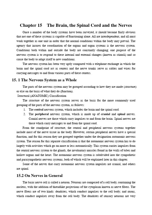
Chapter 15 The Brain, the Spinal Cord and the NervesOnce a number of the body systems have been surveyed, it should become fairly obvious that not one of these systems is capable of functioning alone. All are interdependent, and all must work together as one unit in order that the normal conditions within the body may prevail. The agency that insures the coordination of the organs and organ systems is the nervous system.Conditions both within and outside the body are constantly changing; one purpose of the nervous system is to respond to these internal and external changes (known as stimuli) and so cause the body to adapt itself to new conditions.The nervous system has been very aptly compared with a telephone exchange in which the brain and the spinal cord act as centers and the nerve trunks serve as cables and wires for carrying messages to and from various parts of these centers.15. 1 The Nervous System as a WholeThe parts of the nervous system may be grouped according to how they are made (structure) or else on the basis of what they do (function).Structural (ANATOMIC) ClassificationThe structure of the nervous system serves as the basis for the more commonly used grouping of the parts of the nervous system, as follows:1.The central nervous system, which includes the brain and the spinal cord.2.The peripheral nervous system, which is made up of cranial and spinal nerves.Cranial nerves are those which carry impulses to and from the brain. Spinal nerves arethose which carry messages to and from the spinal cord.From the standpoint of structure, the central and peripheral nervous systems together include most of the nerve tissue in the body. However, certain peripheral nerves have a special function, and for this reason they are grouped together under the designation autonomic nervous system. The reason for this separate classification is that the autonomic nervous system has to do largely with activities which go on more or less automatically. This system carries impulses from the central nervous system to the glands, the involuntary muscles found in the walls of tubes and hollow organs and the heart. The autonomic nervous system is subdivided into the sympathetic and parasympathetic nervous systems, both of which will be explained later in this chapter.Some of the nerves that carry autonomic nervous system impulses are cranial, and others are spinal.15.2 On Nerves in GeneralThe basic nerve cell is called a neuron. Neurons are composed of a cell body, containing the nucleus, with the addition of threadlike projections of the cytoplasm known as nerve fibers. The nerve fibers are of two kinds: dendrites, which conduct impulses to the cell body; and axons, which conduct impulses away from the cell body. The dendrites of sensory neurons are verydifferent from those of other neuron. They are usually single and they may be very long (as much as 3 feet) or they may be short; but in any case, they do not have the treelike appearance so typical of other dendrites. Each sensory nerve fiber (dendrite) has a special structure called the receptor, or end organ, where the stimulus is received and the sensory impulse begins. Sensations such as pain, touch, hearing and seeing which involve these sensory neurons will be discussed in Chapter 11.Each neuron is a separate unit, and there is no anatomic unity between neurons. It would be logical to ask how it is possible for neurons to be in contact; in other words, how the axon of one neuron can be in functional contact with the dendrite of another neuron. This is accomplished by the synapse, from a Greek word meaning “to clasp.” Synapses, then, are points of junction fro transmission of nerve impulses.Nerve fibers that are connected with receptors (for receiving stimuli) conduct impulses to the brain and cord, and when grouped together form afferent nerves. Those fibers that carry impulses from the centers out to the muscles and glands form efferent nerves. Motor neurons, which carry impulses that lead to the contraction of skeletal muscles, are classified as efferent neurons (Fig. 15.1). Some nerves contain a mixture of afferent and efferent nerve fibers and are often referred to as mixed nerves.Fig. 15.1 Diagram of a motor neuron15.3 The Central Nervous SystemTHE BRAINMain parts of the brainThe largest part of the brain is the cerebrum, which is divided into the two cerebral hemispheres, a right and a left one. The brainstem includes the deeper parts that comprise the interbrain (thalamus, etc.) that cannot be seen unless the brain is sectioned, and a series of smaller parts that extend downward. Starting at the upper part of this series, we may see a small part of the midbrain. Below it and plainly visible from the under view of the brain are the pons and the medulla oblongata, which connects with the spinal cord through a large opening in the base of the skull. The pons connects the midbrain and the medulla. Next in size to the cerebralhemispheres is the cerebellum, a word meaning :little brain.” It is located immediately below the back part of the cerebral hemispheres and is connected with the other parts of the brain only by means of the bridgelike pons.Structure of the cerebral hemispheresThe outer nerve tissue of the cerebral hemispheres is gray matter and is called the cerebral cortex. This gray cortex is arranged in folds forming elevated portions known as convolutions, separated by depressions or grooves called fissures, or sulci (Fig. 15.2). Internally the cerebral hemispheres are made largely of white matter and a few islands of gray matter. Inside the hemispheres are two spaces extending in a somewhat irregular fashion. These are the lateral ventricles, which are filled with a watery fluid common to both the brain and the spinal cord called cerebrospinal fluid, to be discussed later.Although there are many fissures (sulci), a few are especially important landmarks. These include the:1. Longitudinal fissure, which is a deep groove that separates the upper parts of the cerebralhemisphere from each other.2. Central fissure, which extends from the top of the brain near the center downward alongthe side at right angles to the longitudinal fissure.3. Lateral fissure, which curves somewhat along the side of the brain and separates thetemporal lobe from the rest of the cerebral hemisphere.Let us examine the cerebral cortex, the layer of gray matter which forms the surface of each cerebral hemisphere. It is within the cerebral cortex that all impulses are received and analyzed. These form the basis of knowledge; the brain “stores”knowledge, much of which can be produced on demand by means of the phenomenon which we call memory. It is in the cerebral cortex that all thought takes place, all association, judgment and discrimination. It is from the cerebral cortex, too, that the orders originating from conscious deliberation emanate; that is, the voluntary movements are controlled here.Fig. 15.2 External surface of the brain showing the main partsand some of the lobes and fissures of the cerebrum.Division and functions of the cerebral cortexThe cerebral cortex of each hemisphere is divided into four lobes, areas named from the overlying cranial bones. It has been found that each area controls a certain category of functions. The four lobes, with some of their characteristic functions, follow:1. The frontal lobe, which is relatively much larger in the human being than in any otherorganism. This contains the motor cortex which controls the voluntary muscles. The left side of the brain governs the right side of the body and the right side of the brain governs the left side of the body. The upper portion of the center controls the lower parts of the body. The frontal lobe also contains two areas used in speech (the speech centers will be discussed later).2. The parietal lobe, which occupies the upper part of each hemisphere, just behind thecentral fissure. This contains the sensory area, in which the general senses such as pain, touch and temperature are interpreted. Also, such interpretations as the determination of distances, sizes and shapes take place here.3. The temporal lobe, which is lateral (at the side) and folds under the hemispheres on eachside. This contains the auditory center for interpreting impulses from the ear.4. The occipital lobe, which is the most posterior, and extends over the cerebellum. Thiscontains the visual area for interpreting messages from the retina of the eye.Speech centersThe speech centers are among the most interesting groups of areas in the cerebral hemispheres (Fig. 15.3). Their development and use are closely connected with the processes of learning. These areas are called the:1. Auditory speech center, located in the temporal lobe near the auditory center. While theauditory center enables a person to interpret sounds, it does not have anything to do with the understanding of words. Such an understanding of language requires the development of the auditory speech center. Often, this is the first speech center to develop in the child.Babies often seem to understand what is being said long before they do any talking themselves. It is usually several years before children learn to read or write words. In many parts of the world people never learn to read or write their language.2. Visual speech center, which is somewhat above and in front of the visual center. In thisarea the ability to read with understanding is developed. You may see the writing in the Japanese language, for example, but this would involve only the visual center in the occipital lobe unless you could read the words.3. Motor speech center, located just in front of the lowest part of the motor cortex in thefrontal lobe. Since the lower part of the motor cortex controls the muscles of the head and the neck, it seems logical to think of the speech area as an extension forward to make possible the control of the muscles of speech in the tongue, the soft palate and the larynx.4. Written speech center, located above the motor speech center, and in front of the corticalarea that controls the muscles of the arm and the hand. Again this center is an extension forward from the motor cortex. The ability to write words usually is one of the last phases in the development of learning words and their meaning, although occasionally a person may write words more readily than he can vocalize them.Fig. 15.3. Functional areas of cerebrumOther parts of the cerebral hemispheresBeneath the gray matter of the cerebral cortex is the white matter, consisting of nerve fibers which connect the cortical areas with each other and with other parts of the nervous system. Among the most important of these large collections of nerve fibers is the internal capsule, a crowded strip of white matter where any injury is apt to cause extensive damage. At the base of each hemisphere are the nerve cell groups called basal ganglia, which regulate the body movements originating in the cerebral cortex. On the underside of each cerebral hemisphere is the olfactory area, concerned with the sense of smell, which is stimulated by the impulses arising in the nerve receptors of the nose.The interbrainThe interbrain, or diencephalon, can be seen only by cutting into the central section of the brain. It includes the thalamus and the hypothalamus. The two masses of gray matter that form the thalamus are relay centers and act to monitor sensory stimuli, suppressing some and magnifying others. The hypothalamus is located in the midline area below the thalamus and contains cells that control body temperature, water balance, sleep, appetite and some of our emotions, such as fear and pleasure. Both divisions of the autonomic nervous system are under the control of the hypothalamus. Thus it influe nces the heart‟s beating, the contractions of the walls of the bladder and other vital body functions.The midbrainThe midbrain is located just below the center of the cerebrum. It forms one of the forward parts of the brainstem. Four rounded masses of gray matter that are hidden by the cerebralhemispheres form the upper part of the midbrain. These four bodies, the corpora quadiigemina, act as relay centers for certain eye and ear reflexes. The ventral white matter of the midbrain conducts impulses between the higher centers of the cerebrum and the lower centers in the pons, cerebellum, medulla and spinal cord.The cerebellumThe cerebellum is made up of three parts: the middle portion, called the vermis (meaning …'wormlike‟‟), and two lateral hemisphere s at the sides. As in the case of the cerebral hemispheres, the cerebellum has an outer area of gray matter and an inner portion that is largely white matter. The functions of the cerebellum are:1. To aid in the coordination of voluntary muscles so that they will function smoothly and inan orderly fashion. Disease of the cerebellum causes muscular jerkiness and tremors.2. To help maintain balance in standing, walking and sitting, as well as during morestrenuous activities. Messages from the internal ear and from the tendon and muscle sensory end organs aid the cerebellum.3. To aid in maintaining muscle tone so that all muscle fibers are slightly tensed and readyto produce necessary changes in position as quickly as may be necessary.The ponsThe pons is white in color because it is composed largely of myelinated nerve fibers. These fibers in the pons carry messages from one side of the cerebellum to the other, from the cerebellum to the higher centers in the cerebrum and midbrain, and from the cerebellum to the lower centers, including the medulla and the spinal cord. Not only is the pons an important connecting link between the cerebellum and the rest of the nervous system, but it also contains connections with four pairs of cranial nerves. Further it contains nerve fibers that carry impulses to and from the centers located above and below it. Certain reflex (involuntary) actions are controlled in the pons; namely, some occurring in respiration.The medulla oblongataThe medulla of the brain is located between the pons and the spinal cord. It appears white externally because, like the pons, it contains many covered (myelinated) nerve fibers. Internally it contains collections of cell bodies (gray matter), which are called centers or nuclei. Among these are the very important vital centers including:1. The respiratory center, which controls the muscles of respiration in response to chemicaland other stimuli.2. The cardiac center, which tends to slow the heart rate so that it will not beat too rapidly tobe effective.3. The vasomotor center, which affects the muscles in the blood vessel walls and hencehelps to determine blood pressure.The last four pairs of cranial nerves are connected with the medulla. The nerve fibers that carry messages up through the spinal cord to the brain continue through the medulla also, as do similar descending or motor fibers. These groups of nerve fibers form tracts (bundles) and are grouped together according to function. The motor fibers from the motor cortex of the cerebral hemispheres .extend down through the medulla, and most of them cross from one side to the other (decussate) while going through this part of the brain. It is in the medulla that the shifting of nerve fibers occurs which causes the right cerebral hemisphere to control muscles in the left side of the body, and the upper portion of the cortex to control of muscles in the lower portions of the person. The medulla is an important reflex centfer, and it is here that certain neurons end and impulses are relayed to other neurons.Ventricles of the brainWithin the brain are four fluid-filled spaces called the ventricles. These extend into the various parts of the brain in a somewhat irregular fashion. We have already mentioned the largest, the lateral ventricles in the two cerebral hemispheres. Their extensions into the lobes of the cerebrum are called horns. These paired ventricles communicate with a midline space, the third ventricle, by means of the openings called foramina. The third ventricle is bounded on each side by the two parts of the thalamus, while the floor is occupied by the hypothalamus. Continuing down from the third ventricle a small canal, called the cerebral aqueduet, extends through the midbrain into the fourth ventricle. The latter is continuous with the near microscopic neural, or central, canal of the spinal cord. Do not confuse this tiny canal inside the cord with the much larger vertebral, or spinal, canal that is a part of the dorsal cavity enclosing the entire cord, together with its membranes and surrounding fluid. .In the roof of the fourth ventricle are three openings that allow the escape of fluid to the area that surrounds the brain and spinal cord. This will be discussed later.After removal of some of the fluid, air or other substances may be injected, and x-rays called encephalograms or ventriculograms are taken. Tumors or other brain disorders may sometimes be located by this means.Another kind of study of the brain and other soft tissues involves the use of high frequency sound impulses. This is called echoencephalography.BrainwavesThe interactions of the billions of nerve cells in the brain give rise to measurable electric currents. These may be recorded by an instrument called the electroencephalograph, which was Mentioned in Chapter 3. Tfie recorded tracings dr brain waves produce an electroencephalogram, not to be confused with the encephalogram mentioned earlier.Disorders of the brainSince the scientific name for the brain is encephalon, infection of, the brain is known asencephalitis. There are many causes of such disease, but the two chief pathogens are:1. Viruses, which cause some of the epidemic types of, sleeping sickness sometimes foundin the United States and in other parts of the world.2. Certain one-celled animals (protozoa) called trypanosoma, which cause the so-calledAfrican sleeping sickness. These protozoa are carried by a kind of fly (tsetse) and are capable of invading the cerebro-spinal fluid of man.Other infections that may involve the brain and related parts include abscesses and meningitis (explained later). As in the case of encephalitis a variety of organisms may cause these disorders. Viruses, bacteria, protozoa and fungi may travel from the centers of infection in the teeth, the sinuses, the tonsils and in the middle ear.Stroke, or cerebral apoplexy, is by far the most common kind of brain disorder. Rupture of a blood vessel (with a consequent cerebral hemorrhage), thrombosis or embolism may cause destruction of brain tissue. Such disorders are more frequent in the presence of artery wall disease, and hence are more common after the age of 40. The onset may seem to be sudden and often is referred to as a cerebrovascular accident. The effects of a stroke will depend on the extent and location of the artery involvement. A hemorrhage into the white matter of the internal capsule in the lower part of the cerebrum may cause extensive paralysis on the side opposite to the affected area. Such a paralysis is called hemiplegia, and the person so afflicted is known as a hemiplegic.Cerebral palsy is a disorder present at birth. It is characterized by diverse disorders of muscles varying in degree from weakness to complete paralysis, and in extent from a slight disorder of the lower extremity muscles to a multiplicity of paralyses involving all four extremities and the speech muscles as well. With patient and continuous muscle reeducation, speech training and other corrective procedures these victims may be helped considerably.Epilepsy, or so-called falling sickness, is a chronic disorder in which there is abnormality of brain function without apparent changes in tlte nerve tissues. In most cases the cause is not known. The study of brain waves obtained with the electroencephalograph usually shows abnormalities and is helpful both in diagnosis and treatment. Research is constantly improving and increasing the knowledge concerning the various forms of epilepsy. Many of these sufferers can be helped to live a normal active life if they follow a very careful hygienic regimen and use appropriate medication as outlined by a physician.Tumors of the brain may develop at any age, but are somewhat more common in young and middle-aged adults. The majority of brain tumors originate from the neuroglia and are called gliomas. The symptoms produced depend on the location of the growth, its destructiveness and the amount it compresses the brain tissue. Involvement of the frontal portions of the cerebrum often causes mental symptoms, such as changes in personality, disordered conduct and drowsiness. Early surgery offers hope of cure in some cases.Aphasia is a term that refers to the loss of the ability to speak or write, or the loss of the understanding of written or spoken language. There are several different kinds of aphasia,depending on what part of the brain is affected. Usually damage to a speech center causes more disturbance in the well-educated person than it does in the illiterate. It also has been noted that there is a tendency for the last language to be acquired to be the first to be lost; and conversely, the speech concepts that were obtained first (in childhood) remain the longest. The lesion that causes aphasia is likely to be in the left cerebral hemisphere in the right-handed person. Often much can be done for these people by patient retraining and much.understanding. The brain is an organ that has a marvelous capacity for adapting itself to different conditions, and its resources are tremendous. Often some means of communi¬cation can be found even though speech areas are damaged.THE SPINAL CORDLocation of the spinal cordIn the embryo the spinal cord occupies the entire spinal canal and so extends down into the tail portion of the vertebral column. However, the column of bone grows much more rapidly than the nerve tissue of the cord, so that the end of the cord soon fails to extend into the lower part of the spinal canal. This disparity in growth increases so that in the adult the cord ends in the region just below the area to which the last rib attaches (between the first and second lumbar vertebrae).Structure of the spinal cordExamination of the spinal cord reveals that it has a small irregularly shaped internal section made of gray matter (nerve cell bodies), and a larger area surrounding this gray part that is made of white matter (nerve fibers). A cross section of the cord shows that the gray matter is so arranged that a column of cells extends up and down dorsally, one on each side; another column is found in the ventral region; while a third less conspicuous part is situated on each side. These three pairs of columns of gray matter give this cross section an H-shaped appearance. The white matter can be seen to be made of thousands of nerve fibers arranged in three areas external to the gray matter on each side.Functions of the spinal cordThe functions of the cord may be divided into three aspects;1. Reflex activities, which involve the transfer and integration of messages that enter thecord, so that a sensory (afferent) impulse entering the center will become a motor (efferent) message leaving the cord.2. A pathway for conducting sensory impulses from afferent nerves upward throughascending tracts to the brain.3. A pathway for conducting motor impulses from the brain down through descending tractsto the nerves that will supply muscles or glands.The reflex pathway through the spinal cord usually involves three or more nerve cellstogether with their fibers, including:1. The sensory neuron, which has its beginning in a receptor and its nerve fiber in a nervethat leads to the cord.2. One or more central neurons, which are entirely within the cord.3. The motor neuron, which receives the impulse from a central neuron and then carries itvia its long axon through a nerve to a muscle or a gland.The knee jerk is an example of a spinal reflex. The pathway for the impulses that make this reflex possible includes a sensory neuron which has its receptor in the tendon just below die knee, its sensory nerve fiber in the nerves that extend to the spinal cord, central neurons inside the lower part of the cord and motor neurons that send processes through nerves from the cord to the effectors in the thigh‟s kicking muscle.Disorders involving the spinal cordAn acute virus disease affecting both the spinal cord and the brain is poliomyelitis, which is most commonly found in children. The polio virus is spread from the nose and the throat; from here it travels to the central nervous system, possibly by way of the respiratory passages and the blood. The virus may destroy the motor nerve cells in the spinal cord, in which case paralysis of one or more limbs results. The virus also can attack some of the cells of the brain and cause death. Prevention of poliomyelitis by means of the oral Sabin vaccine (which was preceded by the Salk vaccine) is one of the many significant advances in preventive medicine.Injuries to the spinal cord occur in traffic accidents in which bones of the spinal column are broken or dislocated. In wars gunshot or shrapnel wounds may damage the cord in varying degrees. Since the nerve tissue of the brain and the cord cannot repair itself, severing the cord causes paralysis below the injury, together with loss of all sensation from this area. Loss of sensation and of motion in the lower part of the body is called paraplegia.Other disorders of the spinal cord include tumors that grow from within the cord or that compress the cord from outside.Multiple sclerosis (sclerosis means hardening) involves the entire spinal cord as well as the brain. In this disease the myelin, a fatlike substance that forms a sheath around certain nerve fibers, disappears and the nerve axons themselves degenerate. It is an extremely disabling disease; however, it usually progresses very slowly so that the patient may have many years of relatively comfortable life remaining to him.Amyotrophic lateral sclerosis is a motor disorder of the nervous system in which certain cells are destroyed. The progressive destruction causes muscle atrophy and loss of motor control, until finally the victim is unable to swallow, or to talk.Spinal puncture and anesthesiaFluid may be removed from the space below the spinal cord. Since the cord is only about 18 inches long and ends some distance above the level of the hip line, a spinal puncture is usuallydone between the third and fourth lumbar vertebrae, about the level of the top of the hip bone. This cerebro-spinal fluid may be studied in the laboratory for evidence of disease or injury.Anesthetics are sometimes dissolved in. cerebrospinal fluid and then injected into the space below the cord, thus temporarily removing all sensation from the lower part of the body. In the hands of a trained operator this method of giving an anesthetic may have a number of advantages for certain types of surgery in selected patients. Under this type of anesthetic the patient is “awake” during the operation, but of course feels nothing in his lower body.COVERINGS OF THE BRAIN AND THE SPINAL CORDThe meninges are three layers of connective tissue that surround the brain and the spinal cord to form a complete enclosure; The outermost of these membranes is called the dura mater. It is the thickest and toughest of these meninges. Inside the skull, the dura mater splits in certain places to provide channels for the blood coming from the brain tissue. The second layer around the brain and the spinal cord is the arachnoid membrane (so-called because it resembles the webs produced by spiders, which belong to a group of animals called the Arachnida). The arachnoid membrane is loosely attached, to the deepest of the meninges by weblike fibers allowing a space for fluid located between the arachnoid and the innermost membrane. The third layer around the brain, the pia mater, is attached to the .nerve tissue of the brain and spinal cord and dips into all the depressions, unlike the other two meninges. It is made of a delicate Connective tissue in which there are many blood vessels. The blood supply to the brain is carried, to a large extent, by the pia mater (Fig. 15.4).Fig. 15.4 Frontal (coroal) section of top of head, showing meninges andrelated parts.。


分析:設極值發生在 P (x0 , y0 ,f, g 在此點附近∈ C 1 ,並設= 0 不妨假定= 0,依隱函數定理,存在 x0 的某鄰域Iδ = − δ + δ 及函數 y (x ∈ C 1 Iδ 滿足 y (x0 = y0 且 g (x, y (x = 0, ∀ x ∈ Iδ 上式對 x 偏導得 gx (x, y (x + · ′ = 又 f (x, y (x 在 x0 取極值⇒ fx (x0 , y (x0 + fy · ′ = 特別地,(20 在 x0 點成立,即 gx (x0 , y (x0 + gy (x0 , y· ′ = 將 (21,(22 視為 1, ′ 的聯立方程式,它有非 0 解,故 fx fy gx gy =0 (x0 ,y0 (21 (22 (23 換言之,< fx (x0 , y0 , fy (x0 , y0 > 與 < gx (x0 , y0 , gy (x0 , y0 > 線性相依,故∃λ使∇f = λ∇g (x0 , y0 b. 三變數 (i 單一限制條件在 g (x, y, z = 0 的條件下,求 f (x, y, z 的極值。
分析:設極值點發生在 P (x0 , y0 ,z0 ,f, g 在此點附近∈ C 1 ,且= 0 不妨假設= 0,依隱函數定理,∃ (x0 , y0 的某鄰域Bδ 及函數 z (x, y ∈ C 1 Bδ 滿足 z (x0 , y0 = z0 且 g (x, y, z (x, y = 0, ∀ (x, y ∈ Bδ 上式分別對 x, y 偏導得gx (x, y, z (x, y + gz (x, y, z (x, y · zx (x, y = 0 gy (x, y, z (x, y + gz (x, y, z (x, y · zy (x, y = 0 ∀ (x, y ∈ Bδ y0 (24 (25又 f (x, y, z (x, y 在 (x0 , y0 取極值⇒ fx (x0 , y0 , z0 + fz (x0 , y0 , z0 · zx (x0 , y0 = 0 fy (x0 , y0 , z0 + fz (x0 , y0 , z0 · zy (x0 , y0 = 0 特別地,(23,(24 對 (x0 , y0 成立:gx (x0 , y0 , z0 + gz (x0 , y0 , z0 · zx (x0 , y0 = 0 gy (x0 , y0 , z0 + gz (x0 , y0 , z0 · zy (x0 , y0 = 0 將 (25,(27 看成 1, zx (x0 , y0 的聯立方程式,它有非 0 解,故 fx fz gx gz 同理,(26,(28⇒ fy fz gy gz (29,(30⇒∃ λ 使∇f = λ∇g (x0 , y0 , z0 (ii 雙限制條件在 g (x, y, z = 0 及 h(x, y, z = 0 的條件下,求 f (x, y, z 的極值。


Part Three formula mattersChapter 15 GreetingsUnit One feeling goodBasic ExpressionsHow’s it going?How are you doing?How are you today?How was your date today, my dear?How about you and your family?I’m wonderful today, thanks for asking.My date was fine.Dialogues<One>How’s it going?Pretty good, and yourself?Good, thanks.<Two>How are you doing?Very well, and you?I’m fine, thank you!<Three>Hi, how are you today?Good, how about you?I’m good, thank you!<Four>How are you doing today?Oh, I’m wonderful today, thanks for asking!That’s good to hear! How about your family?Everybody is fine. My son started college last month and my daughter is engaged.That’s great news.Thank you! How about you and your family?Everything is going good for us too. My wife just started a new job and I just got promotion at work.How wonderful!<Five>How was your day today, my dear?My day was fine. No problems at work.How’s your big project going?Oh, excellent, we finally got approval to put the contract out for beats. It’s a good day. Now what’sfor supper? I’m starved.Unit Two Feeling OkBasic ExpressionsHow’s everything?Everything is Ok.How are you?Not too bad.So far so good!Dialogues<One>How’s everything?Everything is Ok.That’s good but you don’t sound very excited.No, I guess I’m not but I don’t have any major problems. Thanks for asking.<TWO>How are you?Not too bad. Nothing much changes. It’s just the same old thing.Yes, it’s the same for me.<Three>How are you?So far so good!Unit Three Feeling BadBasic ExpressionsHow are you doing?I’m not doing very well.Hi, there, what’s up?I’m not doing so good. I’ve got a cold.How’s your day going?How have you been?Not very well!Dialogues<One>How are you doing?I’m not doing very well.How come?I’m sick.I’m sorry to hear that.I’ll be better soon.You take care of yourself.I will, thanks.<Two>Hi, there, what’s up?I’m not doing so good. I’ve got a cold.Oh, that’s too bad.Well, I’ll be all right soon.Take it easy.Thank you!<Three>How are you doing today?I’m surviving.What happened?My back bothered me a lot last night.I’m sorry to hear that.It’s ok. I’m feeling better now.<Four>How’s your day going?I’m a little down, depressed.Are you sad?Sort of, but I’ll get all right.<Five>Hi, how have you been?Not very well.I can tell. You look so down. What’s the problem? My mum is very sick.I’m sorry to hear that. Is there anything I can do? Not really but thanks for asking.Please take care of yourself and call me if I can help. Thanks.<Six>How are you doing?I’m still here.Try to be happy.I’ll try but it’s hard to do. Thanks for your concern.<Seven>How’s your day?Oh, my day suck so far.What happened? How did you got that mark on your face? Oh, my God, I forgot all about it.Are you all right?Yes, the mark was caused by an accident this morning.Unit Four Social ContactBasic ExpressionsHow do you do! Mr. Brown.I’m please to meet you.Hi, it’s nice to meet you.Hi, how are you?It’s nice to see you!I’m so glad to see you!It’s really nice to see you again.Dialogues<One> formalHow do you do! Mr. Brown. I’m please to meet you. How do you do! Mrs. Harris, I’m please to meet you too.<Two>Hi, it’s nice to meet you.It’s nice to meet you, too.How are you?I’m fine, thanks, and you?I’m fine too, thanks.<Three>Hi, how are you?I’m just fine, and you?I’m fine too.That’s great.<Four>It’s nice to see you.Thank you.So how’s everything going?Everything is fine.<Five>I’m so glad to see you. Come on in.Me too, long time no see.Yeah, how’s everything going?Not bad, at least I’m still alive.Just alive, I heard you’re going out with a beautiful girl.Are you kidding? Who said that? That girl is my sister.Oh, my God!<Six>It’s really nice to see you again.It’s nice to see you too.What’s new?Nothing new, just like usual.That’s good but I heard a lot about you.I hope it was all good news.Of cause it was good news.<Seven>Hi, guys, what’s going on here?Nothing, we are just watching a movie.Unit Five meeting old friendsBasic ExpressionsMartin Black, is that you?It’s you?So how’s the family?I hope everything is going well.What a surprise!You look exactly the same.Oh, my, you have changed.Dialogues<One>Excuse me, Martin Black, is that you?Yes, why? Do I know you?Of cause, I’m Karen Latten. We went to college together. You haven’t changed a bit. Don’t say that. It’s been 20 years.Seriously, you look wonderful.So do you, Karen.Thank you so much!<Two>So how’s the family?Oh, you didn’t know? My parents got divorced.They got divorced?Yeah, my dad shacking up with a young woman, she’s almost 20 years younger than him. She’s just a little older than me.Oh, that’s terrible. I am so sorry to hear this news.I know. It’s ugly.<Three>Hello, David, is that you?Yes, my name is David.David, it’s me, Jennifer Clammily. I used to work with you at the old auto plant.Of cause, now I remember you. It’s been ten years since I worked there. Where have you been for ten years?I got tired of working for the car company. So I joined the army. Then when I was discharged from the service, I went into business for myself.Good for you. I hope everything is going well.Oh, it is.<Four>July? It’s nice to see you!Jennifer? It’s nice to see you too!What a surprise! God, I haven’t seen you for over 15 years.I know, it’s been far too long.You look amazing.Thank you! You look great too.<Five>Peter, my God, is that you?Hey, Magritte, you look exactly the same.Thank you very much! What have you been doing with your life?I went back to school. I’m a computer programmer now. How about you?Oh, I work for a bank as a loan officer.That’s great! It is so good to see you again.Maybe we could get together and have lunch some time.I would love that. How about next Wednesday?Great! I’ll call you later to set a place and time.<Six>Hi, Marty!Hi, Tracy! Oh, my, you have changed. What’s going on in your life?I’m married! I’m great! I’m so great!That’s wonderful! So how’s your new life?Well, everything is changing but in a good way.Unit Six Greetings during ChristmasBasic ExpressionsAre you ready for the Christmas? Almost!Merry Christmas! The same to you! Merry Christmas!Did you have a good Christmas?Oh, yes, I had a very good Christmas.Dialogues<One> a week before ChristmasHow are you?I’m fine! Thanks for asking!Are you ready for the Christmas?Almost, I’ve done a lot of shopping for Christmas. I just have a few more things to buy.。


海底两万里第一部分l15章批注English Answer:Chapter 15: The Nautilus.In this chapter, the narrator provides a detailed description of the Nautilus, the submarine that will take him on his journey to the depths of the sea. The narrator describes the Nautilus as a marvel of engineering, a vessel that is both powerful and graceful. He notes that the Nautilus is made of a metal that is stronger than steel, and that it is capable of withstanding the immense pressure of the ocean depths. The narrator also describes the Nautilus's interior, which is spacious and well-equipped. He notes that the Nautilus has a library, a dining room, and a laboratory, as well as a number of other amenities.The narrator also provides some information about the crew of the Nautilus. He notes that the crew is small, consisting of only a few men, and that they are all loyalto Captain Nemo. The narrator also notes that the crew is highly skilled, and that they are capable of operating the Nautilus in even the most difficult conditions.In this chapter, the narrator also provides some information about Captain Nemo himself. He notes that Nemo is a mysterious figure, and that little is known about his past. However, the narrator does note that Nemo is a brilliant scientist and engineer, and that he is dedicated to exploring the depths of the sea.The narrator's description of the Nautilus and its crew provides the reader with a sense of the wonder and excitement of the journey that lies ahead. The Nautilus is a vessel that is capable of taking the narrator to places that no human has ever been before, and the crew is a group of skilled and dedicated individuals who are committed to helping Nemo achieve his goals.Chapter 16: The Black River.In this chapter, the narrator and Captain Nemo ventureinto the Black River, a mysterious underwater river that flows through the depths of the ocean. The Black River is a dangerous place, and the narrator and Nemo must navigateits treacherous currents and avoid its hidden dangers.The narrator describes the Black River as a vast and powerful river, with currents that are strong enough to sweep away even the largest ships. He also notes that the river is home to a variety of dangerous creatures,including giant squid and electric eels.The narrator and Nemo travel down the Black River in the Nautilus, and they encounter a number of challenges along the way. They are attacked by giant squid, and they must navigate through a narrow and treacherous gorge. However, the narrator and Nemo are able to overcome these challenges, and they eventually reach the end of the Black River.The journey down the Black River is a dangerous and exciting adventure, and it provides the reader with a sense of the wonder and danger of the ocean depths. The narratorand Nemo are able to overcome the challenges that they face, and they emerge from the Black River with a renewed senseof determination.Chapter 17: The Coral Forest.In this chapter, the narrator and Captain Nemo visit a coral forest, a vast and beautiful underwater ecosystem.The narrator describes the coral forest as a world of its own, with its own unique plants and animals.The narrator and Nemo spend several days exploring the coral forest, and they encounter a variety of creatures, including colorful fish, giant clams, and sea turtles. The narrator also notes that the coral forest is home to a number of predators, including sharks and moray eels.The narrator and Nemo are fascinated by the beauty and diversity of the coral forest, and they spend many hours observing its inhabitants. The narrator also notes that the coral forest is a fragile ecosystem, and that it is important to protect it from damage.The visit to the coral forest is a peaceful andrelaxing experience, and it provides the reader with a sense of the wonder and beauty of the ocean depths. The narrator and Nemo are able to observe the coral forest in all its glory, and they emerge from it with a renewed sense of appreciation for the beauty of the natural world.中文回答:第15章,鹦鹉螺号。

