Optical Low Coherence Reflectometry
- 格式:pptx
- 大小:1.90 MB
- 文档页数:27

《JournalofLightwaveTechnology》期刊第2页50条数据《Journal of Lightwave Technology》期刊第2页50条数据https:///doc/8514081923.html,academic-journal-foreign_journal-lightwave-technology_info_11_1/1.《Dynamic Provisioning of Self-Organized Consumer Grid Services Over Integrated OBS/WSON Networks》原文链接:https:///doc/8514081923.html,/academic-journal-foreign_journal-lightwave-technology_thesis/020417921.html2.《Experimental Demonstration of Highly Resilient Wavelength-Switched Optical Networks With a Multivendor Interoperable GMPLS Control Plane》原文链接:https:///doc/8514081923.html,/academic-journal-foreign_journal-lightwave-technology_thesis/020417922.html3.《Overlapped-Subcarrier Multiplexing for WDM Passive Optical Networks: Experimental Verification and Mathematical Analysis》原文链接:https:///doc/8514081923.html,/academic-journal-foreign_journal-lightwave-technology_thesis/020417923.html4.《A Highly Efficient Computational Model for FDTD Simulations of Ultrafast Electromagnetic Interactions With Semiconductor Media With CarrierHeating/Cooling Dynamics》原文链接:https:///doc/8514081923.html,/academic-journal-foreign_journal-lightwave-technology_thesis/020417924.html5.《Demonstration of Wireless Backhauling Over Long-Reach PONs》原文链接:https:///doc/8514081923.html,/academic-journal-foreign_journal-lightwave-technology_thesis/020417925.html6.《All Single-Mode Fiber Mach–Zehnder Interferometer Based on Two Peanut-Shape Structures》原文链接:https:///doc/8514081923.html,/academic-journal-foreign_journal-lightwave-technology_thesis/020417926.html7.《Cross-Diabolo Nanoantenna for Localizing and Enhancing Magnetic Field With Arbitrary Polarization》原文链接:https:///doc/8514081923.html,/academic-journal-foreign_journal-lightwave-technology_thesis/020418029.html8.《A 25 Gbit/s Transmitter Optical Sub-Assembly Package Employing Cost-Effective TO-CAN Materials and Processes》原文链接:https:///doc/8514081923.html,/academic-journal-foreign_journal-lightwave-technology_thesis/020418030.html9.《Reconfigurable All-Optical OTDM-to-WDM ConversionUsing a Multiwavelength Ultrashort Pulse Source Based on Raman Compression》原文链接:https:///doc/8514081923.html,/academic-journal-foreign_journal-lightwave-technology_thesis/020418031.html10.《A General Characterizing Method for Ring Resonators Based on Low Coherence Measurement》原文链接:https:///doc/8514081923.html,/academic-journal-foreign_journal-lightwave-technology_thesis/020418032.html11.《Network-Coding-Based Energy Management for Next-Generation Passive Optical Networks》原文链接:https:///doc/8514081923.html,/academic-journal-foreign_journal-lightwave-technology_thesis/020418033.html12.《Green Packet Optical Transport Networks (P-OTNs) Based on Photonic PBB-TE Switches and Minimized EEE Overhead》原文链接:https:///doc/8514081923.html,/academic-journal-foreign_journal-lightwave-technology_thesis/020418034.html13.《PMD Vector Estimation Through Time-Resolved Waveform Analysis Basedon Ultrafast xy-Field Sampling》原文链接:https:///doc/8514081923.html,/academic-technology_thesis/020418035.html14.《End-to-End Multicore Multimode Fiber Optic Link Operating up to 120 Gb/s》原文链接:https:///doc/8514081923.html,/academic-journal-foreign_journal-lightwave-technology_thesis/020418036.html15.《Modal Birefringence Analysis of Strained Buried-Core Waveguides》原文链接:https:///doc/8514081923.html,/academic-journal-foreign_journal-lightwave-technology_thesis/020418037.html16.《Investigation on the Phase Noise and EVM of Digitally Modulated Millimeter Wave Signal in WDM Optical Heterodyning System》原文链接:https:///doc/8514081923.html,/academic-journal-foreign_journal-lightwave-technology_thesis/020418038.html17.《Mode Classification and Calculation in All-Solid Photonic Bandgap Fibers》原文链接:https:///doc/8514081923.html,/academic-journal-foreign_journal-lightwave-technology_thesis/020418039.html18.《Synergetic Effects of Humidity and Temperature on PMMA Based Fiber Bragg Gratings》原文链接:https:///doc/8514081923.html,/academic-technology_thesis/020418040.html19.《Broadband Chromium-Doped Fiber Amplifiers for Next-Generation Optical Communication Systems》原文链接:https:///doc/8514081923.html,/academic-journal-foreign_journal-lightwave-technology_thesis/020418041.html20.《Resonant Fiber Optic Gyroscope Using an Air-Core Fiber》原文链接:https:///doc/8514081923.html,/academic-journal-foreign_journal-lightwave-technology_thesis/020418091.html21.《Auto Bias Control Technique Based on Asymmetric Bias Dithering for Optical QPSK Modulation》原文链接:https:///doc/8514081923.html,/academic-journal-foreign_journal-lightwave-technology_thesis/020418092.html22.《Broadband Mach–Zehnder Switch for Photonic Networks on Chip》原文链接:https:///doc/8514081923.html,/academic-journal-foreign_journal-lightwave-technology_thesis/020418093.html23.《Ferroelectric Liquid Crystal Mixture Integrated Into Optical Waveguides》原文链接:https:///doc/8514081923.html,/academic-journal-foreign_journal-lightwave-technology_thesis/020418094.html24.《Fabrication of High Glass Transition Temperature Graded-Index Plastic Optical Fiber: Part 2—Fiber Fabrication and Characterizations》原文链接:https:///doc/8514081923.html,/academic-journal-foreign_journal-lightwave-technology_thesis/020418095.html25.《Demonstration of a Remotely Dual-Pumped Long-Reach PON for Flexible Deployment》原文链接:https:///doc/8514081923.html,/academic-journal-foreign_journal-lightwave-technology_thesis/020418096.html26.《Performance Analysis for Optical OFDM Transmission in Short-Range IM/DD Systems》原文链接:https:///doc/8514081923.html,/academic-journal-foreign_journal-lightwave-technology_thesis/020418097.html27.《Simultaneous Measurement of Strain and Temperature by Using a Micro-T apered Fiber Grating》原文链接:https:///doc/8514081923.html,/academic-journal-foreign_journal-lightwave-technology_thesis/020418145.html28.《Extending the Sensing Range of Brillouin Optical Time-Domain Analysis Combining Frequency-Division Multiplexing and In-Line EDFAs》原文链journal-foreign_journal-lightwave-technology_thesis/020418146.html29.《Experimental Research of an All-Polarization-Maintaining Optical Fiber Vector Hydrophone》原文链接:https:///doc/8514081923.html,/academic-journal-foreign_journal-lightwave-technology_thesis/020418147.html30.《Photonic Crystal Fiber Interferometer for Dew Detection》原文链接:https:///doc/8514081923.html,/academic-journal-foreign_journal-lightwave-technology_thesis/020418148.html31.《Impact of Loss Variations on Double-Ended Distributed Temperature Sensors Based on Raman Anti-Stokes Signal Only》原文链接:https:///doc/8514081923.html,/academic-journal-foreign_journal-lightwave-technology_thesis/020418149.html32.《Brillouin Spectrum in LEAF and Simultaneous Temperature and Strain Measurement》原文链接:https:///doc/8514081923.html,/academic-journal-foreign_journal-lightwave-technology_thesis/020418150.html33.《Reduction in the Number of Averages Required in BOTDA Sensors Using Wavelet Denoising Techniques》原文链journal-foreign_journal-lightwave-technology_thesis/020418151.html34.《Raman-Assisted Brillouin Distributed Temperature Sensor Over 100 km Featuring 2 m Resolution and 1.2 C Uncertainty》原文链接:https:///doc/8514081923.html,/academic-journal-foreign_journal-lightwave-technology_thesis/020418152.html35.《Lab-on-a-Fiber Device for Trace Vapor TNT Explosive Detection: Comprehensive Performance Evaluation》原文链接:https:///doc/8514081923.html,/academic-journal-foreign_journal-lightwave-technology_thesis/020418153.html36.《Hybrid TDM/WDM-Based Fiber-Optic Sensor Network for Perimeter Intrusion Detection》原文链接:https:///doc/8514081923.html,/academic-journal-foreign_journal-lightwave-technology_thesis/020418154.html37.《The Use of a Fiber Comb Filter Fabricated by a CO Laser Irradiation to Improve the Resolution of a Ratiometric Wavelength Measurement System》原文链接:https:///doc/8514081923.html,/academic-journal-foreign_journal-lightwave-technology_thesis/020418155.html38.《Index Guiding Photonic Liquid Crystal Fibers forPractical Applications》原文链接:https:///doc/8514081923.html,/academic-journal-foreign_journal-lightwave-technology_thesis/020418156.html39.《Etched-Core Fiber Bragg Grating Sensors Integrated With Microfluidic Channels》原文链接:https:///doc/8514081923.html,/academic-journal-foreign_journal-lightwave-technology_thesis/020418157.html40.《Superstructure Fiber Gratings Via Single Step Femtosecond Laser Inscription》原文链接:https:///doc/8514081923.html,/academic-journal-foreign_journal-lightwave-technology_thesis/020418158.html41.《Inspection T echnique for Cleaved Optical Fiber Ends Based on Fabry–Perot Interferometer》原文链接:https:///doc/8514081923.html,/academic-journal-foreign_journal-lightwave-technology_thesis/020418159.html42.《Temperature Fiber Laser Sensor Based on a Hybrid Cavity and a Random Mirror》原文链接:https:///doc/8514081923.html,/academic-journal-foreign_journal-lightwave-technology_thesis/020418160.html43.《High-Sensitivity Coherent Optical Time DomainReflectometry Employing Frequency-Division Multiplexing》原文链接:https:///doc/8514081923.html,/academic-journal-foreign_journal-lightwave-technology_thesis/020418161.html44.《Refractive-Index Sensing With Inline Core-Cladding Intermodal Interferometer Based on Silicon Nitride Nano-Coated Photonic Crystal Fiber》原文链接:https:///doc/8514081923.html,/academic-journal-foreign_journal-lightwave-technology_thesis/020418162.html45.《Miniaturized Long-Period Fiber Grating Assisted Surface Plasmon Resonance Sensor》原文链接:https:///doc/8514081923.html,/academic-journal-foreign_journal-lightwave-technology_thesis/020418163.html46.《Polarization-Dependent In-Line Mach–Zehnder Interferometer for Discrimination of Temperature and Ambient Index Sensitivities》原文链接:https:///doc/8514081923.html,/academic-journal-foreign_journal-lightwave-technology_thesis/020418164.html47.《Long-Range Coherent OFDR With Light Source Phase Noise Compensation》原文链接:https:///doc/8514081923.html,/academic-journal-foreign_journal-lightwave-technology_thesis/020418165.html48.《Polarization Mode Coupling Involved in a Capillary Optical Fiber Sensor: Modeling and Experimental Validation》原文链接:https:///doc/8514081923.html,/academic-journal-foreign_journal-lightwave-technology_thesis/020418166.html49.《Metrological Evaluation of Optical Fiber Grating-Based Sensors: An Approach Towards the Standardization》原文链接:https:///doc/8514081923.html,/academic-journal-foreign_journal-lightwave-technology_thesis/020418167.html50.《Optical Intensity-Type Refractometer for Remote Measurements Via Fiber-Optic Cables》原文链接:https:///doc/8514081923.html,/academic-journal-foreign_journal-lightwave-technology_thesis/020418168.html。
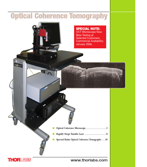
SPECIAL NOTE:OCT Microscope NowBeta Testing atSelected Customers.Commercial Availability-January 2006.■Optical Coherence Microscope . . . . . . . . . . . . . . . . . 2■Rapidly Swept T unable Laser■Spectral Radar Optical Coherence TImaging Specifications2D Cross-sectional OCT Imaging Capability Imaging Speed: 25 Frames Per Second (Based on 500 Axial Scans Per Frame)Maximum Image Size: 800 (W) x 512 (H) pixels Maximum Imaging Width: 6mm OCM1300SS Microscope System: Capable of simultaneous cross-sectional OCT imaging and conventional en-face microscope imaging as well as three-dimensional imaging of the sample.Sales: 973-579-7227The OCT system has sensitivity of 108 dB. The coherence length of the laser is measured to be >7 mm, which supports OCT imagingdepth of > 3 mm. The FWHW of the point-spread function of the interference fringes is measured to be ~12 µm for both forward and backward scan, suggest effective axial resolution of ~ 9 µm in tissue.Forward scan point spread function from seven A-scans each with a different delay.Backward scan point spread function from seven A-scans each with a different delay.Sales: 973-579-7227An OCT signal processing board is developed to accelerate the calibration of fringe signals from time to frequency. The clock boardprocesses the MZI clock signal to generate pulses equally spaced in frequency. The digitizer is configured in external clock mode and uses the clock pulses as time base to sample OCT fringe signals with data points linear in frequency.Backward scan point spread function showing the resolution of the system in air.Forward scan point spread function showing the resolution of the system in air.Real time video-rate imagingReal-time video-rate imaging speed with 17-30 frames/second based on 500 axial scans per frame and 1024 points Fast Fourier T ransform(FFT) can be achieved in the swept source OCT system.Results2D tomographic images of in-vivo human skin(d) palm(e) back of handOCT interferencefringes point spread function diagnosis (1D mode).3D volume imaging mode (500 x 500 x 500pixels) with en-face imaging capability.2D cross-sectional imaging mode (500 x 500 pixels)OCT ChannelCCD ChannelSales: 973-579-7227A 3 mm x 3 mm x 3mm volume containing 500 x 500 x 500 pixels3D imaging of the sample (top to bottom: IR card, screw, leaf, skin )Potential Applications1Biology and medical imaging2.3D optical profilometry3.Material inspection and product quality control4.Thin film test and measurement5.Other non-invasive laser imaging applicationsReferencesR. Huber, M. Wojtkowski, J. G. Fujimoto, J. Y. Jiang, and A. E. Cable, “Three-dimensional and C-mode OCT imaging with a compact, frequency swept laser source at 1300 nm”, submitted to Opt.Exp. 2005.AcknowledgementsWe acknowledge scientific discussions and helpful advice from Dr. Robert Huber and Prof. James G.Fujimoto at Research Laboratory of Electronic of Massachusetts Institute of Technology.Sales: 973-579-7227Swept Source Optical Coherence T omography (SS-OCT) applications require a laser that can be swept over a wide wavelength interval with very high speed. The wide wavelength range is required for obtaining high image resolution, and the high tuning frequency is needed for obtaining video image rates.Thorlabs is pleased to offer a fast sweeping, continuous wavelength, external cavity laser source specifically designed for SS-OCT applications. The standard system sweeps at least 100nm at a 16kHz repetition rate, offers a coherence length of 6mm and delivers more than 12mW of average optical power out of an SMF28 single mode fiber.The laser is available with the following fixed scanning frequency, coherence length and tuning range:■SL1325-P16-16KHz scanning frequency, 6mm coherence length, 120nm tuning rangeThe SL1325 comes with a Mach-Zender interferometer (MZI) with an adjustable free spectral range of 50 to 100 GHz. The MZI is used to digitally resample the raw Optical Coherence T omography (OCT) signal into equally spaced points in frequency. BNC connectors are available to monitor the wavelength sweep direction and laser intensity signals. The SL1325 comes in a 19 inch rack unit configured for 115VAC or 220/240 VAC.OCT is a relatively recent and fundamentally new way of obtaining high-resolution images in turbid media. Various OCT techniques are employed in dermatology, surgery (surgical guidance), and in ophthalmology. One of the driving forces of thedevelopment of the various OCT techniques is to find methods for in-vivo histology. However, the OCT techniques can be used for other types of characterization and visualizing of structures in turbid media, for example, materials research. The development of OCT started in the 1990’s and is a continuously growing field of research and usage.OCT image of in-vivo finger tissue OCT image of in-vivo palm tissueSL1325-P16Sales: 973-579-7227PARAMETERMIN TYPICAL MAX COMMENTSWavelength Range (nm)1265–1385Center Wavelength (nm)131513251340T uning Range (nm) (-10dB cut off point)100–140See models aboveRepetition Rate15–17See models aboveT uning Speed Continuous (nm/ms)–2000–Mean tuning speed around centerwavelengthAverage Optical Output Power (mW)101215Averaged over 1 secondOptical Power Stability (dB)––±0.5Forward to forward scan or backward backward scan during 1 hour usageSignal Source Spontaneous Emission Ratio (SSE) (dB)–25–Optical Isolation (dB)50––Operating Temperature + 10°C –+ 40°C Storage Temperature0°C –+ 70°C Diff. Optical Power at Forward and Back Scan(%)–20–Coherence Length (mm)6–8See models aboveLinear Polarization –>80 : 1–Measured at laser output facet Repetition Rate (kHz)101620Other rates on request Return Loss (dB)–45–Physical Size600 x 450 x 300Width x Depth x HeightSpectrum of the OCT swept laser showing an active wavelength tuning range of 155nm centered around 1325nm.MZI Clock: The frequency clock of the swept laser isfrom a build-in Mach-Zehnder interferometer with balanced detector output. The zero-crossings, as well as maxima and minima, of the interference fringe signals,are equally spaced in frequency and can be used asfrequency clock to synchronize other measurements.For Details on Our Full Lineof OCT ProductsVisit us at An OCT cross-section of a human fingernailSales: 973-579-7227HandheldScanning ProbeImaging Specifications (Other Options Available):■Imaging Speed:. . . . . . . . . . . . . . . . . . . . . . . . . . . . 2-5 Frames per Second Image Depth:. . . . . . . . . . . . . . . . . . . . . . . . . . . . . . . . . . . . . . . . . 1.6 mm Photograph A - Spectral Radar OCT Imaging SystemWith our system in mind, for a fixed position of the reference mirror, and a fixedFigure I -Schematic Device100nm FWHMFFTLD 930nmGratingS pectrometerbalances the desire for low scattering losses within biological samples with the need for operating inside the wavelength range of commercial silicon based linear sensor arrays. Many biological samples provide sufficient transparency at this with our standard system is the handheld application system depicted in Photograph B.Figure V – Solid model, cross sectional view of the handheld scanner probe that contains the free-space interferometer and telecentric imaging optics.Figure IV – Photograph of the handheld scanning probe in use imaging a volunteers arm.Fig. VIV – SR-OCT Surface Measurement of a Sinusoidal Survace.In vitro images from porcine retina and nerve head.SR-OCT Image of a thumb.。


光学断层相干扫描发展史1. 引言光学断层相干扫描(Optical Coherence Tomography,OCT)是一种高分辨率的非侵入性光学成像技术,能够实现对生物组织和材料的三维断层成像。
OCT技术的发展历程可以追溯到20世纪80年代,经过多年的研究和改进,已经成为医学、生物学和材料科学等领域中重要的成像工具。
本文将介绍光学断层相干扫描的发展历史,从早期的概念提出到现在的应用广泛,为读者提供一个全面详细、完整且深入的了解。
2. 早期概念提出光学断层相干扫描的概念最早由美国马萨诸塞州理工学院(MIT)的J.E. Swanson等人于1991年提出。
他们在一篇名为《光学相干域反射显微镜》的文章中,描述了一种利用干涉技术实现高分辨率断层成像的方法。
这篇文章提出了OCT的基本原理,即利用光的相干性实现对样品内部结构的成像。
3. 技术原理的发展在早期的OCT技术中,主要使用光纤光源和干涉仪来实现成像。
光纤光源的发展使得OCT系统的光源变得更加稳定和可靠。
干涉仪的设计和制造也得到了改进,使得相干光的干涉信号可以被准确地检测和分析。
随着技术的进步,OCT的分辨率也得到了提高。
早期的OCT系统分辨率较低,只能实现几十微米的成像分辨率。
然而,随着光源和探测器的改进,现代的OCT系统可以实现亚微米级别的分辨率,使得对生物组织的显微结构进行更加精细的观察成为可能。
4. 临床应用的发展OCT技术在临床应用中的发展也取得了重要的进展。
最早的临床应用是在眼科领域,用于眼底疾病的诊断和治疗。
OCT可以实现对视网膜和视神经的高分辨率成像,帮助医生更好地了解眼部疾病的发展和治疗效果。
随着技术的发展,OCT在其他临床领域也得到了广泛的应用。
例如,在皮肤科领域,OCT可以实现对皮肤组织的三维成像,用于皮肤病的诊断和治疗。
在牙科领域,OCT可以实现对牙齿和牙周组织的高分辨率成像,帮助牙医进行精确的治疗。
5. 生物学研究中的应用除了临床应用,OCT技术在生物学研究中也发挥着重要的作用。

Pentacam眼前节分析系统对早期圆锥角膜前后表面曲率及高度的研究张雪;胡琦;康杨;李雪;周文艳;王珂萌【摘要】Objective To evalute the anterior and posterior corneal curvature and elevation parameters provided by Scheimpflug device , which can effectively discriminated between subclinical keratoconus, keratoconus eyes, and normal eyes and to determin which acts on the diagnosis of keratoconus. Methods Retrospective case series. Sixteen eyes of 16 patients with clinical keratoconus, 19 eyes of 19 patients with subclinical keratoconus, and 56 eyes of 56 normal subjects. One eye from each patient was examined, the anterior and posterior corneal curvature and elevation parameters are provided by Scheimpflug device. The Mann-Whitney test, receiver-operating-characteristic ( ROC ) curves, and Partial Least Squares ( PLS )were used to compare the mean measurements and to evaluate the sensitivity and specificity of the parameters or constructed models. Results All of the parameters which are composed of anterior and posterior corneal curvature and elevation have more significant changes in eyes with subclinical or clinical keratoconus than in normal eyes ( P <0.05 ). The ROC curve analysis showed high overall predictive accuracy of all of the parameters in discriminating ectatic corneas from normal corneas. Partial Least Squares( PLS ) analysis showed that the goodness of fit of a model using a combination of 11 parameters of the corneal anterior surface was best in discriminating keratoconus or subclinical keratoconuseyes from normal eyes. Conclusion Posterior and anterior elevation, and curvature parameters measured by the Pentacam camera can effectively discriminate keratoconus from normal corneas serving as a useful diagnostic tool for disease staging.%目的应用Pentacam眼前节分析系统测得的角膜前后表面曲率、高度参数来探讨正常角膜与临床期圆锥角膜和亚临床期圆锥角膜的关系,以及Pentacam眼前节分析系统在圆锥角膜诊断中的作用.方法回顾性病例系列.选取临床期圆锥角膜患者16例(16只眼),亚临床期圆锥角膜19例(19只眼),对照组56例(56只眼)正常人群.每例患者选取一只眼,将Pentacam眼前节分析系统测得角膜前后表面曲率和前后表面高度参数进行差异检验以及受试者工作特征(ROC)曲线分析,初步筛选出有用参数,进一步进行偏最小二乘(PLS)分析,确定出最终诊断模型,并利用ROC曲线方法对模型作出评价.结果相对于正常患者,临床期患者与亚临床期患者的角膜前后表面曲率、高度参数有明显的变化.ROC曲线分析显示所有参数对于将圆锥角膜从正常眼中区分出来有较高的预测精准度.应用PLS 进行变量筛选并将VIP大于1.0的11个参数建立模型可以有效的将临床期圆锥角膜眼或亚临床期圆锥角膜眼从正常眼中区分出来.结论由Pentacam眼前节分析系统测得的角膜前后表面的曲率、高度参数可以有效地将圆锥角膜从正常角膜中区分出来,并且能作为疾病分期的诊断工具.【期刊名称】《临床眼科杂志》【年(卷),期】2012(020)006【总页数】4页(P496-499)【关键词】圆锥角膜;Pentacam;高度;曲率【作者】张雪;胡琦;康杨;李雪;周文艳;王珂萌【作者单位】150001,哈尔滨医科大学附属第一医院眼科视光学中心;150001,哈尔滨医科大学附属第一医院眼科视光学中心;150001,哈尔滨医科大学附属第一医院眼科视光学中心;150001,哈尔滨医科大学附属第一医院眼科视光学中心;150001,哈尔滨医科大学附属第一医院眼科视光学中心;150001,哈尔滨医科大学附属第一医院眼科视光学中心【正文语种】中文圆锥角膜是不伴有炎症反应的以角膜基质层进行性变薄、向前突出,引起不规则近视散光和不同程度矫正视力下降为特征的疾病。
IntroductionOCT (optical coherence tomography) is a novel noninvasive, noncontact imaging modality which produces high depth resolution (10 microns) cross-sectional tomographs of tissue. It is similar to ultrasound, except that optical rather than acoustic reflectivity is measured. Its value is given by the possibility of achieving pseudo-histological images of the target tissue. It is optically based, analogue to ultrasound B-scan examination and similar to laser reflectometry. Optical coherence tomography involves shining low-level infrared light on a tissue specimen, an interferometer and a computerized imaging system. Because of its high resolution, non-ionizing radiation, etc., in recent years some scholars use it in medical field. This essay will state theory, applications, Advantages and limitations of OCT.System principleLasers transmitting in the chaos medium will be scattering and absorption, and the intensity, coherence and polarization of the light will be changed. According to the number of incidence photons’ scattering, photons can be divided into 3 types: ballistic photon, snake-like photon and scattering photon. Ballistic photons, without scattering through the medium, retain coherence and lots of internal information of scattering medium. Scattering photons have multiple scattering; only bring a small amount of scattering media information. The initial characteristics of the photon are lost, especially coherence. Snake-like photons only have a small number of scattering, transmit with a small incident angle at the direction of axis, retain most of the characteristics of incident photons and have part of structure information of the media. OCT uses the medium information which carried by ballistic photon and the snake-like photon to make image.The core of the OCT system is a fiber-optic Michelson interferometer. Coupling light is transmitted into single-mode fiber. Output is divided by 3dB coupler into two. One of them through the confocal lens system and focused on the sample. The other one through the lens expand and irradiate to the reference mirror. It is used to be reference light. Two beams of light couple and re-join. As Optical path difference less than the coherent light length, the samples of the ballistic light and snake-like light happen to interfere. Interference signal DC circuit transmits through photomultiplier tube and amplifier, collected and processed by a computer. The two-dimensional data we get directly stand for record the organization of the scattering of incident light in samples of the situation regarding the depth and lateral position of function. The results of the data array directly treat as a gray-scale.ApplicationsOptical coherence tomography (OCT) has developed rapidly since its potential for applications in clinical medicine was first demonstrated in 1991. OCT performs high-resolution, cross-sectional tomographic imaging of the internal microstructure in materials and biologic systems by measuring back scattered or back reflected light. Mathematical models have been used to promote understanding of the OCT imaging process and thereby enable development of better imaging instrumentation and data processing algorithms. One of the most important issues in the modeling of OCT systems is the role of the multiple scattered photons, an issue, which only recently has become fully understood; the works of Thrane et al. and Turchin et al. representing the most comprehensive modeling. This technology was used in ophthalmic examination of the retina before. Later, was used for skin and gastrointestinal tract, such as study of incompletely transparent tissues. Until now, we can see the reports of the OCT for the field of stomatology. Colston reported for the first time, in 1998, used OCT technology to get the OCT image of the pig’s premolar dentin and periodontal tissues, Baumgartner in the same year has successful gotten clear OCT images of cementum sector, Amaechi, in 2001, has gotten a typical OCT images of caries tissues. However, these studies are limited to isolated teeth study. Specifically in the future, the success of the OCT detector for nonnasality study will make the OCT images of teeth-in-mouth possible.Results and interpretationsOptical coherence tomography is a new mode of high-resolution tomography, with optical technology and ultra-sensitive detector combined together, application of modern computer image processing, developed into an emerging diagnostic techniques tomography. Since 2001, at the first report of OCT technology in human coronary artery to obtain high-resolution images, the reports on OCT technology in the field of application of percutaneous coronary intervention are gradually increased. Now, the experts all over the world are pay attention to it. OCT has the following advantages: (1) it combines advantages of confocal and weak coherence, optical tomography scan, and has high detection sensitivity and resolution; (2) non-ionizing radiation, safety environmental protection; (3) No damage can be detected; (4) to adopt the optical communications fiber-optic components, it is cheap and portable. However, the shortcomings of the present are: (1) it should not distinguish between the lesion tissues with similar density; (2) incident factors can affect the imaging results; (3) image acquisition time is too long, short term getting a large number of images is still difficult; (4) image analysis software to be further researched and developed, and the lack of different races, different age groups of the normal database.ConclusionBased on the aspects above, it can be concluded that with the rapid development of biomedical Photonics technology, OCT technology will have greater scope for development, technology improvements will focus on the improvement of resolution and imaging depth on the increase. OCT is a young technology. Though it still has little limitations, we can not deny, OCT will become a common and effective means of medical diagnosis.ReferencesFroehly, L & Furfaro, L 2009, ‘Dispersion compensation properties of grating-based temporal-correlation Optical Coherence Tomography systems’, OPTICS COMMUNICATIONS, vol. 282, no. 7, pp1488-1495.Hillman, E &Burgess, S 2009, ‘Sub-millimeter resolution 3D optical imaging of living tissue using laminar optical tomography’, LASER & PHOTONICS REVIEWS, vol. 3, no. 1-2, pp 159-179.Rosenthal, N & Guagliumi, G 2009, ‘Comparison of Intravascular Ultrasound and Optical Coherence Tomography for the Evaluation of Stent Segment Malapposition’, JOURNAL OF THE AMERICAN COLLEGE OF CARDIOLOGY, vol. 53, no. 10, pp A22-A22, NEW YORK.Wolfgang, D & James, G 2008, ‘modeling Light–Tissue Interaction in Optical Coherence Tomography Systems’, Biological and Medical Physics, Biomedical, pp73-115.。
光学相干层析成像随深度变化的色散补偿方法光学相干层析成像(Optical Coherence Tomography, OCT)是一种非侵入式的高分辨率成像技术,在医学、生物学、材料科学等领域有广泛的应用。
然而,OCT成像中的色散效应会导致深度分辨率降低,影响成像质量。
因此,对于OCT成像中的色散补偿技术研究具有重要意义。
本文简要介绍几种OCT色散补偿方法,特别是基于正交多项式的色散补偿方法,重点阐述其随深度变化的色散补偿方法。
OCT成像中色散效应的原因是由于光在不同材料介质中传播速度不同而引起的,这会导致深度分辨率降低。
因此,为了提高OCT成像的深度分辨率,必须对色散效应进行补偿。
传统的色散补偿方法主要包括物理方法和数值方法。
物理方法包括改变光学系统的结构以减小色散效应、使用光学元件进行色散补偿等。
虽然这些方法可以有效减小色散效应,但是它们需要改变光学系统的结构,增加了系统的复杂度和成本。
数值方法则是基于数字信号处理技术进行色散补偿,其中包括后处理法和前处理法。
前处理法主要是通过加入折射率线性变化的模型来消除色散效应。
后处理法则是在成像过程中进行数据处理,利用信号的自相关性去除色散引起的谐波产生的影响。
但是,这些方法的精度和稳定性都受到限制。
近年来,一种基于正交多项式的新型色散补偿方法逐渐引起人们的关注。
这种方法可以快速准确地进行色散补偿,同时也可以随着成像深度的变化自适应地进行调整。
基于正交多项式的方法是一种数值方法,它基于光学相干检测信号的谐波公式,将光路径差与光的传输速度之间的关系表示为一个正交多项式展开式。
正交多项式与傅里叶变换类似,可以将时域信号转化为频域信号,从而实现色散补偿。
在这种方法中,光学相干检测信号首先进行二次谐波波形重建,其中第一个谐波代表检测光的中心波长,第二个谐波则代表了色散效应。
然后,使用正交多项式展开式对第二个谐波进行展开,得到每个深度处的色散系数和对应的光程延迟,利用这些参数对光路进行补偿。
㊃文献综述㊃Lenstar LS900的临床应用进展沈政伟1,薛林平1,2,莫 婷2,尹 禾1,姜 黎1作者单位:1(430070)中国湖北省武汉市,广州军区武汉总医院眼科医院眼科;2(430070)中国湖北省武汉市,湖北中医药大学临床医学院作者简介:沈政伟,男,主任医师,硕士研究生导师,研究方向:屈光学㊂通讯作者:薛林平,男,湖北中医药大学在读硕士研究生,研究方向:屈光学.Linping.235@收稿日期:2012-06-05 修回日期:2012-10-16 Research advances in clinical application of Lenstar LS900Zheng-Wei Shen1,Lin-Ping Xue1,2,Ting Mo2,He Yin1,Li Jiang11Department of Ophthalmology,Eye Hospital,Wuhan General Hospital of Guangzhou Military Command,Wuhan430070,Hubei Province,China;2Clinical Medical College,Hubei University of Traditional Chinese Medicine,Wuhan430070,Hubei Province,ChinaCorrespondence to:Lin-Ping Xue.Clinical Medical College, Hubei University of Traditional Chinese Medicine,Wuhan430070, Hubei Province,China.Linping.235@ Received:2012-06-05 Accepted:2012-10-16Abstract•There are many new instruments used to measure biometric measurements in recent years,such as intraocular len-master(IOL Master),ultrasound biomicroscope(UBM),Pentacam,optical coherence tomography(OCT),Orbscan,Lenstar LS900,Galilei, specular microscope,confocal microscope,and so on. Among these instruments mentioned above,the principle and clinical application are different.This article will review the principle,usage,clinical application of the Lenstar which is a new optical low coherence reflectometry device.•KEYWORDS:Lenstar;optical low coherence reflectometry;principle;biometric measurementsCitation:Shen ZW,Xue LP,Mo T,et al.Research advances in clinical application of Lenstar LS900.Guoji Yanke Zazhi(Int Eye Sci)2012;12(11):2123-2125摘要近年来,各种眼部生物参数测量仪不断改进和更新,如非接触式光学相干生物测量仪(intraocular len-master,IOL Master)㊁超声生物显微镜(ultrasound biomicroscope, UBM)㊁Pentacam眼前节综合分析系统(Pentacam)㊁光学相干断层成像术(optical coherence tomography,OCT)㊁Orbscan裂隙扫描角膜地形图(Orbscan)㊁Lenstar LS900光学生物测量仪(Lenstar LS900)㊁伽利略双通道Scheimpflug眼前节分析仪(Galilei)㊁角膜内皮显微镜(specular microscope)㊁共聚焦显微镜(confocal microscope)等,每一种测量仪的测量原理及临床应用范围各有不同㊂本文主要对新型的基于低相干光反射(optical low coherence reflectometry,OLCR)原理设计的非接触式的光学生物测量仪Lenstar LS900的测量原理㊁使用方法及临床应用进展进行综述㊂关键词:Lenstar;低相干光反射;原理;眼部生物参数DOI:10.3969/j.issn.1672-5123.2012.11.22引用:沈政伟,薛林平,莫婷,等.Lenstar LS900的临床应用进展.国际眼科杂志2012;12(11):2123⁃21250引言 测量眼部生物参数最常用的测量仪是超声(尤其是A超),随着超声测量仪的临床应用日益推广以及国内外的研究,超声测量仪的缺点及局限性被公认㊂在这样的背景下,临床医师和光学工程师开始寻求新的测量方法,先后出现了各种超声和光学测量仪㊂1999年上市的非接触式光学相干生物测量仪(IOL-Master)是目前被较为广泛地应用于测量眼部生物参数的测量仪之一㊂近几年上市的新型眼部生物测量仪 Lenstar LS900是由瑞士Haag-Streit公司和德围Wavelight公司联合研制的基于低相干光反射(optical low coherence reflectometry,OLCR)原理设计的非接触式的光学生物测量仪,可以一次测量角膜中央厚度(CCT)㊁前房深度(ACD)㊁晶状体厚度(LT)㊁眼轴长度(AL)㊁角膜曲率(K1㊁K2㊁AXIS)㊁角膜白到白的距离(W-W)㊁瞳孔直径(PO)㊁视网膜厚度(RT)等㊂本文主要对Lenstar LS900的测量原理㊁使用方法及临床应用进展进行综述㊂1测量原理 Lenstar LS900采用特殊的光学装置 迈克尔逊(Michelson)干涉仪,基于低相干光反射(optical low coherence reflectometry,OLCR)原理设计,采用820nm长的超辐射发光二极管(superluminescent diode,SLED)激光为光源,光谱宽度20~30nm,相干长度大约30μm,理论上具有良好的分辨率和精确性,这一特征使其优于其它光学测量技术[1]㊂眼部不同结构(角膜㊁晶状体和视网膜)的光反射与参考臂的光反射相干的叠加在一起,当患者注视测量光束,同时光束与反射界面垂直时,反射界面3212Int Eye Sci,Vol.12,No.11,Nov.2012 Tel:029⁃82245172 82210956 Email:IJO.2000@就形成干涉信号㊂由于干涉波的时差分离,角膜厚度㊁前房深度(包括或不包括角膜厚度)㊁晶状体厚度及眼轴可以一次测出,不需要重新对视轴进行定位调整[2,3]㊂Lenstar LS900是一种双区自动角膜曲率计,它测量分析投射在角膜表面直径大约为1.65mm 和2.3mm 的两个圆环光学区内32个光点的反射,计算出扁平K 值㊁陡峭K 值和平均K 值(屈光指数1.3375)[4]㊂系统内置有计算人工晶状体度数的各种公式及A 常数,自动计算出人工晶状体度数供临床医师选择㊂此外,Lenstar LS900能够自动监测受检者的固视情况和眨眼睛,只有好的结果才会被分析,进一步确保了测量结果的可靠性及准确性㊂2测量方法 受检者下颌置于仪器的下颌托上㊂令受检者注视仪器中闪烁的光束,以确保所有数据来源于视轴,当仪器的探头离受检者大约6.8cm 时,检查者按电脑屏幕提示进行对焦,系统自动测出眼部生物参数㊂每次的测量由快速连续的16次扫捕组成,通常测量5~6次,取平均值㊂3临床应用 通常情况下,评估新的测量仪的准确性是通过比较新旧两种测量仪测量结果的一致性,如果新的测量仪与相对旧的测量仪之间的一致性好,那么旧的测量仪就可能被替代㊂而评价两种测量仪之间的一致性之前,需要评价每一种测量仪的可重复性[5]㊂Shammas 等[4]认为Lenstar 测量眼部生物参数的可重复性好㊂Ronˇc evic 等[1]通过Lenstar 测量22例32眼患者的CCT,ACD,LT,AL,RT,K(K1,K2,AXIS),W -W,PO 共8个眼部生物参数,认为W-W 和AXIS 可重复性差,其余参数不同检查者的检查结果非常接近,具有良好的重复性㊂3.1角膜厚度 角膜厚度的准确测量具有重要的临床意义,如角膜屈光手术患者的筛选㊁高眼压患者应行角膜厚度检查,以排除角膜厚度的影响㊂Koktekir 等[6]使用光学法(Lenstar)与超声法测量65例130眼正视眼角膜厚度认为两者之间的相关性好,Lenstar 的重复性也好㊂Gursoy 等[7]也有类似的报道㊂而Chehab 等[8]的研究认为Lenstar LS900测量的角膜厚度大于超声测量值,差异有统计学意义㊂此外,Odonnell 等[9]和Huang 等[10]认为Lenstar 与Pentacam 测量角膜厚度的一致性较好,两者测量角膜厚度值可以替代㊂因此,还需要进一步研究Lenstar LS900测量角膜厚度的准确性㊂3.2前房深度 前房深度的定量测量对白内障㊁青光眼和有晶状体眼人工晶状体植入手术术前方案的选择和术后效果的评估具有十分重要意义[11]㊂Lenstar LS900可以测量真实的前房深度,即角膜内皮至晶状体前表面的距离㊂Gursoy 等[7]对565例受试者研究认为Lenstar LS900与超声测量前房深度一致性好,这与Salouti 等[12]的研究结果相反㊂黄锦海等[11]认为Lenstar LS900与IOL Master 测量前房深度的一致性较好,而Mylonas 等[13]认为Lenstar LS900测量值较IOL Master 大㊂Odonnell 等[9]通过使用Lenstar LS900,Pentacam,Visante AS-OCT 测量27眼的前房深度认为Lenstar LS900与Pentacam 的一致性较好,Lenstar LS900与Visante 的一致性较差㊂而Huang 等[10]认为Pentacam 和Lenstar LS900测量的前房深度值可以相互替代㊂研究结果的差异可能与研究对象的调节状态㊁屈光状态及样本大小不同有关,因此需要更进一步大样本研究㊂3.3晶状体厚度 人工晶状体度数的计算公式Holladay 2需要晶状体厚度这一参数㊂Gursoy 等[7]认为Lenstar LS900与超声测量晶状体厚度的一致性差,与Buckhurst 等[3]的研究结果相反㊂这种差异性可能与研究对象的调节状态有关㊂3.4眼轴长度 眼轴长度主要应用于人工晶状体度数的计算,其测量准确性是影响白内障术后屈光状态的重要因素之一㊂Gursoy 等[7]认为Lenstar LS900与超声测量眼轴一致性差,不可以相互代替㊂Lenstar LS900与IOL Master 测量眼轴差异无统计学意义[13-15]㊂宋慧等[16]也有类似的报道㊂叶向或等[17]认为Lenstar LS900与IOLMaster㊁A 超三者测量眼轴之间差异无统计学意义,具有良好的相关性㊂研究结果的差异性可能与超声法和光学法测量距离不同有关㊂超声测量眼轴是角膜表面至视网膜前界膜的距离,光学法测量眼轴是泪膜前表面至视网膜色素上皮之间的距离,是真正意义上的视轴㊂3.5角膜曲率 Mylonas 等[13]使用IOL Master 和Lenstar LS900对51例51眼白内障患者测量角膜曲率,差异有统计学意义㊂黄锦海等[11]也有类似的报道㊂由于Lenstar LS900和IOL Master 测量角膜曲率的原理不同,选择的屈光指数也不同,因此两种测量仪器测量的角膜曲率不可以相互替代[15]㊂3.6视网膜厚度 Read 等[18]使用Lenstar LS900和SDOCT 测量20例年轻受试者的视网膜厚度,两种测量仪测量值高度相关,一致性也好㊂3.7剥脱综合征 Bosnar 使用Lenstar LS900测量白内障患者224眼眼部生物参数(其中并发假性剥脱综合征47眼,无明显并发症177眼),认为假性剥脱综合征组具有明显的前房浅㊁晶状体厚㊁瞳孔小,Lenstar LS900能够在白内障术前发现剥脱综合征患者的悬韧带薄弱㊁晶状体稳定性差[19]㊂4总结 作为一种新型的非接触式眼部生物测量仪,国外学者对其研究较多,而国内关于Lenstar LS900的临床应用报道相对较少㊂Lenstar LS900一次测量就可以提供较多的临床参数供医师参考,具有测量过程非接触㊁方便快捷㊁节约时间等优点,同时也提高了患者和检查者的舒适度,具有广泛的应用前景㊂然而,Lenstar LS900测量的整个过程所需时间是IOL Master 的两倍[14]㊂作为一种光学测量仪,存在固有的弊端,对于各种原因引起的屈光介质混浊明显者,Lenstar LS900无法进行测量㊂对于硅油眼A 超无法测量,Lenstar LS900可以顺利测量㊂在临床使用中,我们需要掌握每种生物测量仪的实用范围以及测量生物参数的准确性㊂参考文献1Ronˇc evic BM,Buˇs ic M,ˇCima I,et al .Intraobserver and interobserver repeatability of ocular components measurement in cataract eyes using a new optical low coherence reflectometer.Graefes Arch Clin ExpOphthalmol 2011;249(1):83-872Rohrer K,Frueh BE,Wälti R,et al .Comparison and Evaluation of4212国际眼科杂志 2012年11月 第12卷 第11期 电话:029⁃82245172 82210956 电子信箱:IJO.2000@Ocular Biometry Using a New Noncontact Optical Low-Coherence Reflectometer.Ophthalmology2009;116(11):2087-20923Bunkhurst PJ,Wolfsohn JS,Shah S,et al.A new optical low coherence reflectometry device for ocular biometiy in cataract patients. Br J Ophthalmol2009;93(7):949-9534Shammas HJ,Hoffer KJ.Repeatability and Reproducibility of Biometry and Keratometry Measurements Using a Noncontact Optical Low-Coherence Reflectometer and Keratometer.Am J Ophthalmol 2012;153(1):55-615Bland JM,Altman DG.Statistical methods for assessing agreement between two methods of clinical ncet1986;1(8476): 307-3106Koktekir BE,Gedik S,Bakbak parison of Central Corneal Thickness Measurements With Optical Low-Coherence Reflectometry and Ultrasound Pachymetry and Reproducibility of Both Devices.Cornea 2012;31(11):1278-12817Gursoy H,Sahin A,Basmak H,et al.Lenstar versus ultrasound for ocular biometry in a pediatric population.Optom Vis Sci2011;88(8): 912-9198El Chehab H,Giraud JM,Le Corre A,et parison between Lenstar LS900non-contact biometry and OcuScan RXP contact biometry for task delegation.J Fr Ophthalmol2011;34(3):175-180 9Odonnell C,Hartwig A,Radhakrishnan parison of Central Corneal Thickness and Anterior Chamber Depth Measured Using LenStar LS900,Pentacam,and Visante AS-OCT.Cornea2012;31(9):983-988 10Huang J,Pesudovs K,Wen D,et parison of anterior segment measurements with rotating Scheimpflug photography and partial coherence reflectometry.J Cataract Refract Surg2011;37(2):341-34811黄锦海,陈世豪,温岱宗,等.Biograph/Lenstar与IOL Master测量眼轴㊁角膜曲率及前房深度的比较.中华眼视光学与视觉科学杂志2011;13(2):126-13012Salouti R,Nowroozzadeh MH,Zamani M,et parison of the ultrasonographic method with2partial coherence interferometry methods for intraocular lens power calculation.Optometry2011;82(3):140-147 13Mylonas G,Sacu S,Buehl W,et al.Performance of three biometry devices in patients with different grades of age-related cataract.Acta Ophthalmol2011;89(3):237-24114Chen YA,Hirnschall N,Findl O.Evaluation of2new optical biometry devices and comparison with the current gold standard biometer.J Cataract Refract Surg2011;37(3):513-51715Kolodzieiczyk W,Galecki T,Lazicka-Galecka M,et al. Comparison of the biometric measurements obtained using noncontact optical biometers LenStar LS900and IOL Master V.5.Klin Oczna 2011;113(1-3):47-5116宋慧,刑晓杰,汤欣.两种生物测量仪对人工晶状体度数测量准确性的比较.中国实用眼科杂志2011;29(7):713-71517叶向或,张文斌,王乐,等.白内障术前测量人工晶状体屈光度不同方法的对比研究.中华眼外伤职业眼病杂志2011;33 (12):905-90818Read SA,Collins MJ,Alonso-Caneiro D.Validation of optical low coherence reflectometry retinal and choroidal biometry.Optom Vis Sci 2011;88(7):855-86319Bosnar D,Kuzmanovic EB,Busic M,et al.Optical low-coherence reflectometry enables preoperative detection of zonular weakness in pseudoexfoliation syndrome.Graefes Arch Clin Exp Ophthalmol2012; 250(1):87-935212Int Eye Sci,Vol.12,No.11,Nov.2012 Tel:029⁃82245172 82210956 Email:IJO.2000@。
Covis ST 与 Pentacam 眼前节分析仪测量近视眼患者中央角膜厚度的差异李欣宇;赵海霞【摘要】目的:比较可视化角膜生物力学仪( Corvis ST )与Pentacam眼前节分析仪(下称Pentacam )测量近视眼患者中央角膜厚度的差异。
方法选择近视眼患者40例80眼,分别用Pentacam和Corvis ST测量角膜厚度。
结果40例近视眼患者角膜中央厚度Pentacam测量值为(535.79±30.76)μm,Corvis ST测量值为(531.16±30.29)μm, P>0.05。
相关分析显示,两种仪器测量结果密切相关(r=0.960,P<0.01)。
结论两种方法测量的中央角膜厚度值无差别,可以相互替换。
【期刊名称】《山东医药》【年(卷),期】2015(000)012【总页数】2页(P29-30)【关键词】近视眼;角膜厚度;眼前节分析系统;可视化角膜生物力学仪【作者】李欣宇;赵海霞【作者单位】内蒙古医科大学附属医院,呼和浩特010050;内蒙古医科大学附属医院,呼和浩特010050【正文语种】中文【中图分类】R778.1近年来,越来越多的近视眼患者选择手术来矫正视力,术前中央角膜厚度的测量对手术设计、术后预测及并发症预防具有重要意义。
目前,常用的测量方法有A型超声测厚仪、Pentacam眼前段分析仪(下称Pentacam)和Orbscan-Ⅱ眼前节分析仪等,其结果稍有不同。
可视化角膜生物力学分析仪(Corvis ST) 是一种联合Scheimpflug高速摄像和气冲印压技术测量角膜生物力学的新设备。
2014年7~10月,我们对比观察了Pentacam与Corvis ST测量近视眼患者中央角膜厚度的差异。
现报告如下。
1.1 临床资料选择内蒙古医科大学附属医院近视眼激光治疗中心就诊的患者40例80眼, 男25例50眼,女15例30眼;年龄18~36岁,平均25岁;术前屈光度(等效球镜)为(-1.25~-11.00)D,球镜(-0.75~-4.25) D,柱镜(2.75±0.61) D。
白内障术后前房深度的变化对术后屈光漂移的影响杨光耀; 张佳晴; 罗莉霞【期刊名称】《《国际眼科杂志》》【年(卷),期】2019(019)010【总页数】3页(P1676-1678)【关键词】白内障; 前房深度; 人工晶状体【作者】杨光耀; 张佳晴; 罗莉霞【作者单位】510060 中国广东省广州市中山大学中山眼科中心眼科学国家重点实验室【正文语种】中文0引言白内障是全球首位致盲眼病,手术是目前唯一有效的治疗方法。
随着技术的不断发展,白内障手术已经由复明手术时代转向屈光手术时代,白内障手术前准确的生物测量直接影响着人工晶状体(intraocular lens,IOL)度数计算以及患者术后的屈光状态。
白内障术后早期屈光状态的波动与IOL位置相关,术后IOL的位置变化会造成视网膜上的成像焦点的改变,从而影响患者的未矫正视力以及患者术后满意度,而前房深度(anterior chamber depth,ACD)被认为是白内障手术后影响实际IOL位置的重要因素[1-2] 。
本综述将从以下几个方面探讨术后ACD的变化和原因,以及ACD变化对术后早期屈光漂移的影响。
1 ACD的定义及对术后屈光的影响关于ACD的定义各不相同。
以往研究关于ACD的定义主要有2种,Holladay[3]将其定义为角膜后表面到有效晶状体平面的距离,而Olsen等[4]将其定义为角膜前表面至晶状体前表面的距离。
目前临床上,我们将有晶状体眼的ACD定义为从角膜后表面到晶状体前表面的距离。
同时,不同测量仪器对ACD的测量定义也略有差别。
目前临床常用的仪器中,LenStar测量的ACD是从角膜内皮到晶状体前表面的距离,IOL Master测量的ACD是从角膜上皮到晶状体前表面的距离,Pentacam则可以通过选择模式进行上述两种测量。
因此,IOL Master不能测量解剖意义上的ACD,正如使用手册所说不应该用于测量解剖意义上的IOL眼的ACD[5];比较IOL Master和Pentacam测量ACD(角膜上皮到晶状体前表面的距离)的准确性,发现这两种仪器测量得出ACD值的差异没有统计学意义[6]。