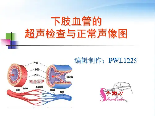
The anatomy of the peripheral veins
2. The main deep veins of the lower limb: Anterior tibial veins, Posterior tibial veins, Peroneal veins, Popliteal vein, Superficial femoral vein, Profunda femoris vein, Common femoral vein.
2. Patient position: in supine and erect position, prone.
the limb is supinated and slightly abducted.
Technical Aspects
3. Equipment requirement A high-capacity scan converter the upper limb: 5~10MHz the lower limb: 5~7MHz 4. Methods : longitudinal scan plane and transverse plane.
Atherosclerosis
1.B-mode images:The earliest ultrasound sign is thickening of the intima and media (>1.0mm) , in the bifurcation (>1.2mm).
Atherosclerosis
A longitudinal B-mode image of the superficial femoral artery, profunda femoris artery