A robust cut-cell method for
- 格式:pdf
- 大小:1.52 MB
- 文档页数:12
全自動活細胞影像分析測定系統 (IN Cell Analyzer)細胞水平功能測定及高內涵藥物篩選1. 緣由基於基因組學及蛋白質組學的發展及藥物篩選的需要,近一年來高內涵細胞及亞細胞水平的分析技術(High Content Screen, or HCS)發展神速。
大量的未知功能的基因及蛋白質的出現急需能有快速,定量定性的功能測定的技術,並且從DNA及蛋白質分子的研究轉入細胞中基因及蛋白質的功能研究。
藥物開發中也急需細胞水平的篩選來得到更多信息從而加快及降低藥物開發的成本。
全自動活細胞影像分析系統就是這一技術的代表。
從基因組及蛋白質組的研究將得到一系列與疾病有關的特定基因或蛋白,利用全自動活細胞影像分析測定系統,可快速定量的觀察到它們在細胞內的功能及作用機制,從而為疾病的產生,預防及治療提供依據,完成從基因,蛋白到細胞生物體內的完整資訊。
全自動活細胞影像分析測定系統結合高內涵細胞水平分析技術是近年來在藥物篩選中應用最廣的技術。
例如:治療SARS 中藥篩選,可透過全自動活細胞影像分析測定系統結合高內涵細胞水平分析技術篩選出對非典型肺炎不同病理環節和改善其臨床症狀有效的中成藥。
藥物篩選急需有新的方法來降低成本,提高效率,讓無效的候選藥物儘快在臨床測定前就被剔除,而在細胞水平層面的篩選完全符合這些要求。
觀察候選藥物在單個細胞中的活動情景能清晰的告訴我們藥物能否進入細胞,在細胞內如何分佈,候選藥物對細胞的作用如何,對細胞的毒性如何等多重資訊。
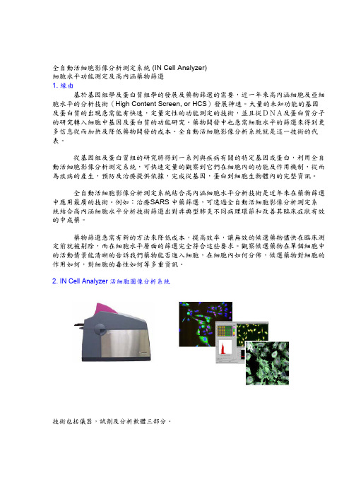
2. IN Cell Analyzer 活細胞圖像分析系統技術包括儀器,試劑及分析軟體三部分。
(1) 儀器儀器包括一個自動升降平台,高解析度CCD 相機,鐳射自動聚焦系統以滿足多螢光成像。
對於一個常規雙色成像,可在5 分鐘內或15 分鐘內完成96 孔板或384 孔板樣品分析。
IN Cell Analyzer 1000 提供多種圖像軟體後處理應用功能。
該系統也是一個開放平台,可容納多種實驗室自動系統,同時也可升級,增加如溫度控制或線上加樣等功能。

Sf-900™ II SFMDescriptionSf-900™ II SFM is a complete, serum-free, protein-free, ready-to-use insect cell culture medium developed for high cell-density growth and high-level recombinant protein expression using the Baculovirus Expression Vector System (BEVS). Sf-900™ II SFM has optimized amino acid, carbohydrate, vitamin, and lipid components, as well as a biologically active raw material providing significant improvement in cell growth, virus production and recombinant protein expression over other serum-free or serum-supplemented media. Sf-900™ II SFM supports long term growth (>20 passages) of Spodoptera frugiperda (Sf9, Sf21), Trichoplusia ni (Tn-368), and Lymantia dispar (Ld) cells in both suspension and monolayer culture.Product Catalog No. Amount Storage Shelf life*Sf-900™ II SFM (1X), liquid 10902-09610902-15310902-08810902-104500 mL10 × 500 mL1000 mL6 × 1000 mL2°C to 8°C; Protect from light 12 monthsSf-900™ II SFM (1X), liquid, Universal Bag 10902-16110902-17910902-187 5 L10 L20 L2°C to 8°C; Protect from light 12 monthsSf-900™ II SFM (1X), liquid w/out methionine or cysteine 21012-026 500 mL 2°C to 8°C; Protect from light 12 months * Shelf life duration is determined from Date of Manufacture.Product useFor Research Use Only. Not for use in diagnostic procedures. Important informationSf-900™ II SFM is a complete, ready to use medium. Do not addL-Glutamine or surfactants such as Pluronic® F-68.Safety informationRead the Safety Data Sheets (SDSs) and follow the handling instructions. Wear appropriate protective eyewear, clothing, and gloves.Culture ConditionsMedia: Sf-900™ II SFMCell line: Sf9, Sf21, Ld, Tn-368 cellsCulture type: Suspension or AdherentCulture vessels: Shake flask, spinner bottle or T-flask. Temperature range: 27°C to 28°CIncubator atmosphere: Non-humidified, air regulated non-CO2 atmosphere. Ensure proper gas exchange and minimize exposure of cultures to light.Recovery1.Rapidly thaw (<1 minute) frozen cells in a 37°C water bath.2.Transfer entire contents of the cryovial into theappropriately sized vessel so that the cells are seeded at3–5 × 10⁵ cells/mL of pre-warmed Sf-900™ II SFM.3.Incubate at 28°C in a non-humidified, air regulated non-CO2atmosphere, on an orbital shaker platform rotating at120–140 rpm. Loosen flask caps to allow for gas exchange. 4.Subculture when cells reach >2 × 10⁶ viable cells/mL. Werecommend subculturing cells a minimum of 3 passagesbefore using in downstream applications. Subculture suspension culturesInsect cells are sensitive to physical shearing. Ensure that impeller mechanisms rotate freely and do not contact vessel walls or base (adjust prior to autoclaving).1.Determine viable cell density using a Countess® AutomatedCell Counter or alternative automated or manual method. 2.Seed cells at 3–5 × 10⁵ viable cells/mL in sterile culturevessels containing pre-warmed Sf-900™ II SFM. (30 mL per125-mL shake flask, 75–100 mL per 100-mL spinner bottle).3.Incubate at 27°C to 28°C in a non-humidified, air regulatednon-CO2 atmosphere. Loosen caps to allow for gas exchange.4.Rotate shake flask cultures on an orbital shaker platform at120–140 rpm, set impeller stirring rate to 85–95 rpm forspinner bottles (optimum impeller speed must be empirically determined for each spinner apparatus for robust cell growth and viability).5.Subculture cells when viable cell density reaches>2 × 10⁶ viable cells/mL (about twice a week) into clean,sterile flask(s) with fresh pre-warmed Sf-900™ II SFM. Note: If cell debris is observed, gently centrifuge the cell suspension at 100 × g for 5–10 minutes and resuspend the cell pellet in fresh Sf-900™ II SFM to reduce accumulation of cell debris and metabolic waste by-products.Note: We recommend thawing a fresh low-passage vial of cells every 3 months or 30 passages.Subculture monolayer cultures1.Observe cell monolayer to ensure 80–90% confluence.Aspirate medium and floating cells from a confluentmonolayer.2.Add 4 mL (per 25 cm2) pre-warmed Sf-900™ II SFM to theflask and resuspend cells by repeatedly pipetting themedium across the monolayer.3.Observe cell monolayer to ensure cell detachment from thesurface of the flask. Firmly rap the side of the flask on thepalm of your hand or a hard flat surface if necessary.Publication Number MAN0007285 Rev. 1.002.4. Transfer entire cell suspension to a sterile conical tube; anycell clumps quickly settle to the bottom after 1–2 minutes. Pipet the clumps into a 10-mL pipette and gently break up the clumps by pressing the pipette tip against the bottom of the tube and gently expell the cells back into the medium, repeat if necessary to break up remaining clumps. Pipetting too harshly will decrease cell viability due to sensitivity of cells to shear force.5. Determine viable cell density using a Countess ® AutomatedCell Counter.6. Inoculate 2–5 × 10⁴ viable cells/cm 2 into new culture flaskscontaining pre-warmed Sf-900™ II SFM (5 mL/25 cm 2). 7. Incubate at 27°C to 28°C in a non-humidified, air regulatednon-CO atmosphere. Loosen caps to allow for gas exchange. 28. Three days post-plating, aspirate medium from the cellmonolayer and re-feed the culture with an equal volume of fresh medium gently added to the side of the flask. Note: Sf9 cells are not anchorage dependent and may be transferred between monolayer and spinner/shaker culture repeatedly without noticeable change in viability, morphology, or growth rate.For additional technical information such as Safety Data Sheets (SDS), Certificates of Analysis, visit /support For further assistance, email ************************All trademarks are the property of Thermo Fisher Scientific and its subsidiaries unless otherwise specified. Pluronic is a trademark of BASF Corporation. ©2014 Thermo Fisher Scientific Inc. All rights reserved.DISCLAIMER - LIFE TECHNOLOGIES CORPORATION AND/OR ITS AFFILIATE(S) DISCLAIM ALL WARRANTIES WITH RESPECT TO THIS DOCUMENT, EXPRESSED OR IMPLIED, INCLUDING BUT NOT LIMITED TO THOSE OF MERCHANTABILITY, FITNESS FOR A PARTICULAR PURPOSE, OR NON-INFRINGEMENT. TO THE EXTENT ALLOWED BY LAW, IN NO EVENT SHALLLIFE TECHNOLOGIES AND/OR ITS AFFILIATE(S) BE LIABLE, WHETHER IN CONTRACT, TORT, WARRANTY, OR UNDER ANY STATUTE OR ON ANY OTHER BASIS FOR SPECIAL, INCIDENTAL, INDIRECT, PUNITIVE, MULTIPLE OR CONSEQUENTIAL DAMAGES IN CONNECTION WITH OR ARISING FROM THIS DOCUMENT, INCLUDING BUT NOT LIMITED TO THE USE THEREOF.Adapt cells to Sf-900™ II SFMIt is critical that cell viability be ≥90% and the growth rate be in mid-logarithmic phase prior to initiating adaptation procedures.Direct adaptationMonolayer cultures need only have the culture media exchanged with prewarmed Sf-900™ II SFM as described in Subculture monolayer cultures.Transfer suspension cultures into Sf-900™ II SFM as follows: 1. Centrifuge the cell suspension at 100 × g for 5–10 minutes.Aspirate and discard the supernatant.2. Resuspend the cell pellet in pre-warmed Sf-900™ II SFM at aviable cell density of >5 × 10⁵ cells/mL and transfer to appropriate culture vessel.3.Return to incubator and monitor cell growth.Note: If suboptimal performance is achieved using the direct adaptation method, use the sequential adaptation method.Sequential adaptationFollow the procedures for subculture of suspension or monolayer cultures with the following modifications.1. During the adaptation procedure use a seeding density of>5 × 10⁵ viable cells/mL.2. Subculture cells into stepwise increasing ratios of Sf-900™ IISFM to original medium with each subsequent passage (25:75, 50:50, 75:25, 90:10 followed by 100% Sf-900™ II SFM). Multiple passages at each step may be needed.After several passages in 100% Sf-900™ II SFM, the viable cell count should exceed 2–4 × 10⁶ cells/mL with a viability exceeding 85% within 4–6 days of culture.Cryopreservation1. Prepare the desired quantity of cells, harvesting in mid-logphase of growth with viability >90%. Reserve theconditioned medium to prepare cryopreservation medium.Determine the viable cell density and calculate the requiredvolume of cryopreservation medium to give a final cell density of >1 × 10⁷ cells/mL.3. Prepare the required volume of cryopreservation medium of92.5% Sf-900™ II SFM (50:50 ratio of fresh to conditioned media) + 7.5% DMSO on day of intended use, store at 4°C until use.4. Centrifuge cell suspension at 100 × g for 5–10 minutes.Resuspend the cell pellet in the pre-determined volume of 4°C cryopreservation medium.5. Dispense aliquots of this cell suspension into cryovialsaccording to the manufacturer’s specifications.6. Cryopreserve in an automated or manual controlled ratefreezing apparatus following standard procedures (1°C decrease per minute).7. Transfer frozen cells to liquid nitrogen, (vapor phase)°C to –125°C.storage at –200Related productsProductCatalog no. Sf9 Cells Adapted in Sf-900™ II SFM 12659 Sf21 Cells Adapted in Sf-900™ II SFM 12682 BaculoDirect ™ N-Term Expression Kit 12562-054 BaculoDirect ™ N-Term Transfection Kit 12562-062 BaculoDirect ™ C-Term Expression Kit 12562-013 BaculoDirect ™ C-Term Transfection Kit 12562-039 Bac-N-Blue ™ Transfection KitK855-01 Bac-to-Bac ® Baculovirus Expression System 10359 Bac-to-Bac ® Vector Kit10360Countess ®Automated Cell CounterC10227Explanation of symbols and warningsThe symbols present on the product label are explained below:Temperature LimitationManufacturerBatch codeUse By:Catalog numberCaution, consult accompanying documentsConsult instructionsfor useKeep away from lightSterilized using aseptic processing techniquesLimited product warrantyLife Technologies Corporation and/or its affiliate(s) warrant their products as set forth in the Life Technologies’ General Terms and Conditions of Sale found on Life Technologies’ website at /termsandconditions . If you have any questions, please contact Life Technologies at /support .Important licensing informationThese products may be covered by one or more Limited Use Label Licenses. By use of these products, you accept the terms and conditions of all applicable Limited Use Label Licenses.。

cells 7,8. The method successfully detected thousands of genes expressed in mouse oocytes,and showed increased sensitivity compared with microarrays 7. However, this first single-cell mRNA-Seq experiment lacked technical controls, making it impossible to distinguishbiological variation between different cells from the technical variation that is intrinsic to cDNA amplification protocols when starting with low amounts of RNA. Therefore, thequestion remained whether single-cell transcriptomes faithfully represent the RNApopulation before amplification and how technical variation limits the power to finddifferential expression. This initial mRNA-Seq method also preferentially amplified the 3′ends of mRNAs, and hence the data could only be used to identify distal splicing events.Recently, a method for multiplexed single-cell RNA-Seq was introduced that quantifiestranscripts through reads mapping to mRNA 5′ ends 9. Neither of these methods generates read coverage across full transcripts. Since most mammalian multi-exons genes are subject to alternative RNA processing 4,5, there is a need for a single-cell transcriptome method that can both quantify gene expression and provide the coverage for efficient detection oftranscript variants and alleles.In this study, we introduce a single-cell RNA-Sequencing protocol with markedly improved transcriptome coverage, which samples cDNAs from more than just the ends of ing this protocol, we have sequenced the mRNAs from a large number of individualmammalian cells, as well as well-defined dilution series of purified total RNAs, tocomprehensively assess how sensitivity, variability and detection of differential expression vary with different amounts of starting material. Our results demonstrate the power ofsingle-cell RNA-Seq for both transcriptional and post-transcriptional studies, and provide valuable insights into the design of experiments that start from few or single cells. Todemonstrate the biological importance of this method, we have applied this new assay to putative circulating tumor cells (CTCs) captured from the blood of a melanoma patient to demonstrate how Smart-Seq enables high-quality transcriptome mapping in individual,clinically important cells.ResultsEfficient and robust single-cell RNA-Sequencing using Smart-SeqFor Smart-Seq, first we lysed each cell in hypotonic solution and converted poly(A)+ RNA to full-length cDNA using oligo(dT) priming and SMART template switching technology,followed by 12-18 cycles of PCR preamplification of cDNA. To enable gene and mRNA isoform expression analyses in single cells, a novel full-transcriptome mRNA-Seq protocol (Smart-Seq) was developed. Smart-Seq makes use of SMART ™ template switchingtechnology for the generation of full-length cDNAs and only 12 to 18 cycles of PCRfollowing the initial cDNA synthesis steps. The amplified cDNA was used to constructstandard Illumina sequencing libraries using either Covaris shearing followed by ligation of adaptors (PE) or Tn5-mediated “tagmentation” using the Nextera technology (Tn5). Both of these library preparation methods enable random shotgun sequencing of cDNAs(Supplementary Fig. 1). We successfully generated Smart-Seq libraries from 42 individual human or mouse cells, and in addition we generated 64 libraries from dilution series of total RNA derived from human brain (16 samples), mouse brain (28 samples) and universalhuman reference RNA (UHRR, 20 samples). Each sequencing library was sequenced on the Illumina platform, typically generating over 20 million uniquely mapping reads(Supplementary Table 1). For comparison, several standard mRNA-Seq libraries were also made from 100 ng to a few micrograms of total RNA.NIH-PA Author Manuscript NIH-PA Author ManuscriptNIH-PA Author ManuscriptSmart-Seq improves coverage across transcriptsIn previous single-cell mRNA-Sequencing studies 7,8, the data suffered from a pronounced 3′-end bias that limited analysis across full-length transcripts. We sequenced single-celltranscriptomes from mouse oocytes to enable a direct comparison with published mouse oocyte single-cell data 7. Analyses of read coverage across transcripts demonstrated thatSmart-Seq has significantly improved full-length coverage of all transcripts longer than 1kb (Fig. 1a and Fig. S2d–k). Smart-Seq analyses of mouse brain RNA at different dilutionsshowed that even better coverage was obtained with increased starting amounts, withnanogram dilutions reaching close to the coverage observed using standard mRNA-Seq from 100 ng to 1 ug total RNA (Fig. 1b). From only 10 pg input amounts (the estimated amount of RNA in a small eukaryotic cell, Supplementary Table 2) we achieved close to 40%coverage at the 5′ end. Analyses of single-cell transcriptomes from cancer cell lines (four cells each from LNCaP, PC3 and T24) obtained equally good read coverage (Fig. 1c) and,indeed, for 25% of all expressed, multi-exon genes our read coverage enabled full-length transcript reconstruction (exemplified in Fig. S3). We conclude that Smart-Seq hassignificantly improved read coverage compared to previous single-cell transcriptomemethods.Quantitative assessment of single-cell transcriptomicsAnalyses of gene expression from millions of cells using mRNA-Seq is highly reproducible and has low technical variation 1,4. So far, no single-cell mRNA-Seq study has measured the technical variation intrinsic to the cDNA pre-amplification components of single-cellmethods. We therefore diluted microgram amounts of reference total RNA down to nano-and picogram levels and applied Smart-Seq to assess sensitivity, technical variability and detection of differentially expressed transcripts of Smart-Seq on low amounts of total RNA.For comparison, standard mRNA-Seq libraries were generated from 100 ng to microgram levels of reference total RNA.First, we addressed the sensitivity of the method in detecting transcripts present at different expression levels. Starting with 10 ng or 1 ng of total RNA, we found no or minimal decline in sensitivity compared with standard mRNA-Seq. However, lowering the starting amounts to single-cell levels decreased the detection rate of less abundant transcripts (Fig. 2a).Analyses of the twelve cancer cell line cells (four cells each from the LNCaP, PC3 and T24lines) showed that ~76% of transcripts expressed at 10 RPKM (reads per kilobase exonmodel and million mappable reads), an expression level that roughly equals the medianexpression level for detected transcripts, were reproducibly detected in all single-cellprofiles (Fig. 2b). We found that the sensitivity of gene detection for the individual cancer cells was similar to that of about 20 pg of starting total RNA (Fig. 2b). Furthermore, weobserved that the starting amount of total RNA had a larger impact on sensitivity than the number of PCR cycles used (Supplementary Fig. 5) and that the sequence depth had little effect on transcript detection at levels above a million uniquely mapping reads per cell, with expression levels stabilizing after 3 million uniquely mapped reads (Supplementary Fig.4b,c). Comparisons of Smart-Seq and previous mouse oocyte data 7 demonstrated similar sensitivity (Supplementary Fig. 2a,b). We conclude that transcript detection sensitivity is affected by limiting starting amounts of RNA that lead to random loss of low abundance transcripts, but still the majority of low abundance and the vast majority of highly expressed transcripts are reliably detected even in single cells.Second, we determined the reproducibility in expression levels generated from diluted RNA and individual cells. Comparison of Smart-Seq and previous mouse oocyte data 7demonstrated improved expression level estimation with Smart-Seq (lower oocyte to oocyte variability) across the whole range of expression levels (Supplementary Fig. 2c). Correlation NIH-PA Author Manuscript NIH-PA Author Manuscript NIH-PA Author Manuscriptanalyses between technical replicates of diluted RNA showed increasing concordance with larger amounts of RNA. Comparing the single cells against the RNA dilution, we observed higher correlations (Pearson correlations of 0.75–0.85) among individual cells of the same type than among dilution replicates at 10 pg (Pearson correlations of 0.65–0.75)(Supplementary Fig. 6). Since variability in measurements of expression levels depends on transcript expression levels, we computed the variability as a function of the expressionlevel (Fig. 2c,d). This analysis showed that Smart-Seq on 10 ng total RNA had the same technical variability as standard mRNA-Seq and that Smart-Seq on 1 ng total RNA showed only a modest increase in technical noise (Fig. 2c). When lowering input amounts down to picogram levels, there was a clear increase in technical variability, particularly for lessabundantly expressed transcripts (Fig. 2c). The levels of technical variability at picogram levels of total RNA were compared to the biological variation found in comparisons ofhuman brain and UHRR using standard mRNA-Seq (Fig. 2c, green line). Interestingly,analyses of variation between individual cancer cells of different origin revealed extensive biological variation in highly expressed genes (Fig. 2d).Finally, we assessed whether pre-amplified single-cell expression profiles wererepresentative of the original expression profiles. Comparing relative gene expression levels (UHRR - brain) estimated using standard mRNA-Seq to those estimated from Smart-Seq with different amounts of input RNA, we again found a high concordance (Fig. 2e–g).Starting with 1 ng or 100 pg total RNA, the relative expression in Smart-Seq and standard mRNA-Seq respectively had Spearman correlations of 0.87 and 0.77 (Fig. 2e,f).Comparisons with 10 pg input RNA showed overall good correlation (Fig. 2g), butidentified two populations of transcripts with distorted expression in Smart-Seq data from either human brain or UHRR, reflecting stochastic losses, mostly of low abundancetranscripts when starting with such minute RNA of levels (Fig. 2g and Fig. 2a). Pre-amplification of cDNA could also lead to disproportionate amplification of short transcripts,but we found no systematic bias (Supplementary Fig. 7). A previous microarray studyanalyzed PCR amplified cDNA (from picogram levels) and found the transcriptome overall preserved, but skewed 10. Our data from 1 ng and 100 pg total RNA show no skewing, i.e.the loess slopes estimated from the data approximated 1 (Fig. 2e–g). Together, these results demonstrated that transcriptome analyses from few or single cells, in general, preservedrelative expression level differences for detected transcripts.Analyses of transcriptional and post-transcriptional differences from single-cellsHaving demonstrated the improved performance of Smart-Seq on low amounts of RNAcompared to previously published methods, we focused our analyses on single-celltranscriptomes from prostate (PC3, LNCaP) and bladder (T24) cancer cell line cells. The twelve individual cells (four from each cell line) clustered according to cell line of origin using their global gene expression levels and we identified hundreds of differentiallyexpressed genes among the three cell lines (Fig. 3a; q<0.05 ANOVA; p<0.05 post-hoc test).The pronounced 3′-end bias of previous single-cell mRNA-Seq studies has hampered the ability to identify significant alternative splicing differences in single cells. We used theBayesian mixture of isoforms framework (MISO)11 to infer exon inclusion levels for known alternatively spliced exons in the twelve individual cells. The improved read coverage with Smart-Seq resulted in a two-fold increase in the number of alternatively spliced exons that could be assessed, compared to previously published single-cell mRNA-Seq data (Fig. 3b),significantly improving our ability to detect alternative splicing. Cell-type specificalternative splicing could be inferred from single-cell transcriptomes, as seen in readcoverage across the differentially included exon 13 of the NEDD4L gene (Fig. 3c). This exon was frequently included in LNCaP cells (93% mean inclusion level) but was included at much lower levels in T24 cells (15% mean inclusion levels) whereas low expression ofNIH-PA Author Manuscript NIH-PA Author Manuscript NIH-PA Author ManuscriptNEDD4L in PC3 cells precluded inclusion level estimation. In total, in this comparison of three cancer cell lines, we found one hundred exons with differential exon inclusion levels among the three cell lines, with a less than 1% false discovery rate (Fig. 3d; Supplementary Table 3). We conclude that Smart-Seq significantly improves our ability to detect alternative RNA processing in single cells.Analyses of circulating tumor cell transcriptomes Having demonstrated that Smart-Seq generates quantitative and reproducible single-cell transcriptomes, we asked whether global transcriptome analyses of putative circulating tumor cells (CTCs) could reveal their tumor of origin and provide data to support the use of this method for unbiased cancer-specific biomarker identification. To this end, we generated transcriptomes from NG2+ putative melanoma circulating tumor cells (CTCs) isolated from peripheral blood drawn from a patient with recurrent melanoma using immunomagnetic purification with a MagSweeper instrument (Illumina Inc.)12. For comparison, we also generated Smart-Seq libraries from single cells derived from primary melanocytes (PMs,n=2), melanoma cancer cell line (SKMEL5, n=4 and UACC257, n=3) cells and from human embryonic stem cells (ESCs, n=8). Since the NG2+ putative CTCs were isolated from blood,it was important to compare them to blood cells. The putative CTCs were distinct from lymphoma cell lines (BL41 and BJAB)13 and immune tissues (lymphnode and white blood cell samples), as well as embryonic stem cells, and instead showed high similarity to PMs and melanoma cell line cells. Unsupervised hierarchical clustering and correlation analyses of gene expression levels showed a clear clustering of cells according to cell type of origin (Fig. 4a and Supplementary Fig. 8), and separation from the human brain RNA samples that were previously analyzed with Smart-Seq or mRNA-Seq (data not shown). Further support for the melanocytic origin of the putative melanoma CTCs came from analyses of melanocyte lineage specific markers, as all NG2+ cells expressed high levels of MLANA 14,TYR 15 and the melanocyte specific m-form of MITF 16 but not immune markers such as PTPRC (Fig. 4b), in contrast to peripheral blood lymphocytes (Supplementary Fig. 9).Furthermore, NG2+ cells expressed high levels of melanoma-associated genes (based on our unbiased selection of the 100 transcripts most strongly associated with melanoma, seeMethods), but not immune cell-associated genes selected in a similar manner (Fig. 4c,p<3.7e-15, Wilcoxon rank sum test). Thus, both their global transcriptomes and expression patterns of melanoma-associated transcripts clearly support a melanocyte origin for theNG2+ cells putative melanoma CTCs.We next investigated whether the NG2+ putative CTCs showed signs of originating from a melanoma tumor. Comparison of their gene expression profiles with those of individual PMs identified 289 genes with higher expression in the putative CTCs than the PMs, and 436genes with significantly lower levels (Supplementary Table 4). The up-regulated genes were significantly enriched (FDR<0.05) for melanoma-associated antigens (Fig. 4d andSupplementary Fig. 10) that have been repeatedly found to be upregulated in cancer 17,mitotic cell cycle genes and additional categories (Supplementary Table 5). Down-regulated genes showed enrichment for regulators of cell death and MHC class I genes. Interestingly,the preferentially expressed antigen in melanoma (PRAME) was found highly expressed in NG2+ cells, which together with elevated expression of known melanoma tumor antigens (MAGEs), provides strong support for the conclusion that the NG2+ cells were CTCs that have originated from a melanoma.In recent years, there has been a strong interest in identifying CTCs from different tumors using the a priori assumption that plasma-membrane proteins would be good diagnosticbiomarkers. We used the CTC transcriptome analysis to screen for membrane proteinsselectively expressed in melanoma-derived CTCs compared to PMs and immune cells. We identified 9 up-regulated plasma membrane-associated transcripts in the CTCs comparedNIH-PA Author ManuscriptNIH-PA Author ManuscriptNIH-PA Author Manuscriptwith primary melanocytes (q<0.05 ANOVA; p<0.05 post-hoc test), many of which are not expressed in immune cells and have not been previously associated with melanomas (Fig.4e). Similarly, screening for loss of expression of plasma-membrane proteins identified 37genes with significantly lower expression in the CTCs than PMs (Fig. 4f). Of note, epithelial Cadherin 1 (CDH1) showed no expression in the CTCs, and loss of CDH1 is thought tocontribute to cancer progression by increasing proliferation, invasion and metastasis 18. We also found downregulation of genes associated with the escape from immune surveillance,including five HLA genes (Fig. 4f), and TRPM1, suggesting that these gene expressionchanges might enable the CTCs to escape from immune surveillance. Notably, lowexpression of TRPM1 has been shown to correlate with melanoma aggressiveness andmetastasis 19. Future studies of these membrane proteins will likely enhance ourunderstanding of CTC migration and invasiveness, and these results highlight the utility of studying single CTC cells with RNA-Seq.Lastly, we investigated whether Smart-Seq transcriptome data could be mined for SNPs and other genetic variants associated with melanomas or other cancers. With the improved read coverage provided by the Smart-Seq method, we were able to identify 4,312 high-confidence genomic sites with support for an alternative allele in at least two CTCs, whereas genotype calls only supported by a single cell showed an excess of novel, likely artifactual,sites (Supplementary Fig. 11) together with a smaller subset (9%) of A-to-G RNA editing sites (data not shown). Ninety-two percent of the high-confidence sites coincided withdocumented SNPs, for example the melanoma-associated SNP in the TYR gene(rs1126809)20 (Fig. 4g). We conclude that Smart-Seq enables screening for SNPs andmutations in transcribed regions using only few cells.Discussion Generating high-coverage transcriptomes from single cells and small numbers of cells will have many applications for studying rare cells; such cells can be either individually picked or identified through cell sorting or laser capture techniques. Our results demonstrate that using Smart-Seq on 10 ng of total RNA is practically indistinguishable from a standard mRNA-Seq, whereas starting with 1 ng (corresponding roughly to 50–100 cells) shows only a minor increase in expression level variability. Therefore, this method could easily be applied to studies on homogeneous cell populations. However, many biologically and clinically important cell types exist in rare quantities and often in heterogeneous milieus,which necessitates single-cell approaches. Smart-Seq generates robust and quantitative transcriptome data from single cells. We found hundreds of differentially expressed genes using only a few individual cells per cell type, e.g. comparing only two primary melanocytes to six melanoma CTCs identified biologically meaningful differences. Even sequencing of a single cell yields useful information, as we, in each cell, detected most of the genes active in a culture of LNCaP cells. Importantly, Smart-Seq has significantly improved read coverage across transcripts, which enables detailed analyses of alternative splicing and identification of SNPs and mutations. Based on our CTC transcriptome results, single-cell analyses are highly informative for identifying candidate biomarkers, SNPs and mutations. In conclusion,datasets obtained with the Smart-Seq protocol provide significantly improved representation of the transcriptomes of individual cells, with relevance for both basic and clinical studies.Methods Generation and amplification of Smart-Seq cDNAWe have developed a new method for the generation of cDNA from total RNA or fromsingle cells, called Smart-Seq. Briefly, polyA + RNA was reverse transcribed through tailed oligo-dT priming directly in total RNA or a whole cell lysate using Moloney MurineNIH-PA Author Manuscript NIH-PA Author ManuscriptNIH-PA Author ManuscriptLeukemia Virus Reverse Transcriptase (MMLV RT). Once the reverse transcription reaction reaches the 5′ end of an RNA molecule, the terminal transferase activity of MMLV adds a few non-templated nucleotides to the 3′ end of the cDNA. The carefully designedSMARTer II A oligo then base-pairs with these additional nucleotides, creating an extended template. The reverse transcriptase then switches templates and continues transcribing to the end of the oligonucleotide. The resulting full-length cDNA contains the complete 5′ end of the mRNA, as well as an anchor sequence that serves as a universal priming site for second strand synthesis. The cDNA is then amplified using 12 cycles for 1 ng of total RNA, 15cycles for 100 pg of total RNA, and 18 cycles for 10 pg total RNA or from single cells. The exact number of cycles for each dilution replicate or single-cell is detailed in Supplementary Table 1. The Smart-Seq cDNA generation and amplification methods developed for this manuscript have recently become available in a kit marketed by Clontech called the“SMARTer Ultra Low RNA Kit for Illumina sequencing”. Although all the libraries in this manuscript were generated before the kit became commercially available, our protocol is reflected in the detailed instructions for generating cDNA from few cells or 100 pg–10 ng of total RNA that is now included in the manual for this kit. For single cell applications, each cell (or control RNA) was added in max 1 λ of media to 4 λ of hypotonic lysis bufferconsisting of 0.2% Triton X-100 and 2 U/μl of ribonuclease (RNase) inhibitors (Clontech,2313B) in RNase free water. The deposition of an intact cell in the hypotonic lysis buffer leads to immediate lysis and stabilization of the RNA through RNase inhibitors. Then,poly(A)+ RNA was reverse-transcribed through tailed oligo(dT) priming using the CDSprimer (5′-AAGCAGTGGTATCAACGCAGAGTACT(30)VN-3′, where V represents A, C or G) directly in total RNA or a whole cell lysate using Moloney murine leukemia virusreverse transcriptase (MMLV RT).Construction and sequencing of Smart-Seq sequencing librariesAmplified cDNA (~5ng cDNA) was used to construct Illumina sequencing libraries using either Illumina’s “Ultra Low Input mRNA-Seq Guide” (the “PE” protocol) or a modification of Epicentre’s Nextera DNA sample preparation protocol (the “Tn5” protocol). With the PE protocol, the amplified cDNA was fragmented using a Covaris acoustic shearing instrument.The resulting fragments were end-repaired, followed by the addition of a single A base,ligation to Illumina PE adaptors, and then amplification with 12–18 cycles of PCR(depending on starting amounts of RNA, see Supplementary Table 1 for detailed instructions of all libraries generated). With the Tn5 protocol, the amplified cDNA was “tagmentated” at 55°C for 5 min in 20μl reaction with 0.25μl of transposase and 4μl of 5x HMW Nextera reaction buffer. 35μl of PB was added to the tagmentation reaction mix to strip thetransposase off the DNA, and the tagmentated DNA was purified with 88μl of SPRI XP beads (sample to beads ratio of 1:1.6). Purified DNA was then amplified by 9 cycles ofstandard Nextera PCR. The libraries were sequenced on either Illumina’s HiSeq 2000,GAIIx or MiSeq instruments and all clusters that passed filter were exported into fastq files.Details on the sequence depth, sequencing platform and library construction method for each dilution replicate and single cell are included in Supplementary Table 1. All data shown in the figures of this manuscript were generated using the PE protocol unless otherwisespecified in the figure legend.Construction and sequencing of standard mRNA-Seq librariesWe generated mRNA-Seq transcriptome data following the Illumina mRNA-Seq kit from 100 ng and 1 microgram of total RNA, as detailed in Supplementary Table 1.Isolation of individual CTCs from peripheral blood10 ml of peripheral blood was collected from a male patient with recurrent, metastaticmelanoma using K2 EDTA blood collection tubes (Becton Dickinson, NJ, USA). The bloodNIH-PA Author Manuscript NIH-PA Author Manuscript NIH-PA Author Manuscriptsample was processed within 3 hours of collection. The erythrocytes in 4.5 ml of the blood sample were lysed with BD Pharm Lyse ™ lysing solution (Becton Dickinson, NJ, USA) for 10 minutes at room temperature. The nucleated cells were pelleted, resuspended in HBSS containing 1% BSA and 5mM EDTA, pelleted, resuspended in 1ml of HBSS containing 1%BSA and 5mM EDTA. The nucleated cells were stained with PE-conjugated anti-human CD45 IgG to label leukocytes. The cells were subsequently reacted with biotinylated anti-human CSPG4 (a.k.a. NG2) mouse IgG at 4 °C for 2 hours, washed with HBSS, and reacted with streptavidin-conjugated MG980A magnetic beads at 4 °C for 2 hours. The cells were captured based on magnetic sweeping to harvest the beads from cell suspension using the MagSweeper instrument (Illumina Inc.) as previously described 12. The harvested cells were stained with 5 μg/ml Calcein AM (Life Technologies, Carlsbad, CA) in HBSS for 20minutes to identify viable cells. Manual picking of viable cells showing desired Calcein-positive/CD45-negative/bead-attached profile was performed to isolate cells for molecular profiling. The individual cells were placed into 2.5 μl of Superblock (Thermo Scientific,Waltham, MA) containing 4000 Unit/ml RNase inhibitor (New England Biolabs, Ipswich,MA) and stored at −80 °C until preparation of Smart-Seq libraries.Isolation of mouse oocytes and human lymphocytesMII oocytes were isolated from 4-week old CAST/EiJ female mice. Mice weresuperovulated by injection of 5 IU PMSG, followed by injection of 5 IU of hCG 48 hrs later.MII oocytes were isolated 14–15 hrs after hCG treatment by dissection of the ampulla of the oviduct and cumulus cells were removed by hyaluronidase digestion. Single oocytes were manually picked, lysed in dilution buffer, and cDNA constructed as described above.Peripheral blood lymphocytes from healthy human volunteers were isolated on Ficollgradients using LymphoPrep (Fresenius Kabi, Norway). Individual cells were manuallypicked into lysis buffer and cDNA constructed as described above.Alignment of short reads to genome and transcriptomeReads were independently aligned using Bowtie 21 against the respective genome assembly (hg19 or mm9) and transcriptome sequences (Ensembl, human and mouse annotationsdownloaded 16th May 2011 and 13th Dec 2010, respectively). Transcriptome mapped reads were converted from transcriptome coordinates to genomic coordinates and thereaftercompared with the genome mapped reads to identify reads that map to a unique genomiclocation. This procedure ensured that mapped reads were unique across both the genome and transcriptome, while allowing for reads to map to different transcripts of the same gene in the initial transcriptome mapping. The uniquely mapped reads were converted to binaryBAM files using Samtools 22. The resulting transcriptome data was visualized using theIntegrated Genome Viewer (IGV, Broad Institute) using the histogram visualization for Fig.1a,d and heatmap visualization for Supplementary Fig. 3.Expression level estimation and technical comparisons of sensitivity and variationGene expression levels for Refseq transcripts were summarized as RPKM values and read counts using rpkmforgenes 23. RefSeq annotations for human and mouse were downloaded on the 31st August 2011 and 13th Dec 2010 respectively. RPKM calculations onlyconsidered uniquely mappable positions for transcript length normalizations using theENCODE Mappability track (wgEncodeCrgMapabilityAlign50mer.bigWig) for human and in-house computed uniqueness files for mouse. Overlapping RefSeq transcripts werecollapsed giving one expression value per gene locus. Only 10 million randomly selected mapped reads were used per sample in order to compare sensitivity and variation in gene and exon levels. Samples with fewer than 10 million uniquely mappable reads (a few ES cells 8) were therefore discarded from analyses. Samples with 20 pg of total RNA (used in Fig. 2b,d) were simulated by using 5 million reads each of two different 10 pg samples.NIH-PA Author Manuscript NIH-PA Author Manuscript NIH-PA Author Manuscript。

Vero Cell Host Cell Proteins Immunoenzymetric Assay for the Measurement of Vero Cell Host Cell ProteinsCatalog # F500Intended UseThis kit is intended for use in determining the presence of host cell protein impurities in products manufactured by expression in Vero cells. The kit is for Research and Manufacturing Use Only and is not intended for diagnostic use in humans or animals.Summary and ExplanationExpression of viral vaccines and other therapeutic proteins in Vero cells is a cost effective method for production of commercial quantities of a drug substance. The manufacturing and purification process of these products leaves the potential for impurities by host cell proteins (HCPs) from Vero cells. Such impurities can reduce the efficacy of the therapeutic agent and result in adverse toxic or immunological reactions and thus it is desirable to reduce HCP impurities to the lowest levels practical.Immunological methods using antibodies to HCPs such as Western Blot and ELISA are conventionally accepted. While Western blot is a useful method aiding in the identity of HCPs, it suffers from a number of limitations. Western blot is a complex and technique dependent procedure requiring subjective interpretation of results. Furthermore, it is essentially a qualitative method and does not lend itself to obtaining quantitative answers. The sensitivity of Western blot is severely limited by the volume of sample that can be tested and by interference from the presence of high concentrations of the intended product. While Western Blot may be able to detect HCPs in samples from upstream in the purification process, it often lacks adequate sensitivity and specificity to detect HCPs in purified downstream and final product. The microtiter plate immunoenzymetric assay (ELISA) method employed in this kit overcomes the limitations of Western blots providing on the order of 100 fold better sensitivity. This simple to use, objective, and semi-quantitative ELISA is a powerful method to aid in optimal purification process development, process control, routine quality control, and product release testing. This kit is “generic” in the sense that it is intended to react with essentially all of the HCPs that could pollute the product independent of the purification process. The antibodies have been generated against and affinity purified using mild lysate of Vero cells. The resulting antibodies have then been characterized against four commercial cell lines used to produce various viral and protein products. This analysis indicated the vast majority of HCPs are conserved among multiple Vero cell lines and product purification processes. If you have a need of a more sensitive method to demonstrate coverage to HCPs in your process Cygnus Technologies recommends a method that is superior to Western blot called Antibody Affinity Extraction (AAE). AAE is has greatly increased sensitivity and specificity to Western blot which makes it a better predictor of how the antibodies will perform in the ELISA. For additional information on AAE please visit our website and read the posted articles under Technical Documents or contact our Technical Services Department.Special procedures were utilized in the generation of these antibodies to ensure that low molecular weight and less immunogenic impurities as well as high molecular weight components would be represented. As such, this kit can be used as a process development tool to monitor the optimal removal of host cell impurities as well as in routine final product release.This highly sensitive ELISA kit has been qualified for testing of final product HCPs using actual in-process and final drug substance samples from 4 vaccine products all with somewhat different growth and purification processes. Each user of this kit is encouraged to perform a similar qualification study to demonstrate it meets their analytical needs. Provided this kit can be satisfactorily qualified for your samples, the application of a more process specific assay may not be necessary, in that such an assay would only provide information redundant to this generic assay. However, if your qualification studies indicate the antibodies in this kit are not sufficiently reactive with your process specific HCPs it may be desirable to also develop a more process specific ELISA. This later generation assay may require the use of a more specific and defined antisera. Alternatively, if the polyclonal antibody used in this kit provides sufficient sensitivity and broad antigen reactivity, it may be possible to substitute the standards used in this kit for ones made from the impurities that typically co-purify through your purification process and thus achieve better accuracy for process specific HCPs. The use of a process specific assay with more defined antigens and antibodies in theory may yield better specificity, however such an assay runs the risk of beingtoo specific in that it may fail to detect new or atypical impurities that might result from some process irregularity or change. For this reason it is recommendedth at a broadly reactive “generic” host cell protein assay be used as part of the final product purity analysis even when a process specific assay is available. If you deem a more process specific assay is necessary, Cygnus Technologies is available to apply its proven technologies to develop such antibodies and assays on custom basis.The Vero cell assay is a two-site immunoenzymetric assay. Samples containing Vero cell HCPs are reacted simultaneously with a horseradish peroxidase (HRP) enzyme labeled anti-Vero cell antibody (goat polyclonal) in microtiter strips coated with an affinity purified capture goat polyclonal anti-Vero cell antibody. The immunological reactions result in the formation of a sandwich complex of solid phase antibody-HCP-enzyme labeled antibody. The microtiter strips are washed to remove any unbound reactants. The substrate, tetramethylbenzidine (TMB) is then reacted. The amount of hydrolyzed substrate is read on a microtiter plate reader and is directly proportional to the concentration of Vero cell HCPs present.Storage & Stability•All reagents should be stored at 2︒C to 8︒C for stability until the expiration date printed on the kit. •After prolonged storage, you may notice a salt precipitate and/or yellowing of the washconcentrate. These changes will not impact assayperformance. To dissolve the precipitate, mix thewash concentrate thoroughly and dilute asdirected in the ‘Preparation on Reagents’ section. •Reconstituted wash solution is stable until the expiration date of the kit. •Microtiter plate reader spectrophotometer with dual wavelength capability at 450 & 650nm. (Ifyour plate reader does not provide dualwavelength analysis you may read at just the450nm wavelength.)•Pipettors - 50μL and 100μL•Repeating or multichannel pipettor - 100μL •Microtiter plate rotator (400 - 600 rpm) •Sample Diluent (recommended Cat # I028) •Distilled water• 1 liter wash bottle for diluted wash solution•For Research or Manufacturing use only. •Stop reagent is 0.5M H2SO4. Avoid contact with eyes, skin, and clothing.•This kit should only be used by qualified technicians.•Bring all reagents to room temperature. •Dilute wash concentrate to 1 liter in distilled water, label with kit lot and expiration date, and store at4︒C.Assay Protocol•The assay is very robust such that assay variables like incubation times, sample size, and othersequential incubation schemes can be altered tomanipulate assay performance for moresensitivity, increased upper analytical range, orreduced sample matrix interference. Beforemodifying the protocol from what is recommended,you are advised to contact Technical Services forinput on the best way to achieve your desiredgoals.•The protocol specifies use of an approved orbital microtiter plate shaker for the immunologicalsteps. These can be purchased from mostlaboratory supply companies. If you do not havesuch a device, it is possible to incubate the platewithout shaking however, it will be necessary toextend the immunological incubation step in theplate by about one hour in order to achievecomparable results to shaking protocol. Do notshake during the 30-minute substrateincubation step, as this may result in higherbackgrounds and worse precision.800-F500, Rev. 3, 26DEC2019 Vero Cell HCP ELISA Product Insert 2•Bring all reagents to room temperature. Set-up plate spectrophotometer to read dual wavelengthat 450nm for the test wavelength and ~650nm forthe reference.•Thorough washing is essential to proper performance of this assay. Automated platewashing systems or other vacuum aspirationdevices are not recommended. The manualmethod described in the assay protocol ispreferred for best precision, sensitivity andaccuracy. A more detailed discussion of thisprocedure can be obtained from our TechnicalServices Department or on our web site. Inaddition, a video demonstration of proper platewashing technique is available in the ‘TechnicalHelp’ section of our we b site.•All standards, controls, and samples should be assayed at least in duplicate.•Maintain a repetitive timing sequence from well to well for all assay steps to ensure that all incubationtimes are the same for each well.•Make a work list for each assay to identify the location of each standard, control, and sample. •It is recommended that your laboratory assay appropriate quality control samples in each run toensure that all reagents and procedures arecorrect. You are strongly urged to makecontrols in your typical sample matrix usingHCPs derived from your cell line. Thesecontrols can be aliquoted into single use vialsand stored frozen for long-term stability.•If the substrate has a distinct blue color prior to assay it may have been contaminated. If theabsorbance of 100μL of substrate plus 100μL ofstop against a water blank is greater than 0.1 itmay be necessary to obtain new substrate or thesensitivity of the assay may be compromised. •Strips should be read within 30 minutes after adding stop solution since color will fade over time.Limitations•Before relying exclusively on this assay to detect host cell proteins, each laboratory should qualifythat the kit antibodies and assay procedure yieldacceptable specificity, accuracy, and precision. Asuggested protocol for this qualification can beobtained from our Technical Services Departmentor our web site.•The standards used in this assay are comprised of Vero cell HCPs solubilized by methods commonlyused in initial harvesting steps for vaccineproducts. 1D Western blot analysis of theantibodies used in this kit demonstrates that theyrecognize the majority of distinct PAGE separatedbands seen using sensitive protein stainingmethods like silver stain or colloidal gold. Becausethe majority of HCPs will show sufficient antigenicconservation among all lines of Vero cells this kitshould be adequately reactive to HCPs from yourcell line. However, there can be no guarantee thatthis assay will detect all proteins or proteinfragments from your process. If you desire a much800-F500, Rev. 3, 26DEC2019 Vero Cell HCP ELISA Product Insert 3more sensitive method than western blot to detectthe reactivity of the antibodies in this kit to yourindividual HCPs Cygnus Technologies is pleasedto perform AAE as a service to provide coverageinformation of the antibodies to the HCPs in yourprocess samples.•Certain sample matrices may interfere in this assay. The standards used in this kit attempt tosimulate typical sample protein and matrices.However, the potential exists that the product itselfor other components in the sample matrix mayresult in either positive or negative interference inthis assay. High or low pH, detergents, urea, highsalt concentrations, and organic solvents are someof the known interference factors. It is advised totest all sample matrices for interference by dilutingthe 200ng/mL standard, 1 part to 4 parts of thematrix containing no or very low HCP impurities.This diluted standard when assayed as anunknown, should give an added HCP value in therange of 30 to 50 ng/mL. Consult CygnusTechnologies Technical Service Department foradvice on how to quantitate the assay inproblematic matrices.•Avoid the assay of samples containing sodium azide (NaN3) which will destroy the HRP activity ofthe conjugate and could result in the under-estimation of HCP levels.1. Complete washing of the plates to remove excess unreacted reagents is essential to good assay reproducibility and sensitivity. We advise against the use of automated or other manually operated vacuum aspiration devices for washing plates as these may result in lower specific absorbances, higher non-specific absorbance, and more variable precision. The manual wash procedure described below generally provides lower backgrounds, higher specific absorbance, and better precision. If duplicate CVs are poor, or if the absorbance of the 0 standard is greater than 0.200, evaluate plate washing procedure for proper performance.2. High Dose Hook Effect or poor dilutional linearity may be observed in samples with very high concentrations of HCP. High Dose Hook Effect is due to insufficient excess of antibody for very high concentrations of HCPs present in samples upstream in the purification process. Samples greater than 1 mg/mL may give absorbances less than the 200 ng/mL standard. It is also possible for samples to have certain HCPs in concentrations exceeding the amount of antibody for that particular HCP. In such cases the absorbance of the undiluted sample may be lower than the highest standard in the kit, however these samples will fail to show acceptable dilutional linearity/ parallelism as evidenced by an apparent increase in dilution corrected HCP concentration with increasing dilution. High Dose Hook and poor dilutional linearity are most likely to be encountered from samples early in the purification process. If a hook effect is possible, samples should also be assayed diluted. If the HCP concentration of the undiluted sample is less than the diluted sample this may be indicative of the hook effect. Such samples should be diluted at least to the minimum required dilutions (MRDs) as established by your qualification studies using your actual final and in-process drug samples. The MRD is the first dilution at which all subsequent dilutions yield the same HCP value within the statistical limits of assay precision. The HCP value to be reported for such samples is the dilution corrected value at or greater than the established MRD. The diluent used should be compatible with accurate recovery. The preferred diluent is our Cat# I028 available in 100mL, 500mL, or 1 liter bottles. This is the same material used to prepare the kit standards. As the sample is diluted in I028, its matrix begins to approach that of the standards, thus reducing any inaccuracies caused by dilutional artifacts. Other prospective diluents must be tested for non-specific binding and recovery by using them to dilute the 200ng/mL standard, as describ ed in the “Limitations” section below.•Precision on duplicate samples should yield average % coefficients of variation of less than10% for samples in the range of 8-200ng/mL. CVsfor samples less than 8 ng/mL may be greaterthan 10%.•It is recommended that each laboratory assay appropriate quality control samples in each run toensure that all reagents and procedures arecorrect.The standards may be used to construct a standard curve with values reported in ng/m L “total immuno-reactive HCP equivalents”. This data reduction may be performed through computer methods using curve fitting routines such as point-to-point, cubic spline, or 4 parameter logistic fit. Do not use linear regression analysis to interpolate values for samples as this may lead to significant inaccuracies! Data may also be manually reduced by plotting the absorbance values of the standard on the y-axis versus concentration on the x-axis and drawing a smooth point-to-point line. Absorbances of samples are then interpolated from this standard curve.800-F500, Rev. 3, 26DEC2019 Vero Cell HCP ELISA Product Insert 4800-F500, Rev. 3, 26DEC2019 Vero Cell HCP ELISA Product Insert 5Performance CharacteristicsCygnus Technologies has qualified this assay by conventional criteria as indicated below. A copy of this qualification report can be obtained on our web site or by request. This qualification is generic in nature and is intended to supplement but not replace certain user and product specific qualification and qualification that should be performed by each laboratory. At a minimum each laboratory is urged to perform a spike and recovery study in their sample types. In addition, any of your samples types containing process derived HCPs within or above the analytical range of this assay should be evaluated for dilutional linearity to ensure that the assay is accurate and has sufficient antibody excess for your particular HCPs. Each laboratory and technician should also demonstrate competency in the assay by performing a precision study similar to that described below. A more detailed discussion of recommended user qualification protocols can be obtained by contacting our Technical Services Department or at our web site.SensitivityThe lower limit of detection (LOD ) is defined as that concentration corresponding to a signal two standard deviations above the mean of the zero standard. LOD is ~0.7 ng/mL.The lower limit of quantitation (LOQ ) is defined as the lowest concentration, where concentration coefficients of variation (CVs) are less than 20%. The LOQ is less than 2 ng/mL.PrecisionBoth intra (n=20 replicates) and inter-assay (n=10 assays) precision were determined on 3 pools with low (~8ng/mL), medium (~25ng/mL), and high concentrations (~75ng/mL). The % CV is the standard deviation divided by the mean and multiplied by 100.Specificity/Cross-Reactivity1D Western blot and ELISA analysis against 4commercial Vero cell strains indicate that most of the proteins are conserved among all cell lines. Therefore, this assay should be useful for detecting HCPs from other Vero cell lines. Western blot, both 1 & 2dimensional, is highly orthogonal to ELISA and to non-specific protein staining methods such as silver stain or colloidal gold. As such, the lack of identity between silver stain and western blot does not necessarily mean there is not antibody to that protein or that the ELISA will not detect that protein. If you desire a much more sensitive and specific method than western blot to detect the reactivity of the antibodies in this kit to your individual HCPs Cygnus Technologies is pleased to perform AAE as a service to provide coverage information of the antibodies to the HCPs in your process samples. This method has been shown to be much more sensitive and specific than Western blots in detecting antibody reactivity to individual HCPs. The same antibody as is used for both capture and HRP label can be purchased separately as Cat# VC 807-AF.Cross reactivity to non-HCP components has not been extensively investigated with this kit. You should evaluate components in your samples for positive interferences such as cross reactivity and non-specific binding. Negative interference studies are described below.Recovery/ Interference StudiesVarious buffer matrices commonly used in purification and final formulation of drug substances expressed in Vero cells were evaluated by adding known amounts of Vero cell HCP preparation used to make the standards in this kit. Because this assay is designed to minimize matrix interference most of these buffers yielded acceptable recovery defined as between 80-120%. The standards used in this kit contain 8mg/mL of bovine serum albumin intended to simulate non-specific protein affects of most sample proteins or virus products. However very high concentrations of some products may interfere in the accurate measurement of HCPs. In general, extremes in pH (less than 5.0 and greater than 8.5), high salt concentration, high polysaccharide concentrations, and most detergents can cause under-recovery. Each user should qualify that their sample matrices yield accurate recovery. Such an experiment can be performed, by diluting the 200ng/mL standard provided with this kit, into the sample matrix in question as described in the “Limitations” section. CygnusTechnologies offers a more concentrated form of theHCP (Cat # F503H at 100μg/mL) used to prepare thekits standards for your spike recovery and preparation ofanalyte controls.Hook CapacityIncreasing concentrations of HCPs greater than 200ng/mL were assayed as unknowns. The hook capacity,defined as that concentration yielding an absorbancereading less than the 200 ng/mL standard was greaterthan 1000 μg/mL.Cygnus Technologies also offers kits for the extractionand detection of CHO Host Cell DNA. The following kitsare available:•Residual Host Cell DNA extraction:Cat # D100W, DNA Extraction Kit in 96 deep well plateCat # D100T, DNA Extraction Kit in microfuge tubesTo place an order or to obtain additional productinformation contact Cygnus Technologies:Cygnus Technologies, LLC4332 Southport Supply Rd. SESouthport, NC 28461 USATel: 910-454-9442Email for all Order inquiries:*****************************Email for Technical Support:**********************************_____________________________________________800-F500, Rev. 3, 26DEC2019 Vero Cell HCP ELISA Product Insert 6。


南通市人民政府关于公布南通市第十二届自然科学优秀学术论文的通知文章属性•【制定机关】南通市人民政府•【公布日期】2021.08.27•【字号】通政发〔2021〕28号•【施行日期】2021.08.27•【效力等级】地方规范性文件•【时效性】现行有效•【主题分类】科技成果与知识产权正文市政府关于公布南通市第十二届自然科学优秀学术论文的通知各县(市、区)人民政府,市各直属园区管委会,市各委、办、局,市各直属单位:近年来,全市上下深入学习贯彻习近平总书记关于科技创新的重要论述,着力构建“如鱼得水、如鸟归林”的一流创新生态,注重发挥科技人员的积极性和创造性,鼓励科技人员进行理论创新和实践创新,取得了较好成绩。
2019~2020年度,全市科技人员结合南通实际,撰写并在省级以上刊物发表了一大批基础科学和工程技术科学领域的学术论文,为推进科技创新工程作出了积极贡献。
经南通市自然科学优秀学术论文奖评审委员会认真评审,并向社会公示,共评出南通市第十二届自然科学优秀学术论文119篇,现将获奖论文名单予以公布。
希望各地、各部门、各单位和全市广大科技工作者切实担负起新时代科技创新责任与使命,自觉投身我市高质量发展生动实践,奋力谱写无愧于时代的壮丽篇章,为“强富美高”新南通建设贡献智慧和力量。
附件:南通市第十二届自然科学优秀学术论文奖获奖名单南通市人民政府2021年8月27日附件南通市第十二届自然科学优秀学术论文奖获奖名单(共119篇)一等奖(共12篇)1.紫菜番茄红素环化酶的功能研究阐明了红藻的叶黄素合成过程(Functional characterization of lycopene cyclases illustrates the metabolic pathway toward lutein in red algal seaweeds)邓银银(江苏省海洋水产研究所)、程璐、王齐2.串联式细纱机短车集落改造技术探讨及应用效果分析吉宜军(南通双弘纺织有限公司)、夏春明、吕兴明3.靶向递送siVEGF的仿病毒壳聚糖胶束和FRET技术追踪下的酸触发释药过程(Virus Mimetic Shell-Sheddable Chitosan Micelles for siVEGF Delivery and FRET-Traceable Acid-Triggered Release)张胜喻(南通市海门区人民医院)、干烨、邵兰兰4.精神应激-糖皮质激素-tsc22d3信号通路抑制肿瘤治疗诱导的抗肿瘤免疫应答(Stress–glucocorticoid–TSC22D3 axis compromises therapy-induced antitumor immunity)陈健(南通市肿瘤医院)、马瑜婷、杨衡5.1ncRNA Gm10451靶向miR-338-3p调控PTIP促进胰岛类β细胞体外分化的机制研究(1ncRNA Gm10451 regulates PTIP to facilitate iPSCs-derived β-like cell differentiation by targeting miR-338-3p as a ceRNA)黄(南通大学附属医院)、徐阳、陆玉华6.大跨度钢桥沥青混凝土面层疲劳寿命损伤演化新规律(New damage evolution law for modeling fatigue life of asphalt concrete surfacing of long-span steel bridge)徐勋倩(南通大学)、杨霄、黄卫7.元麦麸皮羧甲基β-葡聚糖的制备及其对金黄色葡萄球菌的抗菌活性和机理研究(Synthesis of carboxymethylated β-glucan from naked barley bran and its antibacterial activity an d mechanism against Staphylococcus aureus)宋居易(江苏沿江地区农业科学研究所)、陈惠、魏亚凤8.基于最近邻模因组量子粒子群算法的深度神经-感知模糊属性协同约简(Deep neuro-cognitive co-evolution for fuzzy attribute reduction by quantum leaping PSO with nearest-neighbor memeplexes)丁卫平(南通大学)、Chin-Teng Lin、Zehong Cao9.血糖响应控制释放-红细胞载药平台的构建及解决肿瘤乏氧提高放疗效果研究(Overcoming Hypoxia-Restrained Radiotherapy Using an Erythrocyte-Inspired and Glucose-Activatable Platform)夏栋林(南通大学)10.激光冲击诱发镍基高温合金GH202渗铝涂层高温氧化性能的提升(Laser shock processing improving the high temperature oxidation resistanceof the aluminized coating on GH202 by pack cementation)曹将栋(江苏航运职业技术学院)11.“封城”措施遏制中国黄石市新冠疫情发展——早期流行病学发现(Lockdown Contained the Spread of 2019 Novel Coronavirus Diseas e in Huangshi City,China:EarlyEpidemiological Findings)秦刚(南通市第三人民医院)、纪托、陈海莲12.无单元伽辽金法在船体开孔板格弹性屈曲分析中的应用杨源(南通中远海运川崎船舶工程有限公司)、莫中华、孙启荣二等奖(共24篇)1.通过具有联合稀疏性的堆叠式深度嵌入式回归进行脑电特征选择(EEG Feature Selection via Stacked Deep Embedded Regression Wit h Joint Sparsity)蒋葵(南通大学)、唐嘉茜、王宇龙2.有序介孔五氧化二铌/氮掺杂氧化石墨烯复合材料的制备及光催化性能(Structure Retentively Synthesis of Highly Ordered Mesoporous Nb 2O5/N-Doped Graphene Nanocomposite with Superior Interfacial Contacts a nd Improved Visible Photocatalysis)黄徽(南通职业大学)、周君、周杰3.聚合硅酸铁钛混凝剂的表征及其处理分散和活性印染废水的研究(Characterization and application of poly-ferric-titanium-silicate-sulfate in disperse and reactive dye wastewaters treatment)石健(南通大学)、万杨4.利用锌指介导的蛋白标记方法揭示膜蛋白复合体的亚基几何构型(Zinc-finger-mediated labeling reveals the stoichiometry of membrane proteins )XXX盛(南通大学)、Maximilian H. Ulbrich5.用于下一代设备的功能性2D MXene纳米结构的最新进展(Recent Advances in Functional 2D MXene-Based Nanostructures for Next-Generation Devices)黄卫春(南通大学)、胡兰萍、汤艳峰6.矿物质粉尘诱导基因在肿瘤外在调节作用的新发现(New discoveries of mdig in the epigenetic regulation of cance rs)施军卫(南通市第六人民医院)7.脑卒中患者早期肌力训练的最佳证据总结陈晓艳(南通大学附属医院)、王娅、仲悦萍8.严重创伤患者谵妄发生风险预测模型的构建吉云兰(南通大学附属医院)、徐旭娟、单君9.直流电场干扰对γ-FeOOH向α-FeOOH转变的抑制作用加速碳钢在模拟工业大气环境中的腐蚀速率(The Suppression of transformation of γ-FeOOH to α-FeOOH accelerating the steel corrosion in simulated industrial a tmospheric environment with a DC electric field interference)顾剑锋(南通科技职业学院)、肖轶、戴念维10.养老机构照护服务质量评价指标的构建及信效度检验耿桂灵(南通大学)、高晶、肖玉华11.低频交变电磁疗法结合计算机辅助认知训练对脑卒中患者康复的影响胡永林(南通市第二人民医院)、陈晓磊、华永萍12.代谢相关基因FDFT1和UQCR5在CLM中表达和突变的双重调控机制(Dual Regulatory Mechanisms of Expression and Mutation Involving Metabolism-Related Genes FDFT1 and UQCR5 during CLM)吴徐明(南通市第四人民医院)、刘继斌13.绿色合成具有强磁性的复合石墨烯气凝胶用于有效的水修复(Green Synthesis of Composite Graphene Aerogels with Robust Mag netism for Effective Water Remediation)刘其霞(南通大学)、胡世棋、杨智联14.响应面法优化蒲公英根多糖的提取工艺、结构表征及抗氧化活性(Optimization of extraction of polysaccharide from dandelion roo t by response surface methodology: Structural characterization an dantioxidant activity)蔡亮亮(南通大学附属医院)、陈伯华、易芳莲15.异质结和磷掺杂协同提升氮化碳光催化降解抗生素废水性能的研究(Boosting Photocatalytic Degradation of Antibiotic Wastewater by Synergy Effect of Heterojunction and Phosphorus Doping)周杰(南通职业大学)16.基于弹塑性减震曲线的黏滞阻尼器减震加固结构设计方法研究(Design method of structural retrofitting using viscous dampers based on elastic-plastic response reduction curve)沈华(南通职业大学)、张瑞甫、翁大根17.p-Ag2O/n-Nb2O5分级结构异质结微球制备及其光催化性能研究(Facile fabrication of hierarchical p-Ag2O/n-Nb2O5 heterojunction microspheres with enhanced visible-light photocatalytic activity)王璐(南通职业大学)、李亚、韩萍芳18.益气养阴方联合化疗治疗非小细胞肺癌的荟萃分析与系统回顾(Chinese herbal medicines of supplementing Qi and nourishing Yi n combined with chemotherapy for non–small cell lung cancer: A meta‐analysis and systematic review)沈水杰(南通市中医院)、姜水菊19.B/Bax/Caspase-3通路调控脑出血后神经元凋亡(GATA-4 regulates neuronal apoptosis after intracerebral hemorrhage via the NF- B/Bax/Caspase-3 pathway both in vivo and in vitro GATA-4蛋白通过NF-κ)徐辉(南通市第六人民医院)20.海马PPARα参与文拉法辛对小鼠的抗抑郁样作用(Hippocampal PPARαis involved in the antidepressant-like effects of venlafaxine in mice)陈诚(南通市第六人民医院)、吴中华、沈剑虹21.中国帕金森病患者血清SIRT1下降——一项病例对照研究(Reduced serum SIRT1 levels in patients with Parkinson’s disea se:a cross-sectional study in China)朱向阳(南通市第一人民医院)、朱羽婷、周永22.半潜式起重拆解平台重型吊机基座疲劳损伤分析陈文科〔招商局重工(江苏)有限公司〕、来海华、张时运23.高维分位数回归模型的纠偏和分布式估计(Debiasing and distributed estimation for high-dimensional quantile regression)赵为华(南通大学)、Zhang Fode、Lian Heng24.携带myocilin基因Val25lAla突变的中国青光眼大家系临床表型研究(Glaucoma phenotype in a large Chinese family with myocilin Va l25lAla mutation)陆宏(南通大学附属医院)、徐绘、陈颖三等奖(共83篇)1.低成本且价态丰富的铜-铁-硫-氧多孔纳米簇在碱性或近中性电解质中驱动出色节能的碳酰肼氧化反应(Low-cost valence-rich copper–iron–sulfur–oxygen porous nanocluster that drives an exceptional energy-saving carbohydrazide oxidization reaction in alkali and near-neutral electrolytes)王艳青(南通大学)、李岳濛、丁丽萍2.一个核糖体DNAl来源的microRNA调控斑马鱼胚胎血管新生(A ribosomal DNA-hosted microRNA regulates zebrafish embryonic angiogenesis)石运伟(南通大学)、段旭初、许广敏3.高表达的MIR106A-5p可抑制自噬并促进鼻咽癌的恶性进展(MIR106A-5p upregulation suppresses autophagy and accelerates malignant ph enotype in nasopharyngeal carcinoma)游波(南通大学附属医院)、尤易文、张启成4.可以查询肿瘤表型及免疫微环境相关性的DNA调控元件网络平台(SPACE: a web server for linking chromatin accessibility with clinical phenotypes and the immune microenvironment in pan-cancer analysis)范义辉(南通大学)、吴英成5.各向异性沟脊微结构调节雪旺细胞形态和生物功能的研究(Anisotropic ridge/groove microstructure for regulating morphology and biological function of Schwann cells)李贵才(南通大学)、赵雪莹、张鲁中6.Bi(OH)3修饰Pt纳米框架的精准构筑及其催化乙醇氧化研究(Porous Pt nanoframes decorated with Bi(OH)3 as highly efficien t and stable electrocatalyst for ethanol oxidation reaction)袁小磊(南通大学)、蒋孛、曹暮寒7.BDH2通过促进Nrf2泛素化在胃癌中触发ROS诱导的细胞死亡和自噬(BDH2 triggers ROS-induced cell death and autophagy by promoting Nrf2 ubiquitinatio n in gastric cancer)刘家洲(南通大学附属医院)、毛勤生、薛万江8.LncRNA H19过表达通过miR-29b-3p靶向MCL-1诱导多发性骨髓瘤对硼替佐米耐药(LncRNA H19 overexpression induces bortezomib resistance in mult iple myeloma by targeting MCL-1 via miR-29b-3p)潘亚芳(南通大学附属医院)、丛辉、陈宏梅9.风电接入系统的低碳电力调度策略优化(Optimization of power dispatching strategies integrating managem ent attitudes with low carbon factors)金晶亮(南通大学)、李晨宇、温晴岚10.基于纳米粒修饰的中性粒细胞的高灵敏“活”探针用于精准肿瘤影像诊断(A highly sensitive living probe derived from nanoparticle-remodeled neutrophils for precision tumor imaging diagnosi)邱钱赛(南通市肿瘤医院)、冯峰、温亚11.ABHD6通过调控单酰甘油的脂解影响非小细胞肺癌的发病机制(Enhanced monoacylglycerol lipolysis by ABHD6 promotes NSCLC pat hogenesis)汤志远(南通大学附属医院)、倪松石12.新生对比成年大鼠源性星形胶质细胞对神经干细胞的增殖影响及其机制研究(Effects and Mechanism of Action of Neonatal Versus Adult Astr ocytes on Neural Stem Cell Proliferation After Traumatic Brain Injury)戴勇(南通大学附属医院)、孙非凡、朱慧13.启东:肝癌病因学和预防研究的熔炉(Qidong: A Crucible for Studies on Liver Cancer Etiology and Prevention)陈建国(启东肝癌防治研究所)、朱健、王高仁14.长链非编码RNA ANRIL通过表观抑制ERRFI1基因的表达促进胆管癌恶性进展(Long non-coding RNA ANRIL promotes the malignant progression of cholangiocarcinoma by epigenetically repressing ERRFI1 expression)于洋(南通市肿瘤医院)、陈俏羽、张珣磊15.血清半乳糖凝集素-3可以作为胰腺癌筛查、早期诊断、预后和疗效评价的生物标记物(Serum galectin-3 as a biomarker for screening, early diagnosis, prognosis, and therapeutic effect evaluation of pancreatic cancer)易楠(南通大学附属医院)、赵絮影、江枫16.表观遗传调控机制在丙戊酸抑制肝星状细胞激活中的交互作用:蛋白质组和miRNA表达谱的整合研究(Crosstalk between Epigenetic Modulations in Valproic Acid Deact ivated Hepatic Stellate Cells: An Integrated Protein and miRNA Profiling Study)陆鹏(南通大学)、颜民、何理17.hsa_circ_0005785通过miR-578/APRIL轴促进肝细胞癌的细胞生长和转移的研究(Upregulated hsa_circ_0005785 Facilitates Cell Growth andMetastasi s of Hepatocellular Carcinoma Through the miR-578/APRIL Axis)陈琳(南通市第三人民医院)、王峰、吴安琪18.长非编码RNA NR_027471作为miRNA-8055的竞争性内源RNA通过调节TP53INP1的表达抑制骨肉瘤的生长(LncRNA NR_027471 Functions as a ceRNA for miRNA-8055 Leading to Suppression of Osteosarcoma by Regulating the E xpression of TP53INP1)陈佳佳(南通市第一人民医院)、缪吴军、杨赛帅19.第二代不可逆性表皮生长受体抑制剂——阿法替尼氧化还原敏感脂质聚合物纳米粒用于非小细胞肺癌靶向给药系统的体内外评价(Non-small cell lung cancer-targeted,redox-sensitive lipid-polymer hybrid nanoparticles for the delivery of a second-generation irreversible epidermal growth factor inhibitor—Afatinib: In vitro and in vivo evaluation)王金丽(南通大学附属医院)、苏高星、殷晓芹20.未知控制方向下高阶非线性多智能体系统一致性分布式控制(Consensus control of higher-order nonlinear multi-agent systems with unknown control directions)张智华(江苏航运职业技术学院)、王朝立、蔡轩21.慢性吗啡诱导小鼠脊髓环磷酸腺苷的形成和超极化激活环核苷酸门控通道的表达(Chronic morphine induces cyclic adenosine monophosphate formatio n and hyperpolarization-activated cyclic nucleotide-gated channel expression in the spinal cord of mic)袁林(南通市通州区人民医院)、骆利敏、马霞青22.胰腺癌来源血清外泌体的特征和蛋白质组学分析(Characterization and proteomic profiling of pancreaticcancer-derived serum exosomes)江枫(南通大学附属医院)、倪温慨、朱净23.人类活动背景下江苏近岸海域(中国东部)海洋生物价值评价研究(The evaluation of marine biological value of the Jiangsu coas tal zone (east of China) under the interference of human activities)于雯雯(江苏省海洋水产研究所)、邹欣庆、张东菊24.一种新的逆转录环介导的恒温扩增方法用于快速检测SARS-CoV-2 (A Novel Reverse Tranion Loop-Mediated Isothermal Ampli?cationMethod for Rapid Detection)陆仁飞(南通市第三人民医院)、武秀明、万郑州25.刺激性干预在老年创伤性颅脑损伤昏迷患者中的应用顾宇丹(南通大学附属医院)、费雅雅、秦殊26.丙氨酸乙醛酸-丝氨酸丙酮酸氨基转移酶低表达促进肝细胞肝癌演进和预后不良(Loss of alanine-glyoxylate and serine-pyruvate aminotransferase expression accelerated the progression o f hepatocellular carcinoma and predicted poor prognosis)孙玉风(南通大学)、李文超、沈诗琪27.在非酒精性脂肪性肝中CCN1促进肝脏脂肪变性和炎症(CCN1 promotes hepatic steatosisand inflammation in non-alcoholicsteatohepatitis)居林玲(南通市第三人民医院)、孙燕、薛红28.CD14在结直肠癌中的临床和免疫特征的大样本的分析(The clinical and immune features of CD14 in colorectalcancer identified vialage-scale analysis)陈达天(南通市海门区人民医院)29.用于电池充电的谐振变换器设计及其CC-CV输出特性研究(Resonant Converter for Battery Charging Applications With CC-CV Output Profiles)王书昶〔海迪科(南通)光电科技有限公司〕、刘玉申、王雪峰30.肾母细胞瘤基因(WT1)通过调控E-cadherin和ERK1/2信号通路促进卵巢癌进展(Wilms’tumor 1 (WT1) promotes ovarian cancer progression by regulating E-cadherin and ERK1/2 signaling)韩云(南通市第一人民医院)、宋超、张婷婷31.基于共价组装的荧光探针用于活细胞中hNQO1的检测与成像(Covalent-Assembly Based Fluorescent Probes for Detection of hNQO1 and Im aging in Living Cells)韩佳玲(南通市海门区人民医院)32.黏膜相关恒定T细胞在乙肝病毒相关肝衰竭中的表达(Mucosal-associated invariant T cells in hepatitis B virus-related liver failure)卞兆连(南通市第三人民医院)、薛红、李晗33.以医院为基础的肿瘤登记系统资料收集过程中常见问题辨析潘敏侠(江苏省南通卫生高等职业技术学校)、陈海珍、沈茜34.由华北污染物区域输送引起的一次江苏污染天气分析(Cold fronts transport features of North China pollutant over Jiangsu Province, China)顾沛澍(南通市气象局)、钱俊龙、刘端阳35.一类适用于血浆浓度预测的基于自记忆算法的非线性灰色Bernoulli组合模型(A prediction method for plasma concentration by using a nonli near grey Bernoulli combined model based on a self-memory algorithm)郭晓君(南通大学)、刘思峰、Yingjie Yang36.一种去除细菌生物膜的聚酯基伤口清创材料(A textile pile debridement material consisting of polyester fi bers for in vitro removal of biofilm)付译鋆(南通大学)、安琪、成悦37.基于热刺激驻极的高过滤效率稳定性聚丙烯熔喷非织造材料(Design of Polypropylene Electret Melt Blown Nonwovens with Sup erior Filtration Efficiency Stability through Thermally Stimulated Charging)张海峰(南通大学)、刘诺、曾倩茹38.基于高通量测序的舌癌转录组学研究(Tranome analysis of tongue cancer based on high throughput se quencing)汤明明(南通市肿瘤医院)、韩靓39.玉米苞叶数目和长度的遗传解析及苞叶数目主效QTL的精细定位(Genetic dissection of husk number and length across multiple environments and fine-mapping of a major-effect QTL for husk number in maize (Zea may L.))周广飞(江苏沿江地区农科所)、冒宇翔、薛林40.并行框架下大数据挖掘的改进K-Means聚类算法(Improved K-Means Clustering Algorithm for Big Data Mining under Hadoop Par allel Framework.Hadoop)陆维嘉(南通大学附属医院)41.木板抓取机器人手眼标定方法徐呈艺(南通职业大学)、刘英、贾民平42.考虑应力——锈胀开裂动态相互作用的钢筋混凝土构件耐久性劣(Durability of Reinforced Concrete Members Considering the Dynam ic Interaction of Stress-Corrosion Expansion and Cracking)戴丽(南通理工学院)、吴旭、刘荣桂43.超声辅助双水相萃取虎杖酶解液中的白藜芦醇(Ultrasound-assisted aqueous two-phase extraction of resveratrol from the enzymatic hydrolysates of Poly-gonum cuspidatum)周林芳(江苏工程职业技术学院)、江波、张涛44.Ti3Zr2Sn3Mo25Nb新型β钛合金超声冲击纳米化后的疲劳性能(Effect of Ultrasonic Surface Impact on the Fatigue Properties of Ti3Zr2Sn3Mo25Nb)曹小建(南通大学)、徐小丽45.角度可控性斜坡支架在经皮肾镜手术中的应用(Application of angle controllable slope stent in percutaneousne phrolithotomy)毛秋月(南通市第一人民医院)、陈黎敏46.南通地区住宅使用分户式地源热泵系统设计和运行分析邹丽丽(南通国能制冷空调技术有限公司)、吴志华、杨晓宏47.FGF21通过抑制神经炎症保护帕金森模型中多巴胺能神经元的研究(FGF21 Protects Dopaminergic Neurons in Parkinson’s Disease Mod els Via Repression of Neuroinflammation)连博琳(南通大学)、孙诚、房星星48.基于形态联合约束的结直肠肿瘤病理图像分割研究(Multiple Morphological Constraints-Based Complex Gland Segmentation in Colorectal Cancer Pathology Image Analysis)张堃(南通大学)、付君红、华亮49.针刺配合呼吸训练在慢性阻塞性肺疾病急性加重期病人中的应用王小琴(海安市人民医院)50.卵巢切除诱导大鼠前额叶皮质小胶质细胞活化和炎症反应加速慢性应激介导的焦虑和抑郁机制研究(Ovariectomy Induces Microglial Cell Activation and Inflammatory Response in Rat Prefrontal Cortices to Accelerate the Chronic Unpredictable Stress-Mediated Anxiety and Depression)葛飞(海安市中医院)、刘丽娜、严晶51.个别差异与交通要素对儿童在虚拟交通情境中过马路行为的影响(Roles of individual differences and traffic environment factors on children’s street-crossing behaviour in a VR environment)王华容(南通大学)、高瞻、沈婷52.双面神亲/疏水锌箔制备及其气泡运输特性肖轶(南通职业大学)、孟东、徐呈艺53.中西医结合治疗急性哺乳期乳腺炎并脓肿形成临床疗效观察乔楠(南通市中医院)、丁晓雯、倪毓生54.改良腰腹肌康复锻炼对经皮椎间孔镜髓核摘除术后患者的影响郭玲(海安市中医院)、田春燕、邵月琴55.轴影响阿尔茨海默病的发生发展(LncRNA ZBTB20-AS1靶向miR-132-3p/MAPT)李文玲(南通大学附属医院)、陈伯华、徐新56.水稻种植对沿海滩涂土壤有机碳及碳库管理指数的影响张蛟(江苏沿江地区农业科学研究所)、崔士友、胡帅栋57.文蛤CDK1基因克隆及其在早期生长阶段中的差异表达陈素华(江苏省海洋水产研究所)、吴杨平、陈爱华58.解毒消瘿汤治疗亚急性甲状腺炎热毒壅盛证临床疗效及对血清炎性因子水平的影响张允申(南通市中医院)、方勇、丁晓雯59.C反应蛋白及降钙素原在血流细菌感染诊断中的应用价值沈旭峰(如东县中医院)60.不同形式冷空气侵入台风暴雨过程对比分析张树民(南通市气象局)、吴海英、王坤61.基于第一性原理的锰掺杂二维二硫族化物的电磁学特性研究卿晓梅(南通理工学院)、镇思琦62.污水处理厂达标尾水导流排江可行性研究——以南通市益民污水处理厂为例张云(江苏省水文水资源勘测局南通分局)、蔡彬彬63.有极小边界的非负Bakry-émery Ricci曲率流形(Manifolds with non-negative Bakry-émery Ricci curvature and minimal boundary)杨宁(南通师范高等专科学校)64.del Nido心脏停搏液在成人冠脉动脉旁路移植联合瓣膜置换手术中的安全性姜秀丽(南通市第一人民医院)、顾天玉、刘麟65.固定卡座级进模设计孟玉喜(南通开放大学)、李强66.用好河长制“金钥匙”打造农民身边“幸福河”——江苏省南通市农村治水初探吴晓春(南通市水利局)、卢建均、喻红芬67.医学科研人员科研数据管理的认知调查与分析——以江苏省某地三甲医院医学科研人员为例王玥(南通大学附属医院)、陈飞、徐水珠68.基于认知分析的急诊标准化分诊及质控软件升级与应用刘颖(南通市第一人民医院)、陈建荣、张鹏69.模块化康复训练在车祸致脑外伤偏瘫痪患者中的应用吴莉蓉(如东县人民医院)、季晓平、石利平70.不同拭子和润湿试剂对生物物证的转移释放效果研究高泽华(南通市公安局)、贾东涛、韩海军71.XDA-1大孔树脂吸附处理含苯甲酸废水李珣珣(江苏九九久科技有限公司)、周新基、葛大伟72.南通市农机化发展短板及对策研究姜广林(南通市农业农村局)、周宇、陆锦林73.黄秋葵花的采摘贮运保鲜方法初探唐明霞(江苏沿江地区农业科学研究所)、顾拥建、袁春新74.血清外泌体Annexin A11检测方法学构建及其在胰腺癌中的临床应用肖明兵(南通大学附属医院)、徐伟松、陈晓君75.胸腹部肿瘤手术患者术后重度疼痛的危险因素王迪(南通市肿瘤医院)、缪长虹、陈万坤76.长链非编码RNAATB检测在乳腺癌诊断中的意义洪宏(南通市中医院)、喻海忠、袁建芬77.人工授精前实时三维子宫输卵管超声造影对输卵管通畅性评估的有效性彭琛(南通大学附属医院)、王迪、王霞78.基于心肺交互机制的监测技术对感染性休克患者容量反应性预测价值祁峰(南通市第一人民医院)、曹亮、张玲玲79.新型城镇化背景下土地资源节约集约利用的标准化实践与探索茆根明(海安市自然资源和规划局)、夏晶、崔晓鹏80.互联网医院发热咨询平台在新型冠状病毒肺炎疫情防控中的应用蒋杏茂(南通市第六人民医院)、金琰斐、尹栗81.肝癌患者血清miR-493-5p检测临床应用研究蔡卫华(南通市第三人民医院)、陈琳、居林玲82.如皋市桑树主要害虫的消长规律与防控布局徐祥(如皋市蚕桑技术指导站)、王静、钱小兰83.红木家具雕刻写实手法的应用探析陈加国(江苏翎视界红木艺术品有限公司)。
基因工程的效益和风险英语作文Benefits and Risks of Genetic Engineering.Genetic engineering refers to the process of modifying the genetic makeup of an organism, typically by introducing or deleting genes. It holds immense potential for revolutionizing various fields, including medicine, agriculture, and scientific research. However, it also raises ethical, environmental, and health concerns that necessitate careful consideration.Benefits of Genetic Engineering.Enhanced Crop Production: Genetic engineering allows scientists to develop crops with desirable traits, such as resistance to pests, diseases, and drought. This technology can increase crop yields and reduce the need for chemical pesticides and fertilizers, contributing to global food security.Medical Advancements: Genetic engineering has openedup new avenues for treating and preventing diseases. By manipulating genes, researchers can develop targeted therapies, gene therapies, and personalized medicinetailored to individual genetic profiles.Pharmaceutical Production: Genetic engineering enables the production of complex proteins and other therapeutic substances in microorganisms or cell lines. This method provides a cost-effective and efficient means of manufacturing pharmaceuticals, including vaccines, hormones, and enzymes.Scientific Research: Genetic engineering serves as a powerful tool for investigating the function and regulation of genes. It allows scientists to manipulate genes in model organisms, providing insights into complex biological processes and the development of new therapies.Risks of Genetic Engineering.Ecological Impacts: Genetically modified crops canpotentially crossbreed with wild plants, transferring modified genes to natural populations. This could disrupt ecological balance and biodiversity.Unintended Health Consequences: Altering genetic material can have unintended effects on the modified organism and its offspring. There is a risk of introducing harmful mutations or disrupting essential genetic pathways.Ethical Concerns: Genetic engineering raises ethical questions about the potential for misuse and unintended consequences. It involves altering the genetic code of living organisms, blurring the boundaries between natural and artificial.Regulation and Oversight: The use of genetic engineering requires robust regulation and oversight to ensure safety and minimize risks. There is a need for thorough risk assessments, long-term monitoring, and transparent labeling of genetically modified products.Conclusion.Genetic engineering holds both great promise and potential risks that must be carefully weighed. While it offers the potential to address pressing global challenges, it is crucial to proceed with caution, ensuring that the benefits outweigh the risks. Robust regulation, thorough research, and public dialogue are essential to harness the benefits of genetic engineering while mitigating its potential negative impacts.中文回答。
Chapter19Detection and Quantitative Analysis of Small RNAs by PCR Seungil Ro and Wei YanAbstractIncreasing lines of evidence indicate that small non-coding RNAs including miRNAs,piRNAs,rasiRNAs, 21U endo-siRNAs,and snoRNAs are involved in many critical biological processes.Functional studies of these small RNAs require a simple,sensitive,and reliable method for detecting and quantifying levels of small RNAs.Here,we describe such a method that has been widely used for the validation of cloned small RNAs and also for quantitative analyses of small RNAs in both tissues and cells.Key words:Small RNAs,miRNAs,piRNAs,expression,PCR.1.IntroductionThe past several years have witnessed the surprising discovery ofnumerous non-coding small RNAs species encoded by genomesof virtually all species(1–6),which include microRNAs(miR-NAs)(7–10),piwi-interacting RNAs(piRNAs)(11–14),repeat-associated siRNAs(rasiRNAs)(15–18),21U endo-siRNAs(19),and small nucleolar RNAs(snoRNAs)(20).These small RNAsare involved in all aspects of cellular functions through direct orindirect interactions with genomic DNAs,RNAs,and proteins.Functional studies on these small RNAs are just beginning,andsome preliminaryfindings have suggested that they are involvedin regulating genome stability,epigenetic marking,transcription,translation,and protein functions(5,21–23).An easy and sensi-tive method to detect and quantify levels of these small RNAs inorgans or cells during developmental courses,or under different M.Sioud(ed.),RNA Therapeutics,Methods in Molecular Biology629,DOI10.1007/978-1-60761-657-3_19,©Springer Science+Business Media,LLC2010295296Ro and Yanphysiological and pathophysiological conditions,is essential forfunctional studies.Quantitative analyses of small RNAs appear tobe challenging because of their small sizes[∼20nucleotides(nt)for miRNAs,∼30nt for piRNAs,and60–200nt for snoRNAs].Northern blot analysis has been the standard method for detec-tion and quantitative analyses of RNAs.But it requires a relativelylarge amount of starting material(10–20μg of total RNA or>5μg of small RNA fraction).It is also a labor-intensive pro-cedure involving the use of polyacrylamide gel electrophoresis,electrotransfer,radioisotope-labeled probes,and autoradiogra-phy.We have developed a simple and reliable PCR-based methodfor detection and quantification of all types of small non-codingRNAs.In this method,small RNA fractions are isolated and polyAtails are added to the3 ends by polyadenylation(Fig.19.1).Small RNA cDNAs(srcDNAs)are then generated by reverseFig.19.1.Overview of small RNA complementary DNA(srcDNA)library construction forPCR or qPCR analysis.Small RNAs are polyadenylated using a polyA polymerase.ThepolyA-tailed RNAs are reverse-transcribed using a primer miRTQ containing oligo dTsflanked by an adaptor sequence.RNAs are removed by RNase H from the srcDNA.ThesrcDNA is ready for PCR or qPCR to be carried out using a small RNA-specific primer(srSP)and a universal reverse primer,RTQ-UNIr.Quantitative Analysis of Small RNAs297transcription using a primer consisting of adaptor sequences atthe5 end and polyT at the3 end(miRTQ).Using the srcD-NAs,non-quantitative or quantitative PCR can then be per-formed using a small RNA-specific primer and the RTQ-UNIrprimer.This method has been utilized by investigators in numer-ous studies(18,24–38).Two recent technologies,454sequenc-ing and microarray(39,40)for high-throughput analyses of miR-NAs and other small RNAs,also need an independent method forvalidation.454sequencing,the next-generation sequencing tech-nology,allows virtually exhaustive sequencing of all small RNAspecies within a small RNA library.However,each of the clonednovel small RNAs needs to be validated by examining its expres-sion in organs or in cells.Microarray assays of miRNAs have beenavailable but only known or bioinformatically predicted miR-NAs are covered.Similar to mRNA microarray analyses,the up-or down-regulation of miRNA levels under different conditionsneeds to be further validated using conventional Northern blotanalyses or PCR-based methods like the one that we are describ-ing here.2.Materials2.1.Isolation of Small RNAs, Polyadenylation,and Purification 1.mirVana miRNA Isolation Kit(Ambion).2.Phosphate-buffered saline(PBS)buffer.3.Poly(A)polymerase.4.mirVana Probe and Marker Kit(Ambion).2.2.Reverse Transcription,PCR, and Quantitative PCR 1.Superscript III First-Strand Synthesis System for RT-PCR(Invitrogen).2.miRTQ primers(Table19.1).3.AmpliTaq Gold PCR Master Mix for PCR.4.SYBR Green PCR Master Mix for qPCR.5.A miRNA-specific primer(e.g.,let-7a)and RTQ-UNIr(Table19.1).6.Agarose and100bp DNA ladder.3.Methods3.1.Isolation of Small RNAs 1.Harvest tissue(≤250mg)or cells in a1.7-mL tube with500μL of cold PBS.T a b l e 19.1O l i g o n u c l e o t i d e s u s e dN a m eS e q u e n c e (5 –3 )N o t eU s a g em i R T QC G A A T T C T A G A G C T C G A G G C A G G C G A C A T G G C T G G C T A G T T A A G C T T G G T A C C G A G C T A G T C C T T T T T T T T T T T T T T T T T T T T T T T T T V N ∗R N a s e f r e e ,H P L CR e v e r s e t r a n s c r i p t i o nR T Q -U N I r C G A A T T C T A G A G C T C G A G G C A G GR e g u l a r d e s a l t i n gP C R /q P C Rl e t -7a T G A G G T A G T A G G T T G T A T A G R e g u l a r d e s a l t i n gP C R /q P C R∗V =A ,C ,o r G ;N =A ,C ,G ,o r TQuantitative Analysis of Small RNAs299 2.Centrifuge at∼5,000rpm for2min at room temperature(RT).3.Remove PBS as much as possible.For cells,remove PBScarefully without breaking the pellet,leave∼100μL of PBS,and resuspend cells by tapping gently.4.Add300–600μL of lysis/binding buffer(10volumes pertissue mass)on ice.When you start with frozen tissue or cells,immediately add lysis/binding buffer(10volumes per tissue mass)on ice.5.Cut tissue into small pieces using scissors and grind it usinga homogenizer.For cells,skip this step.6.Vortex for40s to mix.7.Add one-tenth volume of miRNA homogenate additive onice and mix well by vortexing.8.Leave the mixture on ice for10min.For tissue,mix it every2min.9.Add an equal volume(330–660μL)of acid-phenol:chloroform.Be sure to withdraw from the bottom phase(the upper phase is an aqueous buffer).10.Mix thoroughly by inverting the tubes several times.11.Centrifuge at10,000rpm for5min at RT.12.Recover the aqueous phase carefully without disrupting thelower phase and transfer it to a fresh tube.13.Measure the volume using a scale(1g=∼1mL)andnote it.14.Add one-third volume of100%ethanol at RT to the recov-ered aqueous phase.15.Mix thoroughly by inverting the tubes several times.16.Transfer up to700μL of the mixture into afilter cartridgewithin a collection bel thefilter as total RNA.When you have>700μL of the mixture,apply it in suc-cessive application to the samefilter.17.Centrifuge at10,000rpm for15s at RT.18.Collect thefiltrate(theflow-through).Save the cartridgefor total RNA isolation(go to Step24).19.Add two-third volume of100%ethanol at RT to theflow-through.20.Mix thoroughly by inverting the tubes several times.21.Transfer up to700μL of the mixture into a newfilterbel thefilter as small RNA.When you have >700μL of thefiltrate mixture,apply it in successive appli-cation to the samefilter.300Ro and Yan22.Centrifuge at10,000rpm for15s at RT.23.Discard theflow-through and repeat until all of thefiltratemixture is passed through thefilter.Reuse the collectiontube for the following washing steps.24.Apply700μL of miRNA wash solution1(working solu-tion mixed with ethanol)to thefilter.25.Centrifuge at10,000rpm for15s at RT.26.Discard theflow-through.27.Apply500μL of miRNA wash solution2/3(working solu-tion mixed with ethanol)to thefilter.28.Centrifuge at10,000rpm for15s at RT.29.Discard theflow-through and repeat Step27.30.Centrifuge at12,000rpm for1min at RT.31.Transfer thefilter cartridge to a new collection tube.32.Apply100μL of pre-heated(95◦C)elution solution orRNase-free water to the center of thefilter and close thecap.Aliquot a desired amount of elution solution intoa1.7-mL tube and heat it on a heat block at95◦C for∼15min.Open the cap carefully because it might splashdue to pressure buildup.33.Leave thefilter tube alone for1min at RT.34.Centrifuge at12,000rpm for1min at RT.35.Measure total RNA and small RNA concentrations usingNanoDrop or another spectrophotometer.36.Store it at–80◦C until used.3.2.Polyadenylation1.Set up a reaction mixture with a total volume of50μL in a0.5-mL tube containing0.1–2μg of small RNAs,10μL of5×E-PAP buffer,5μL of25mM MnCl2,5μL of10mMATP,1μL(2U)of Escherichia coli poly(A)polymerase I,and RNase-free water(up to50μL).When you have a lowconcentration of small RNAs,increase the total volume;5×E-PAP buffer,25mM MnCl2,and10mM ATP should beincreased accordingly.2.Mix well and spin the tube briefly.3.Incubate for1h at37◦C.3.3.Purification 1.Add an equal volume(50μL)of acid-phenol:chloroformto the polyadenylation reaction mixture.When you have>50μL of the mixture,increase acid-phenol:chloroformaccordingly.2.Mix thoroughly by tapping the tube.Quantitative Analysis of Small RNAs3013.Centrifuge at10,000rpm for5min at RT.4.Recover the aqueous phase carefully without disrupting thelower phase and transfer it to a fresh tube.5.Add12volumes(600μL)of binding/washing buffer tothe aqueous phase.When you have>50μL of the aqueous phase,increase binding/washing buffer accordingly.6.Transfer up to460μL of the mixture into a purificationcartridge within a collection tube.7.Centrifuge at10,000rpm for15s at RT.8.Discard thefiltrate(theflow-through)and repeat until allof the mixture is passed through the cartridge.Reuse the collection tube.9.Apply300μL of binding/washing buffer to the cartridge.10.Centrifuge at12,000rpm for1min at RT.11.Transfer the cartridge to a new collection tube.12.Apply25μL of pre-heated(95◦C)elution solution to thecenter of thefilter and close the cap.Aliquot a desired amount of elution solution into a1.7-mL tube and heat it on a heat block at95◦C for∼15min.Open the cap care-fully because it might be splash due to pressure buildup.13.Let thefilter tube stand for1min at RT.14.Centrifuge at12,000rpm for1min at RT.15.Repeat Steps12–14with a second aliquot of25μL ofpre-heated(95◦C)elution solution.16.Measure polyadenylated(tailed)RNA concentration usingNanoDrop or another spectrophotometer.17.Store it at–80◦C until used.After polyadenylation,RNAconcentration should increase up to5–10times of the start-ing concentration.3.4.Reverse Transcription 1.Mix2μg of tailed RNAs,1μL(1μg)of miRTQ,andRNase-free water(up to21μL)in a PCR tube.2.Incubate for10min at65◦C and for5min at4◦C.3.Add1μL of10mM dNTP mix,1μL of RNaseOUT,4μLof10×RT buffer,4μL of0.1M DTT,8μL of25mM MgCl2,and1μL of SuperScript III reverse transcriptase to the mixture.When you have a low concentration of lig-ated RNAs,increase the total volume;10×RT buffer,0.1M DTT,and25mM MgCl2should be increased accordingly.4.Mix well and spin the tube briefly.5.Incubate for60min at50◦C and for5min at85◦C toinactivate the reaction.302Ro and Yan6.Add1μL of RNase H to the mixture.7.Incubate for20min at37◦C.8.Add60μL of nuclease-free water.3.5.PCR and qPCR 1.Set up a reaction mixture with a total volume of25μL ina PCR tube containing1μL of small RNA cDNAs(srcD-NAs),1μL(5pmol of a miRNA-specific primer(srSP),1μL(5pmol)of RTQ-UNIr,12.5μL of AmpliTaq GoldPCR Master Mix,and9.5μL of nuclease-free water.ForqPCR,use SYBR Green PCR Master Mix instead of Ampli-Taq Gold PCR Master Mix.2.Mix well and spin the tube briefly.3.Start PCR or qPCR with the conditions:95◦C for10minand then40cycles at95◦C for15s,at48◦C for30s and at60◦C for1min.4.Adjust annealing Tm according to the Tm of your primer5.Run2μL of the PCR or qPCR products along with a100bpDNA ladder on a2%agarose gel.∼PCR products should be∼120–200bp depending on the small RNA species(e.g.,∼120–130bp for miRNAs and piRNAs).4.Notes1.This PCR method can be used for quantitative PCR(qPCR)or semi-quantitative PCR(semi-qPCR)on small RNAs suchas miRNAs,piRNAs,snoRNAs,small interfering RNAs(siRNAs),transfer RNAs(tRNAs),and ribosomal RNAs(rRNAs)(18,24–38).2.Design miRNA-specific primers to contain only the“coresequence”since our cloning method uses two degeneratenucleotides(VN)at the3 end to make small RNA cDNAs(srcDNAs)(see let-7a,Table19.1).3.For qPCR analysis,two miRNAs and a piRNA were quan-titated using the SYBR Green PCR Master Mix(41).Cyclethreshold(Ct)is the cycle number at which thefluorescencesignal reaches the threshold level above the background.ACt value for each miRNA tested was automatically calculatedby setting the threshold level to be0.1–0.3with auto base-line.All Ct values depend on the abundance of target miR-NAs.For example,average Ct values for let-7isoforms rangefrom17to20when25ng of each srcDNA sample from themultiple tissues was used(see(41).Quantitative Analysis of Small RNAs3034.This method amplifies over a broad dynamic range up to10orders of magnitude and has excellent sensitivity capable ofdetecting as little as0.001ng of the srcDNA in qPCR assays.5.For qPCR,each small RNA-specific primer should be testedalong with a known control primer(e.g.,let-7a)for PCRefficiency.Good efficiencies range from90%to110%calcu-lated from slopes between–3.1and–3.6.6.On an agarose gel,mature miRNAs and precursor miRNAs(pre-miRNAs)can be differentiated by their size.PCR prod-ucts containing miRNAs will be∼120bp long in size whileproducts containing pre-miRNAs will be∼170bp long.However,our PCR method preferentially amplifies maturemiRNAs(see Results and Discussion in(41)).We testedour PCR method to quantify over100miRNAs,but neverdetected pre-miRNAs(18,29–31,38). AcknowledgmentsThe authors would like to thank Jonathan Cho for reading andediting the text.This work was supported by grants from theNational Institute of Health(HD048855and HD050281)toW.Y.References1.Ambros,V.(2004)The functions of animalmicroRNAs.Nature,431,350–355.2.Bartel,D.P.(2004)MicroRNAs:genomics,biogenesis,mechanism,and function.Cell, 116,281–297.3.Chang,T.C.and Mendell,J.T.(2007)Theroles of microRNAs in vertebrate physiol-ogy and human disease.Annu Rev Genomics Hum Genet.4.Kim,V.N.(2005)MicroRNA biogenesis:coordinated cropping and dicing.Nat Rev Mol Cell Biol,6,376–385.5.Kim,V.N.(2006)Small RNAs just gotbigger:Piwi-interacting RNAs(piRNAs) in mammalian testes.Genes Dev,20, 1993–1997.6.Kotaja,N.,Bhattacharyya,S.N.,Jaskiewicz,L.,Kimmins,S.,Parvinen,M.,Filipowicz, W.,and Sassone-Corsi,P.(2006)The chro-matoid body of male germ cells:similarity with processing bodies and presence of Dicer and microRNA pathway components.Proc Natl Acad Sci U S A,103,2647–2652.7.Aravin,A.A.,Lagos-Quintana,M.,Yalcin,A.,Zavolan,M.,Marks,D.,Snyder,B.,Gaaster-land,T.,Meyer,J.,and Tuschl,T.(2003) The small RNA profile during Drosophilamelanogaster development.Dev Cell,5, 337–350.8.Lee,R.C.and Ambros,V.(2001)An exten-sive class of small RNAs in Caenorhabditis ele-gans.Science,294,862–864.u,N.C.,Lim,L.P.,Weinstein, E.G.,and Bartel,D.P.(2001)An abundant class of tiny RNAs with probable regulatory roles in Caenorhabditis elegans.Science,294, 858–862.gos-Quintana,M.,Rauhut,R.,Lendeckel,W.,and Tuschl,T.(2001)Identification of novel genes coding for small expressed RNAs.Science,294,853–858.u,N.C.,Seto,A.G.,Kim,J.,Kuramochi-Miyagawa,S.,Nakano,T.,Bartel,D.P.,and Kingston,R.E.(2006)Characterization of the piRNA complex from rat testes.Science, 313,363–367.12.Grivna,S.T.,Beyret,E.,Wang,Z.,and Lin,H.(2006)A novel class of small RNAs inmouse spermatogenic cells.Genes Dev,20, 1709–1714.13.Girard, A.,Sachidanandam,R.,Hannon,G.J.,and Carmell,M.A.(2006)A germline-specific class of small RNAs binds mammalian Piwi proteins.Nature,442,199–202.304Ro and Yan14.Aravin,A.,Gaidatzis,D.,Pfeffer,S.,Lagos-Quintana,M.,Landgraf,P.,Iovino,N., Morris,P.,Brownstein,M.J.,Kuramochi-Miyagawa,S.,Nakano,T.,Chien,M.,Russo, J.J.,Ju,J.,Sheridan,R.,Sander,C.,Zavolan, M.,and Tuschl,T.(2006)A novel class of small RNAs bind to MILI protein in mouse testes.Nature,442,203–207.15.Watanabe,T.,Takeda, A.,Tsukiyama,T.,Mise,K.,Okuno,T.,Sasaki,H.,Minami, N.,and Imai,H.(2006)Identification and characterization of two novel classes of small RNAs in the mouse germline: retrotransposon-derived siRNAs in oocytes and germline small RNAs in testes.Genes Dev,20,1732–1743.16.Vagin,V.V.,Sigova,A.,Li,C.,Seitz,H.,Gvozdev,V.,and Zamore,P.D.(2006)A distinct small RNA pathway silences selfish genetic elements in the germline.Science, 313,320–324.17.Saito,K.,Nishida,K.M.,Mori,T.,Kawa-mura,Y.,Miyoshi,K.,Nagami,T.,Siomi,H.,and Siomi,M.C.(2006)Specific asso-ciation of Piwi with rasiRNAs derived from retrotransposon and heterochromatic regions in the Drosophila genome.Genes Dev,20, 2214–2222.18.Ro,S.,Song,R.,Park, C.,Zheng,H.,Sanders,K.M.,and Yan,W.(2007)Cloning and expression profiling of small RNAs expressed in the mouse ovary.RNA,13, 2366–2380.19.Ruby,J.G.,Jan,C.,Player,C.,Axtell,M.J.,Lee,W.,Nusbaum,C.,Ge,H.,and Bartel,D.P.(2006)Large-scale sequencing reveals21U-RNAs and additional microRNAs and endogenous siRNAs in C.elegans.Cell,127, 1193–1207.20.Terns,M.P.and Terns,R.M.(2002)Small nucleolar RNAs:versatile trans-acting molecules of ancient evolutionary origin.Gene Expr,10,17–39.21.Ouellet,D.L.,Perron,M.P.,Gobeil,L.A.,Plante,P.,and Provost,P.(2006)MicroR-NAs in gene regulation:when the smallest governs it all.J Biomed Biotechnol,2006, 69616.22.Maatouk,D.and Harfe,B.(2006)MicroR-NAs in development.ScientificWorldJournal, 6,1828–1840.23.Kim,V.N.and Nam,J.W.(2006)Genomics of microRNA.Trends Genet,22, 165–173.24.Bohnsack,M.T.,Kos,M.,and Tollervey,D.(2008)Quantitative analysis of snoRNAassociation with pre-ribosomes and release of snR30by Rok1helicase.EMBO Rep,9, 1230–1236.25.Hertel,J.,de Jong, D.,Marz,M.,Rose,D.,Tafer,H.,Tanzer, A.,Schierwater,B.,and Stadler,P.F.(2009)Non-codingRNA annotation of the genome of Tri-choplax adhaerens.Nucleic Acids Res,37, 1602–1615.26.Kim,M.,Patel,B.,Schroeder,K.E.,Raza,A.,and Dejong,J.(2008)Organization andtranscriptional output of a novel mRNA-like piRNA gene(mpiR)located on mouse chro-mosome10.RNA,14,1005–1011.27.Mishima,T.,Takizawa,T.,Luo,S.S.,Ishibashi,O.,Kawahigashi,Y.,Mizuguchi, Y.,Ishikawa,T.,Mori,M.,Kanda,T., and Goto,T.(2008)MicroRNA(miRNA) cloning analysis reveals sex differences in miRNA expression profiles between adult mouse testis and ovary.Reproduction,136, 811–822.28.Papaioannou,M.D.,Pitetti,J.L.,Ro,S.,Park, C.,Aubry, F.,Schaad,O.,Vejnar,C.E.,Kuhne, F.,Descombes,P.,Zdob-nov, E.M.,McManus,M.T.,Guillou, F., Harfe,B.D.,Yan,W.,Jegou,B.,and Nef, S.(2009)Sertoli cell Dicer is essential for spermatogenesis in mice.Dev Biol,326, 250–259.29.Ro,S.,Park,C.,Sanders,K.M.,McCarrey,J.R.,and Yan,W.(2007)Cloning and expres-sion profiling of testis-expressed microRNAs.Dev Biol,311,592–602.30.Ro,S.,Park,C.,Song,R.,Nguyen,D.,Jin,J.,Sanders,K.M.,McCarrey,J.R.,and Yan, W.(2007)Cloning and expression profiling of testis-expressed piRNA-like RNAs.RNA, 13,1693–1702.31.Ro,S.,Park,C.,Young,D.,Sanders,K.M.,and Yan,W.(2007)Tissue-dependent paired expression of miRNAs.Nucleic Acids Res, 35,5944–5953.32.Siebolts,U.,Varnholt,H.,Drebber,U.,Dienes,H.P.,Wickenhauser,C.,and Oden-thal,M.(2009)Tissues from routine pathol-ogy archives are suitable for microRNA anal-yses by quantitative PCR.J Clin Pathol,62, 84–88.33.Smits,G.,Mungall,A.J.,Griffiths-Jones,S.,Smith,P.,Beury,D.,Matthews,L.,Rogers, J.,Pask, A.J.,Shaw,G.,VandeBerg,J.L., McCarrey,J.R.,Renfree,M.B.,Reik,W.,and Dunham,I.(2008)Conservation of the H19 noncoding RNA and H19-IGF2imprint-ing mechanism in therians.Nat Genet,40, 971–976.34.Song,R.,Ro,S.,Michaels,J.D.,Park,C.,McCarrey,J.R.,and Yan,W.(2009)Many X-linked microRNAs escape meiotic sex chromosome inactivation.Nat Genet,41, 488–493.Quantitative Analysis of Small RNAs30535.Wang,W.X.,Wilfred,B.R.,Baldwin,D.A.,Isett,R.B.,Ren,N.,Stromberg, A.,and Nelson,P.T.(2008)Focus on RNA iso-lation:obtaining RNA for microRNA (miRNA)expression profiling analyses of neural tissue.Biochim Biophys Acta,1779, 749–757.36.Wu,F.,Zikusoka,M.,Trindade,A.,Das-sopoulos,T.,Harris,M.L.,Bayless,T.M., Brant,S.R.,Chakravarti,S.,and Kwon, J.H.(2008)MicroRNAs are differen-tially expressed in ulcerative colitis and alter expression of macrophage inflam-matory peptide-2alpha.Gastroenterology, 135(1624–1635),e24.37.Wu,H.,Neilson,J.R.,Kumar,P.,Manocha,M.,Shankar,P.,Sharp,P.A.,and Manjunath, N.(2007)miRNA profiling of naive,effec-tor and memory CD8T cells.PLoS ONE,2, e1020.38.Yan,W.,Morozumi,K.,Zhang,J.,Ro,S.,Park, C.,and Yanagimachi,R.(2008) Birth of mice after intracytoplasmic injec-tion of single purified sperm nuclei and detection of messenger RNAs and microR-NAs in the sperm nuclei.Biol Reprod,78, 896–902.39.Guryev,V.and Cuppen,E.(2009)Next-generation sequencing approaches in genetic rodent model systems to study func-tional effects of human genetic variation.FEBS Lett.40.Li,W.and Ruan,K.(2009)MicroRNAdetection by microarray.Anal Bioanal Chem.41.Ro,S.,Park,C.,Jin,JL.,Sanders,KM.,andYan,W.(2006)A PCR-based method for detection and quantification of small RNAs.Biochem and Biophys Res Commun,351, 756–763.。
Henrob self-piercerivetingCustomers in 180countriesTurnover of nearly86B SEK / 9B EUROA decentralized group with 4business areas34000 employees in 90countriesEstablished in 1873Stockholm, Sweden3TighteningSCA adhesive dispensing Henrob self-pierceriveting K-Flow flow drill fasteningPart of the Atlas Copco group since 2014Compressor TechniqueVacuum TechniquePower TechniqueBattery assemblyFlow drill fasteningQuality InspectionHybrid joiningSelf-pierce riveting Sound dampeningSealing & adhesive dispensing#1 Global joining partnerCustomer challenges Increased need for innovative joining▪Lowering of emissions▪Improving energy efficiency ▪Increasing usageof lightweight materialsincar bodies▪Electric vehicles▪Combining manydifferent materialsto a strong andlight car body▪Environmentallegislation▪Innovative joiningtechnologiestailored tocustomer needsGlobal market drivers Trends Customer challenges Our solutionHenrob self-pierce rivetingRivets andriveting systemsfor applicationsin cutting edgeindustriesSCA dispensingIncreasedproductivityin demandingenvironmentsK-Flow fasteningReliable andreversible jointwith one-sidedaccessQuiss vision systemQualityinspection ofdispensingapplicationsCompetence in our customers’ processesPartnership with the automotive industryPaint shop Powertrain Simultaneous engineering Body shopFinal assembly Quality assurance ServiceGlobal presenceHenrob self-pierce rivetingManufacturing facilitiesRivet production capability: 10 billion / yearOver 600employeesIn-houseplant production:Tooling assemblyCold forgingHeat treatmentPlatingOver 8,500systems inoperation globally44,000m2ofmanufacturingspaceNew Hudson, MI, USA and Deeside, UKIndustry segments and applicationsM o t o r v e h i c l eH e a v y t r u c kR e c r e a t i o n a lH V A C d u c t i n g S t e e l f r a m e h o u s i n gS o l a r p a n e l sWhat is self-pierce riveting?Self-pierce riveting (SPR) is a method of joining two or more pieces of material using a rivet without the need for a pre-drilled hole.The basic self-pierce riveting process involves driving a rivet at high force through the material layers to be joined, into a die which causes the tail of the rivet to flare out and form a joint.Starting condition MaterialclampedRivetpenetrationRivet insertion InsertioncompleteSPRAdvantages of SPRAbility to use with coated or painted surfacesCompatible with adhesives and lubricantsFast cycle timesLow energy demandsJoinsmultiple material stacksAluminumPlasticsHigh strength steel (HSS)CompositesOperator and environment friendlyNo heatNo excessive noiseNo fumesNo sparksNo debris or filingsJoins multiple similar or different material combinationsHigh strength with a visibly checkable jointNo pre-drilling requiredComponents of a reliable SPR jointRivet length·Longer rivets required to pierce multiple layers·Determined by thickness of material to be joinedRivet geometry·Body style ·Wall thickness·Tip geometry·Determined by material type being joinedRivet diameter·ø3mm for thin material · ø5mm for structural·Determined by applicationRivet hardness·Influences rivet flare·Determined by hardness of material being joinedRivet head style·Flush heads for mating panels·Thicker heads for UHSS·Determined by applicationDie volume·Increases with rivet size·Influences rivet performance·Determined by material ductilityDie geometry·Influences rivet flare·Determined by material typeLeading the way in rivet designsC, J, and K-Rivets•Used in the widest range of applicationsPG-Rivet•Piercing 1100MPa steel<1.5mm thick•HSS to HSS T-Rivets•Designed for low-ductility thick Al joints•Enables smaller die volumesSelf-piercing studs•High impact resistance •High torque•Low electrical impedance A-Rivets•Designed for low-ductility thin Al joints•Enables smaller die volumes BG-Rivet•Ideal for castings •Optimized for mixedmaterials•HSS / UHSS to AluminumJoining capabilitiesHigh strength steelShock towers, pillars& rails Thick jointsBody sides, Al pillars,rocker panelsThin joints Hood, fender & closuresCompositesSunroofAluminum joiningFloors, dash & cowlpanels, body sides,pillarsAll steps of rivet manufacture in-houseRivet manufacturingWire stockPackagingWire stockCold forgingHeat treatPlating PackagingSPR joint developmentWire stockCustomer materialsAdhesive application(optional)Create test couponsJoint analysisCorrosion testingFacilities to test large (full BIW) or small scale projectsTesting conducted on customer supplied material offers the opportunity to trial and validate riveting parameters. Rivets and dies chosen to join a specific material combination can be assessed for suitability:▪Ensure the rivet correctly flares and engages in the materials▪Determine how strong the joint will be▪Simulate production variation to establish joint design robustnessJointattributes Visual inspection & cross sectionalanalysisJointstrengthTensile testingJointrepeatabilityHead height rangetestingJoint developmentJoint attributes: Visual inspection of head height & button appearanceHead heightHead height is a simple measurementwhich can be useful as a non-destructiveindicator of whether a joint isperforming as expectedDetermine process parametersto operate nominally at 0.00mmButton appearanceVisual inspection of the rivetbutton can indicate any potentialissues and whether further jointanalysis should be conductedHead flushness (HH)•Flush with top sheet •Sealed under-head •No crevice pointsInterlock (IntX & dY)•Engagement of rivet legs into bottom sheet •Indicator of strength •Legs flare evenly without bucklingt min•Minimum bottom layer thickness•Corrosion protection •Bottom sheet completelyencapsulates rivetJoint symmetry•Indication of joint stability •Robust joint performanceHHIntXt mindYJoint attributes: Cross-section analysisJoint strength: Tensile testingLap shear joints rotate during testing, applying a combination of shear and peel loadingLap shear testPeel load is distributed all around the rivet headCross-tension peel testPeel load is concentrated in one area of the rivet head. Also referred to as a “T peel”Coach peel testJoint repeatability: Head height range testingHHIntXdYHHt minHHIntXdY tminChanges in parameters such as material thickness and composition can result in a variation of head height (HH). Our Fixed Energy process allows us to use HH as an indicator of the joint integrity. A simple gauge check is then a robust non-destructive test method of joint performance.Repeatability testing in the lab validates a range of headheights for use on the production lineSystem manufacturingDesign MachiningLean manufacturing Tool build System testUpset die manufacturingIn-house manufacturing of more than 144 die typesWe manufacture upset dies in-house to be able to offercomprehensive solutions for your SPR needs.A two-step operation with robotic loadingand unloading finishes with rigidquality inspection.Optional poka-yoke posts are availablefor your lean manufacturing processesto prevent using an incorrect die.Largest range of application specific systemsSelf-piercing rivet systems•Hydraulic actuation •Fixed or suspended•Manual or auto applications •Double Acting or Pre-clamp •Tape or loose feed RivLiteHydraulicManual Servo ElectricAuto Servo Electric•Inertia system•Fixed or robot-mount •85mm -780mm size •Tape or loose feed (2-4 rivets)•Inertia system •Fixed or manual •Multi axis hangers •Tape feed•Limited size range •2-5 sec cycle time•Battery powered hydraulic pump •Fixed range •Tape feed• 5 sec cycle time•Rivet setting and removalApplication-specific feed systemsRivet feedingTape feedLoose feed•Pneumatic feed system •Sprocket holes in tape •Poka-yoke spools available •Trigger, sensor and anti-pullback make it ideal for low or high actuation automated •Magazine in robot tools •Direct blow to pedestals •T-tube feed for consistency•RFID poka-yoke containers available •Magazine ideal for high actuation robot-mounted systemsThe Henrob Die Changer for robot-mounted riveting tools enables an upset die to be changed out automatically, without the need for operators to enter an automated cell.Automatic die changing allows for flexible manufacturing which lowers the overall cost of operations. Multiple robots for each process are not needed and more processes can be performed at one station.The optional Die Check Camera inspectsdies for integrity. A broken die willtrigger a NOK.Die camera & die changerSPR system accessoriesSystem controls and softwareDDS process monitoring Individual control panels with interconnectivityProprietary controls hardware and software is used tooperate the tools and communicate with a robotand/or the assembly cell controllerVisit an Innovation CenterDeeside, UKNew Hudson, USBretten, GermanyShanghai, ChinaYokohama, JapanSeoul, South KoreaIstanbul, TurkeyPuebla, MexicoYour global service partner>200field servicetechniciansOur experience –your successGlobal network We are whereveryou are.CompetenceBenefit from our process andapplication experience.Innovative solutionsWe can offer a solution tomatch your needs.。