A Root-Knot Nematode Secretory Peptide Functions
- 格式:pdf
- 大小:457.52 KB
- 文档页数:8

青蒿素英语介绍作文英文回答:Artemisinin, a sesquiterpene lactone endoperoxide, is a natural product isolated from the plant Artemisia annua L. It is a potent antimalarial drug that has been used for centuries in traditional Chinese medicine. Artemisinin is effective against all species of Plasmodium, the parasite that causes malaria. It is particularly effective against Plasmodium falciparum, the most deadly species of malaria parasite.Artemisinin has a unique mechanism of action that targets the parasite's heme metabolism. This leads to oxidative stress and death of the parasite. Artemisinin is also highly effective against drug-resistant Plasmodium strains.Artemisinin is a safe and well-tolerated drug. The most common side effects are nausea, vomiting, and dizziness.However, these side effects are usually mild and transient.Artemisinin is used in combination with other antimalarial drugs to treat malaria. This helps to prevent the development of drug resistance. The most common artemisinin-based combination therapies (ACTs) are artemether-lumefantrine (Coartem), artesunate-amodiaquine (ASAQ), and artesunate-mefloquine (ASMQ).ACTs are the first-line treatment for uncomplicated malaria in most malaria-endemic countries. They are also used to treat severe malaria.Artemisinin is a life-saving drug that hassignificantly reduced the burden of malaria around the world. It is a safe and effective treatment that is well-tolerated by patients.中文回答:青蒿素是一种从青蒿植物中分离出的倍半萜内过氧化物内酯。
1. IDENTIFICATION OF THE SUBSTANCE/TREPARATION AND THE COMPANY/UNDERTAKING3.HAZARDS IDENTIFICATION4. FIRST AID MEASURESMATERIAL SAFETY DATA SHEETProduct name:Supplier:Tel:EMERGENCY OVERVIEW: May cause skin irritation and/or dermatitisPrinciple routes of exposure: Inhalation: Ingestion: Skin contact: Eye contact:SkinMay cause irritation of respiratory tract May be harmful if swallowed May cause allergic skin reaction Avoid contact with eyesStatements of hazard MAY CAUSE ALLERGIC SKIN REACTION.Statements of Spill of Leak Label Eliminate all ignition sources. Absorb and/or contain spill with inert materials (e.g., sand, vermiculite). Then place in appropriate container. For large spills, use water spray to disperse vapors, flush spill area. Prevent runoff from entering waterways or sewers.General advice:POSITION/INFORMATION ON INGREDIENTSInhalation:Skin contact:Ingestion:Eye contact:Protection of first – aiders:Medical conditions aggravated by exposure: In the case of accident or if you fell unwell, seek medical advice immediately (show the label where possible).Move to fresh air, call a physician immediately.Rinse immediately with plenty of water and seek medical adviceDo not induce vomiting without medical advice.In the case of contact with eyes, rinse immediately with plenty of water and seek medical advice.No information availableNone knownSuitable extinguishing media:Specific hazards:Special protective equipment for firefighters:Flash point:Autoignition temperature:NFPA rating Use dry chemical, CO2, water spray or “alcohol” foam Burning produces irritant fumes.As in any fire, wear self-contained breathing apparatus pressure-demand, MSHA/NIOSH (approved or equivalent) and full protective gearNot determinedNot determinedNFPA Health: 1 NFPA Flammability: 1 NFPA Reactivity: 0Personal precautions: Environmental precautions: Methods for cleaning up: Use personal protective equipment.Prevent product from entering drains.Sweep up and shovel into suitable containers for disposalStorage:7. HANDLING AND STORAGE5.FIRE-FIGHTING MEASURES6. ACCIDENTAL RELEASE MEASURESRoom temperature Handling:Safe handling advice: Incompatible products:Use only in area provided with appropriate exhaust ventilation.Wear personal protective equipment.Oxidising and spontaneously flammable productsEngineering measures: Respiratory protection: Skin and body protection:Eye protection: Hand protection: Hygiene measures:Ensure adequate ventilation.Breathing apparatus only if aerosol or dust is formed. Usual safety precautions while handling the product will provide adequate protection against this potential effect. Safety glasses with side-shieldsPVC or other plastic material glovesHandle in accordance with good industrial hygiene and safety practice.Melting point/range: Boiling point/range: Density: Vapor pressure: Evaporation rate: Vapor density: Solubility (in water): Flash point:Autoignition temperature:No Data available at this time. No Data available at this time. No data available No data available No data available No data available No data available Not determined Not determinedStability: Stable under recommended storage conditions. Polymerization: None under normal processing.Hazardous decomposition products: Thermal decomposition can lead to release of irritating gases and vapours such as carbon oxides.Materials to avoid: Strong oxidising agents.10. STABILITY AND REACTIVITY9. PHYSICAL AND CHEMICAL PROPERTIES8. EXPOSURE CONTROLS/PERSONAL PROTECTION11. TOXICOLOGICAL INFORMATIONConditions to avoid: Exposure to air or moisture over prolonged periods.Product information Acute toxicityChronic toxicity:Local effects: Chronic exposure may cause nausea and vomiting, higher exposure causes unconsciousness.Symptoms of overexposure may be headache, dizziness, tiredness, nausea and vomiting.Specific effects:May include moderate to severe erythema (redness) and moderate edema (raised skin), nausea, vomiting,headache.Primary irritation: Carcingenic effects: Mutagenic effects: Reproductive toxicity:No data is available on the product itself. No data is available on the product itself. No data is available on the product itself. No data is available on the product itself.Mobility:Bioaccumulation: Ecotoxicity effects: Aquatic toxicity:No data available No data available No data availableMay cause long-term adverse effects in the aquatic environment.12. ECOLOGICAL INFORMATION13. DISPOSAL CONSIDERATIONSWaste from residues/unused products:Contaminated packaging:Waste disposal must be in accordance with appropriate Federal, State and local regulations. This product, if unaltered by use, may be disposed of treatment at a permitted facility or as advised by your local hazardous waste regulatory authority. Residue from fires extinguished with this material may be hazardous.Do not re-use empty containers.UN/Id No:Not regulated14. TRANSPORT INFFORMATIONDOTProper shipping name: Not regulatedTGD(Canada)WHMIS hazard class: Non - controlledIMDG/IMOIMDG – Hazard Classifications Not ApplicableIMO – labels:15. REGULATORY INFOTMATION International Inventories16. OTHER INFORMATIONPrepared by: Health & SafetyDisclaimer: The information and recommendations contained herein are based upon tests believed to be reliable.However, XABC does not guarantee the accuracy or completeness NOR SHALL ANY OF THIS INFORMATION CONSTITUTE A WARRANTY, WHETHER EXPRESSED OR IMPLIED, AS TO THE SAFETY OF THE GOOD, THE MERCHANTABILITY OF THE GOODS, OR THE FITNESS OF THE FITNESS OF THE GOODS FOR A PARTICULAR PURPOSE. Adjustment to conform to actual conditions of usage maybe required. XABC assumes no responsibility for results obtained or for incidental or consequential damages, including lost profits arising from the use of these data. No warranty against infringement of any patent, copyright or trademark is made or implied.End of safety data sheet。
tutorspeptide例子的翻译Gromaconline教程上面的一个关于peptide的模拟的教程:模拟的对象是一种由胰脏分泌的消化酶RibonucleaeA被ubtiliin (枯草杆菌蛋白酶)切割后产生的S多肽复合物,叫RibonucleaeS-peptide,具有催化活性。
由于RibonucleaeS-peptide这种小的肽链在水溶液中含有大量的alpha螺旋结构,所以针对这种小肽链人们展开了很多实验和理论方面的研究。
关于-peptide的所有文件都在目录tutor/peptide之中。
我们可以从蛋白数据文库当中获得所需的关于-peptide的初始结构的文件。
事实上,RibonucleaeS有很多的结构,我们只从中挑选了一个结构,取其头20个残基作为切割后的-peptide的结构存入184peptide.pdb文件中。
可以用命令morepeptide.pdb来浏览pdb文件的内容。
如果有像ramol这样的分子作图程序,还可以将分子在屏幕上显示出来:ramolpeptide.pdb用Gromac进行分子动力学模拟的七个必要步骤:1.将pdb文件转换成一个Gromac结构文件(.gro)和一个Gromac拓扑文件(.top);2.将蛋白(多肽)溶解(inwater);3.能量最小化;4.加上必须的离子(对于peptide而言这步可以省略。
);5.ShortMDrun,目的是为了进行一次位置受限的MD;6.FullMD;7.数据分析。
8.em,pr,md每一模拟都要收敛!逐步模拟方法使模拟过程平稳不易崩溃!以下详细描述如何在peptide的模拟中完成这些步骤。
如何由pdb文件产生gro和top文件?可以由pdb2gm某程序产生gro和top文件,命令如下:pdb2gm某-fpeptide.pdb-ppeptide.top-opeptide.gro注意命令行参数中,输出文件的扩展名必须正确。

GLOBAL JOURNAL OF PURE AND APPLIED SCIENCES VOL 15, NO. 3, 2009: 365-368 COPYRIGHT© BACHUDO SCIENCE CO. LTD PRINTED IN NIGERIA. ISSN 1118-057ANTIBACTERIAL ACTIVITY AND MEDICINALPROPERTIES OF GINGER (zingiber officinale )S. P. MALU, G. O. OBOCHI, E. N. TAWO AND B. E. NYONG(Received 20, June 2008; Revision Accepted 6, November 2008)ABSTRACTThe antibacterial activity and medicinal properties of ginger extracts were studied. Ginger extracts were obtained using solvents, n-hexane, ethyl acetate, ethanolic soxhlet and water. The extracts were assayed for antibacterial activity and bacterial growth inhibition activity. The results showed that all the extracts except the water extract have antibacterial activity and that the inhibition of bacterial growth was dose dependent. The results also showed that ginger extracts possesses antibacterial properties and could be used for the treatment of bacterial infections.KEY WORDS : Ginger, Antibacterial activity, inhibition of bacterial growth, medicinal properties and bacterial infections.INTRODUCTION Ginger (zingiber officinale ), Roscoe belonging to the Family Zingiberaceae, is a perennial herb with thick tuberous rhizomes. The erect leafy aerial stem grows up to approximately 1 meter in height and has purple flowers (fig.1). Its roots are used as spice in cooking throughout the world. The ginger plant has a long history of cultivation known to originate in China and then spread to India, South East Asia, West Africa and the Caribbean (Weiss, 1997; McGee, 2004). Ginger contains up to 3% of an essential oil that causes the fragrance of the spice (O’Hara et al,1998). The main constituents are sesquiterpenoids with (-)-zingiberene as the main component. Other components include β-sesquiphellandrene bisabolene and farnesene which are also sesquiterpenoids,( β-sesquiphellandrene, The pungent taste of ginger is due to non-volatile phenylpropanoid – derived compounds, gingerols and shogaols. The shogaols are formed fromFigure1: An erect stem of ginger zingiber officinale ) Figure 2: A thick tuberous rhizome of ginger root. 365is less pungent and has a sweet aroma (O’Harold, 2004). Ginger is a minor chemical irritant, and has a sialagogue action, stimulating the production of saliva (O’Hara et al, 1998). Mature ginger roots are fibrous and nearly dry. They can be cooked as an ingredient in many dishes. They can be stewed in boiling water to make ginger tea, to which honey is often added as a sweetener; sliced orange or lemon fruit may also be added. The juice of ginger roots is extremely potent and is often used as spice to flavour dishes such as seafood, mutton, snacks or stew. Powdered dry ginger roots (ginger powder) are typically used to add spiciness to ginger bread and other recipes. Ginger is also made into candy and used as flavoring for cookies, crackers and cakes as well as flavour in ginger ale-a sweet, carbonated, non-alcoholic beverage. ginger bread, ginger snaps, ginger cake and ginger biscuits (McGee,2004).Medically ginger is used as a stimulant and carminative, and is used frequently for drypepsia and colic (O’Hara et al, 1998). It has a sialaggogue action, stimulating the production of saliva. It is also used to disguise the taste of medicines. Ginger promotes the release of bile from the gall bladder (Opdyke, 1974; Kato et al, 1993; O’Hara et al, 1998). Ginger may also decrease joint pain from arthritis, may have blood thinning and cholesterol lowering properties and may be useful for the treatment of heart diseases and lungs diseases (Opdyke, 1974; Kato et al, 1993; O’Hara et al, 1998; Kuschener and Stark, 2003). The characteristic odour and flavour of ginger root is caused by a mixture of gingerone, shoagoles and gingerols, volatile oils that make up about 1-3% of the weight fresh ginger.The gingerols increase the motility of the gastrointestinal tract and have analgesic, sedative and antibacterial properties (O’Hara et al, 1998). Ginger has been found effective by multiple studies for treating nausea caused by seasickness, morning sickness and chemotherapy (Ernst and Phittler, 2000). Ginger has been reported to be effective for the treatment of inflammation, rheumatism, cold, heat cramps, and diabetes (Al-Amin, 2006; Afshari, 2007).Allergic reactions to ginger include heartburn, bloating, gas, belching and nausea (particularly if taken in powdered form). Unchewed fresh ginger may result in intestinal blockage, and individuals who have had ulcers, inflammatory bowel diseases or blocked intestines may react badly to large quantities of fresh ginger (Opdyke, 1974; O’Hara et al, 1998). Ginger can also adversely affect individuals with gallstones, and may affect blood pressure, clotting, and heart rhythms (O’Hara et al, 1998).The aim and objective of the present study was to investigate the antibacterial activity and bacterial growth inhibition of ginger extracts.MATERIAL AND METHODSCollection and treatment of sampleThe ginger roots were obtained at the Marian Market (Calabar, Nigeria). The roots were sun dried for seven days and ground into fine powder using an electric grinder. Then 100g of the powdered mass obtained was stored in clean sterile bottles at room temperature and used for the extractions. soxhlet extraction of 20g of ginger powder in 100ml of 95% ethanol at 78o C using soxhlet apparatus. The extract was then concentrated to 20ml on a water bath and dried at room temperature.The n-hexane extract was obtained by dissolving 20g of the powdered ginger in 100ml of n-hexane in a conical flask. The mixture was stirred, covered, and allowed to stand for 24hrs, and filtered using sterile Whitman No.1 filter paper. The filtrate was concentrated to 20ml on a water bath and evaporated to dryness at room temperature.The ethyl acetate and water extracts were obtained by repeating the above procedure for n-hexane. The various extracts were used for the analysis of antibacterial activities and bacterial inhibition assay. Antibacterial activityThe antibacterial activity was determined by the diffusion method of Kirby Bauer described by Duguid et al, (1989). This method determines the antibacterial activity of the extracts.Preparation of the nutrient mediumNutrient agar medium was prepared by dissolving 2.8g of nutrient agar in 100ml distilled water. The solution was sterilized in an autoclave at 121o C (1.1N pressure) for 15 min. The suspension was cooled and poured into sterile Petri-dishes to solidify. The agar depth of the medium was 4.0mm.Preparation of cultures and innoculationPure cultures of coli form bacillus, staphylococcus epidermidis and streptococcus viridans obtained from the Microbiology Laboratory in the Department of Microbiology, Cross River University of Technology, Calabar, Nigeria, were separately used to inoculate the Petri-dishes. This was done by streaking the surface of the plates in a zigzag manner until the entire surface was then covered. The inoculated plates were then incubated at room temperature for 24hours. Assay of antibacterial activityThe extracts were serially diluted to obtain 1.0%, 0.5%, 0.25%, and 0.125% solutions in sterile test tubes. Sterilized 9mm filter paper disc soaked in the diluted extracts were placed on the plates and incubated for 24hours at room temperature.The plates were examined for clear zones of inhibition. Presence of zones of inhibition indicated activity. The zones were measured.RESULTSTable 1 presents the results of antibacterial activity of the ginger extracts. The results showed that the extracts except the water extract have antibacterial activity. The results that ginger roots extracts, viz.n-hexane, ethyl acetate and soxhlet extracts have antibacterial activities on colliform bacillus, staphylococcus epidermidis and streptococcus viridians while the water extract did not have antibacterial activity on these bacterial. The results may suggest that n-hexane; ethyl acetate and soxhlet extract of ginger root could be potent against bacterial infections while the water extract of ginger roots could be ineffective.bacterial growth by the extracts. The results showed that n-hexane, ethyl acetate and soxhlet extracts showed inhibition of bacterial growth of coliform bacillus, strapylococcus epidermidis and streptococcus viridans. However, there was no bacterial growth inhibition with inhibition of bacterial growth appeared to be dose dependent since no activiy was observed at low concentrations. Since no activity was observed at very low concentrations.Table 1.The antibacterial activity of the ginger roots extractsTest organism n-hexane extract Ethyl acetate extractSoxhlet extract Water extractColiform bacillus+ + + - Staphylococcus Epidermidis + + + - Streptococcus viridans+ + + - + = Active - = Not activeTable 2. Inhibition of bacterial growth by the ginger extractsZone of inhibition (mm)n-hexane Ethyl acetate Soxhlet WaterTest organism Dilution (%)1.00 4.0 5.0 5.5 _ 0.50 1.52.53.0 _0.25 _ Coliform bacillus 0.125 _1.00 4.5 5.0 6.5 _ 0.502.53.54.0 _ 0.25 1.0 2.5 _Straphylococcus epidermidis 0.125 _ _ _1.00 5.0 5.6 7.0 _ 0.50 3.0 4.0 4.5 _0.25Streptococcus viridans 0.125DISCUSSION The results for the antibacterial screening have shown that the entire extracts except the water extracts have antibacterial activity. The results of the inhibition of bacterial growth have shown that the extracts are active at high concentration and inactive at very low concentrations. Thus the study may suggest that the inhibition of bacterial growth activity of the extracts is dose dependent. The soxhlet appears to be most active and can be beneficial in the treatment of bacterial infections. The antibacterial activity and inhibition activity of ginger extracts could be attributed to the chemical properties of ginger. The main constituents of ginger are sesquiterpenoids with zingiberene as the main component. Other components include β-sesquiphellandrene, bisabolene and farnesene, which are sesquiterpenoids, and trace monoterpenoidfraction,( β-sesquiphellandrene, cineol and citral)(O’Hara et al,1998). The terpenoids are of important in pharmacy due to their relationship with such compounds as vitamin A and could be of immense medical applications. Terpenoids are reactive compounds (Ekam and Ebong, 2007). Ginger has a sialagogue action, which stimulate the production of saliva, and can be used to disguise the taste of medicines (O’Hara et al,1998). The gingerols could make ginger available for treatment of stomach acidity and may have analgesic and sedative properties (O’Hara et al, 1998). In conclusion, this study has shown that ginger extracts possess medicinal properties, antibacterial activity and that the inhibition of bacterial growth was dose dependent. The results of this may suggest that the n-hexane, ethyl acetate and soxhlet extracts of the ginger roots could be used for treating bacterial infections, drypepsia and colic. these extracts may also be used for treating common cold, digestive disorders, hypercholesterolemia, heart diseases, lung diseases and could also be used as analgesic, particularly, in relieving pains from arthritis.REFERENCES Afshari, A.T., 2007. Effect of ginger on diabetic neuropathy, plasma antioxidant capacity and lipid peroxidation in rats. Food Chemistry, 101(1):148-153. Al-Amin, Z. M., 2006. Antidiabetic and hypolipidaemic properties of zingiber (zingiber officinale) inJournal of Nutrition 96:660-666.Duguid, J.P., Marmoid, B.P., Swain, R. H. A., 1989.Mackie and McCartney’s MedicalMicrobiology.Vol.1.13th Edition. ChurchillLivingstone London.163.Ernst, E and Pittle, M. H., 2000. Efficacy of ginger for nausea and vomiting systematic review ofrandomized clinical trials. British Journal ofAnesthesia.84 (3):367-371.Kato, M, Rocha, M. L, Carvallo, A. B, Chaves, M.E, Rana, M.C and Olverra, F. C., 1993.Occupational exposure to nuerotoxicants. Preliminary surveyin five industries of caricari petrochemicalcomplex.Brazil Environ.Res.61:133-139. Kuschener, W.G., Stark. P., 2003.Occupational toxicants exposure have an important roles inOccupational lungs diseases. Part 1.identifyingwork. Related asthma and otherdisorders.Postgrad.med.113 (4):70-78. McGee, H., 2004. On food and cooking. The science and lore of the kitchen.2nd Edition. HaroldMcGee(Ed).New York.pp.425-426.O’Hara, M., Keifer, D., Farrel, K and Kemper. K., 1998.A review of 12 commonly used medicinal herbs.Archives. Fam. Med. (7)523-536.Opdyke, D. L. J., 1974. Food Cosmet. Toxicology. 12 (Suppl.) PP.901.Weiss, E. A., 1997. Essential Oil Crops, CAB International, Oxon, UK & New York, pp.76.。

热带作物学报2022, 43(1): 165 172 Chinese Journal of Tropical Crops收稿日期 2021-03-04;修回日期 2021-06-01 基金项目 国家自然科学基金地区科学基金项目(No. 31760574);海南省普通高等学校研究生创新科研课题(No. Hys2020-232);海南省自然科学基金项目(No. 321QN189)。
作者简介 田潇潇(1997—),女,硕士研究生,研究方向:蔬菜抗病育种。
*通信作者(Corresponding author ):朱 婕(ZHUJie ),E-mail :********************。
中国辣椒(Capsicum chinense )对象耳豆根结线虫的抗性鉴定及机理研究田潇潇1,姜秉政1,曹振木2,刘子记2,凌 鹏3,谢尚潜3,朱 婕1,3*1. 海南大学园艺学院,海南海口 570228;2. 中国热带农业科学院热带作物品种资源研究所,海南海口 571101;3. 海南大学/热带特色林木花卉遗传与种质创新教育部重点实验室,海南海口 570228摘 要:根结线虫(Meloidogyne spp .)是一类高度专化性的杂食性植物病原线虫。
目前,世界上已报道的根结线虫种类有98种,我国危害农作物最为严重的根结线虫有6种,包括南方根结线虫(M. incognita )、北方根结线虫(M. hapla )、爪哇根结线虫(M. javanica )、花生根结线虫(M. arenaria )、拟禾谷根结线虫(M. graminicola )和象耳豆根结线虫(M. enteroloblii )。
象耳豆根结线虫寄主范围广、致病力强,选育抗病品种是防治该线虫最为经济有效的手段。
由于象耳豆根结线虫在辣椒生产中造成的危害越来越大,急需筛选高抗象耳豆根结线虫的辣椒种质,为抗病育种工作提供物质前提。
本研究采用苗期接种鉴定法,对27份中国辣椒(Capsicum chinense )接种象耳豆根结线虫,60 d 后开展抗病性鉴定工作。

接骨木提取物花青素英文回答:Elderberry extract, also known as Sambucus extract, is derived from the flowers of the elderberry plant. It isrich in a group of antioxidants called anthocyanins, which are responsible for its vibrant purple color. These anthocyanins have been widely studied for their potential health benefits.One of the main advantages of elderberry extract is its immune-boosting properties. It has been shown to enhance the production of cytokines, which are small proteins that play a crucial role in regulating the immune response. By stimulating the immune system, elderberry extract can help defend against viral and bacterial infections.In addition to its immune-boosting effects, elderberry extract also has anti-inflammatory properties. Chronic inflammation is associated with various health conditions,including heart disease, diabetes, and certain types of cancer. The anthocyanins in elderberry extract have been found to inhibit the production of inflammatory markers, reducing the risk of chronic inflammation.Furthermore, elderberry extract has been studied forits potential anti-cancer properties. Some research suggests that the anthocyanins in elderberry extract may help prevent the growth and spread of cancer cells. They are believed to work by inhibiting the enzymes involved in cancer cell proliferation and inducing apoptosis, or programmed cell death, in cancer cells.Moreover, elderberry extract has been shown to haveanti-viral properties, particularly against certain strains of the influenza virus. It has been found to inhibit the entry of the virus into host cells, preventing viral replication and reducing the severity and duration of flu symptoms. This makes elderberry extract a popular natural remedy for colds and flu.In addition to its health benefits, elderberry extractis also used in the food and beverage industry. It is commonly used to flavor various products such as jams, jellies, and wines. The vibrant purple color of elderberry extract also makes it a popular natural food coloring agent.中文回答:接骨木提取物,也被称为接骨木提取物,是从接骨木植物的花中提取得到的。
固定液英汉对照阿皮松(真空润滑脂).................................................Apeizon-八(2-羟内基)蔗糖 .............................................Hyprose SP-80苯磺酸钠乳化剂 ......................................................Otonite-苯喹啉 ...................................................7.8-Benzoquinoline苯乙腈 ........................................................Bebzylcyanide [HIDE]苄基联苯 .....................................................Benzyldiphenyl 丙烷壬酸三甲酯(纤烷丝酯)............................... Celanese Ester No.9NN-一,二(2氰乙基)甲酰胺 ...............N,N-Bis-(2cyanoethyl)formamide(BCEF)N,N-一,二甲基硬脂酸酰胺(混以十四十六烷碱) ......................Hallcomiol M-二甲基双十八烷基皂士 ...............................................Bentone34癸二腈 .......................................................Sebaconitrile癸二酸二(2-乙氧基乙)酯 ...................Bis-(2-ethoxyethyl)Sebacate.BEES癸二酸二辛酯.......................................... Dioctyl Sebacate(DOS)环氧树脂..............................................................EPON-已二酸二(2-甲氧基乙)酯 ....................Bis-(2-moethoxyethyl)adipate(bmea)已二酸二(2-乙氧基乙)酯 ....................Bis-(2-moethoxyethyl)adipate(bmea)甲基硅氧烷与聚乙醇丁二酸共聚物 ........................................ECSS-X甲基三氧苯基(10%)硅氧烷 .......................................Versilube F-50甲基硅油(甲基硅氧烷) (V)角鲨烷 .............................................................Spulane角鲨烯 .............................................................Squlane季戊四醇四氰乙基醚 ..............Tetracyanoethylanted penterythriyriol(tcepe)聚1,4-丁二醇丁二基酸酯 .......................Butanel,1,4-diol suceinate(BDS)聚乙烯基吡咯烷酮 ....................................Polyinylpyrrolidone(PVP)聚苯基二乙醇胺丁二酸酯 .................Phenyldiethanolqnine succinate(PDEAS)聚苯醚 ......................................................Polyhenylether聚苯醚砜 ...........................................................Poly-S-聚丙二醇已二酸酯.................................................. Roplex400聚二乙二醇丁二酸酯 ...............Dienthyeneglycol succinate(DECS,LAC-3R-728)聚二乙二醇已二酸酯 ...............Dienthyeneglycol adipate(DEGA,LAC-1R-296)聚环已烷二甲醇丁二酸酯 ................Cyclohexanedimetheanolsuccinate(CHDMS)聚三氟氟乙烯油 ..................................................Kel-FOil#10聚碳硼烷甲基硅氧烷 ...................................................Dexsil-聚烷撑二醇 ............................................................UCON-聚酰胺 ..............................................................Poly-A-聚新戊二醇丁二酸酯.......................Neeopentyl glyol succinate(NGS)聚亚酰胺110 ..................................................Poly-110-聚乙二醇 ................................................Carbowax-,PEC-聚乙二醇20M与2-硝基对苯二甲酸反应物 .........................EfFAP,OV-351聚乙二醇20M与对苯二甲酯反应物 ............................Carbowax20M-TPA聚乙醇单硬脂酸脂 ...........................................Ethofat60/25聚乙二醇丁二酸脂 ...............Ethylene glycol succinate(EGS,LAC-4-R866聚乙二醇已二酸脂 ...........................Ethylene glycol adipate(EGA)聚乙二醇壬基苯基醚............................................ Lgepal CO邻苯二甲酸二三(2-丁氧基乙)酯 .................Bis(2-butoxyethyl)phthalan邻苯二甲酸二癸酯 ..............................DI-N-decyl phthalate(DDP)邻苯二甲酸二壬酯.................................. Dinonylphthalate(DNP)邻苯二甲酸二辛酯.................................. Diocyl phthalate(DOP)邻苯二甲酸二(2-乙基已)酯 ....................Bis(2-ethylthexyl)phthalate邻苯二甲酸二异癸酯............................ Diisodecyl phthalate(DDP)磷酸邻三甲苯酯 ......................................Tricresy phosphate.β,β一硫代二丙腈 .......................β,β-Thiodipripionitrile(TDPN)卤碳油 ................................................Halocarbon oit-马来酸二正丁酯 .................................Di-n-butyl maleate(DBM)1-羟基-2十七碳烷基噗唑啉..................................... Amine-220氰丙基苯基硅氧烷 ................................................Silar-氰乙基甲基硅氧烷与聚乙二醇丁二酸酯共聚物 ...........................ECNSSM.曲拉通 .........................................................Triton1、2、3-三(2-氰乙氧基)丙烷 .......1.2.3-Tris-(2-Cyanothoxy)pronane(TGEP)三氟丙基(50%)甲基硅氧烷 .............................................QF-三乙醇胺 ..............................................Triethanolamin.e山梨糖醇 ......................................................Sorbitol双甘油 ......................................................Diglycerol四(2-羟乙基)乙二胺............. Tetrahydroxyethyl ethylenediamine(THEND)四乙二醇二甲醚 T ......................eraethylene gycol dimethyl etherβ-β-氧二丙腈 ...........................β,β-Oxydipropionitrile(ODPN)液体石蜡 ..........................................................Nujo液晶 ..........................................................BMBTBMBI有机皂士.................................................. -34Bentone34正十六烷 ..................................................n-Hexadecath聚酰胺树脂................................................. Versamid900聚新戊二醇已二酸酯........................ Neeopentyl glyol adipate(NGA)[/HIDE]...............Ethylene glycol adipate(EGA)聚乙二醇壬基苯基醚............................................ Lgepal CO邻苯二甲酸二三(2-丁氧基乙)酯 .................Bis(2-butoxyethyl)phthalan邻苯二甲酸二癸酯 ..............................DI-N-decyl phthalate(DDP)邻苯二甲酸二壬酯.................................. Dinonylphthalate(DNP)邻苯二甲酸二辛酯.................................. Diocyl phthalate(DOP)邻苯二甲酸二(2-乙基已)酯 ....................Bis(2-ethylthexyl)phthalate邻苯二甲酸二异癸酯............................ Diisodecyl phthalate(DDP)磷酸邻三甲苯酯 ......................................Tricresy phosphate.β,β一硫代二丙腈 .......................β,β-Thiodipripionitrile(TDPN)卤碳油 ................................................Halocarbon oit-马来酸二正丁酯 .................................Di-n-butyl maleate(DBM)1-羟基-2十七碳烷基噗唑啉..................................... Amine-220氰丙基苯基硅氧烷 ................................................Silar-氰乙基甲基硅氧烷与聚乙二醇丁二酸酯共聚物 ...........................ECNSSM.曲拉通 .........................................................Triton<BR< span>。
【英文说明书】雅思敏抗衰产品使用说明书有效补水,增加面部肌肤弹性【产品简介】雅思敏抗衰产品由皮下注射器、不锈钢注射针和预装在注射器中的15ml玻尿酸凝胶融合磷酸盐缓冲生理盐水组成。
雅思敏抗衰产品经严格过滤消毒并盛放于玻璃质一次性注射器中;该注射器包装内部存有2支30mm注射针头,注射器顶端安置用以保证针头与注射器安全接头。
玻尿酸玻尿酸是一种高分子的多醣体,是由葡萄醛酸-N-乙酸氨基葡萄糖为双糖分子单位组成的直链高分子多醣,是一种皮肤组织中自然存在的物质。
雅思敏抗衰产品的Ph值以及渗透压度与皮肤数值一致,不相互排斥。
雅思敏抗衰产品不含乳胶成分,不属于动物源性产品。
CE0344雅思敏抗衰产品存放于一次性玻璃注射器内,打开包装即可正常使用。
CE0086 注射器泡沫包装内置放规格为30mm注射针2支(已经放射消毒)。
【剂量管理】医患间沟通保证透明,令患者明确注射部位、方法,并告知其产品性能、禁忌、不良影响以及副作用等。
注射疼痛感需要做出评定,视情况做局麻处理。
注射部位保证清洁无感染。
药物注射需以30口径针头进行皮下注射,针头与针管连接牢固并在针头顶端出现小滴液体后予以注射。
皮下注射建议分次进行。
针头变形立即替换更新。
注射剂量视情况而定,不可过量。
首次注射可作为修正治疗。
治疗部位经注射后,轻轻按摩局部,使进入皮内的药物均匀分布到皱纹及凹陷下。
告知患者以正确的化妆方式,并令其防止太阳直射、紫外线辐射,术后两周内避免蒸气浴。
【产品用途】雅思敏抗衰产品可对皮肤有效补水,增加面部肌肤弹性。
该产品对因年龄增长、恶劣环境所致的皮肤质酸流失致使面部皮肤衰老问题,可进行高效增补。
【禁忌症】对质酸有过敏反应的患者禁用。
【注意事项】该产品仅限于在国家正式批准的医疗机构中由具有相关专业医师资格的人员,严格按照产品使用说明书的要求进行使用。
由于玻尿酸是具黏性的胶状物质,故该产品不得注入于血管,以防血管堵闭造成栓塞或梗死。
如患者面部待注射部位出现肿胀、感染、过敏或具有慢性疾病,勿对其治疗。
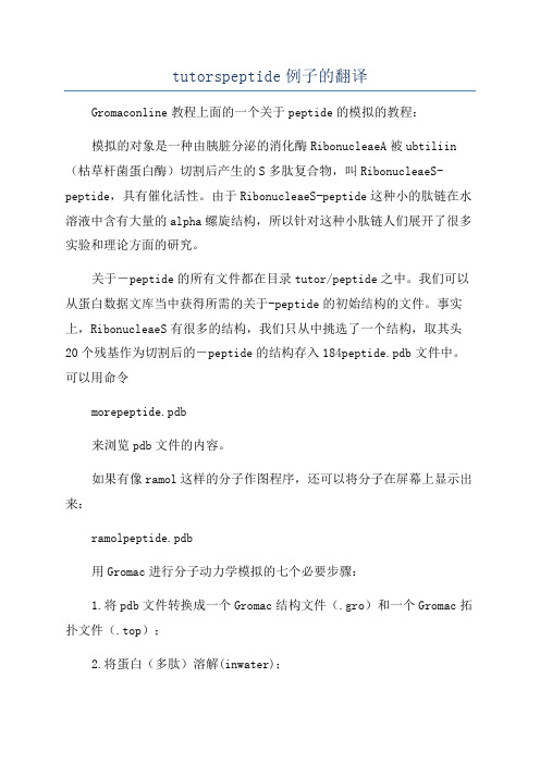
MPMI Vol. 19, No. 5, 2006, pp. 463–470. DOI: 10.1094/MPMI-19-0463. © 2006 The American Phytopathological SocietyA Root-Knot Nematode Secretory Peptide Functionsas a Ligand for a Plant Transcription FactorGuozhong Huang,1 Ruihua Dong,1 Rex Allen,1 Eric L. Davis,2 Thomas J. Baum,3 andRichard S. Hussey11Department of Plant Pathology, University of Georgia, Athens 30602-7274, U.S.A.; 2Department of Plant Pathology, North Carolina State University, Raleigh 27695-7616, U.S.A.; 3Department of Plant Pathology, Iowa State University, Ames 50011, U.S.A.Submitted 4 October 2005. Accepted 16 January 2006.Parasitism genes expressed in the esophageal gland cells of root-knot nematodes encode proteins that are secreted into host root cells to transform the recipient cells into enlarged multinucleate feeding cells called giant-cells. Expression of a root-knot nematod e parasitism gene which encod es a novel 13-amino-acid secretory peptid e in plant tissues stimulated root growth. Two SCARECROW-like tran-scription factors of the GRAS protein family were id enti-fied as the putative targets for this bioactive nematode pep-tide in yeast two-hybrid analyses and confirmed by in vitro and in vivo coimmunoprecipitations. This discovery is the first d emonstration of a d irect interaction of a nematod e-secreted parasitism peptide with a plant-regulatory protein, which may represent an early signaling event in the root-knot nematode–host interaction.Additional keywords: stylet secretion, transgenic plants.Root-knot nematodes (RKNs; Meloidogyne spp.) are among nature’s most successful parasites. These highly spe-cialized biotrophic parasites attack more than 3,000 plant species from diverse plant families and represent a tremen-dous threat to crop production worldwide (Sasser 1980). A successful host–parasite relationship requires molecular sig-nals from RKNs to transform, directly or indirectly, cells within the vascular tissue of susceptible plant roots into elaborate feeding cells, called giant-cells, that are required for RKN development and reproduction. Giant-cell formation represents one of the most complex responses elicited in plant tissue by any parasite or pathogen (Davis and Mitchum 2005). Infective RKN second-stage juveniles (J2) penetrate near the root tip and migrate intercellularly to a site near the differentiating vascular tissue. Secretory parasitism proteins are injected through the nematode’s protrusible stylet (oral feeding spear) to transform five to seven root vascular cells into the metabolically active multinucleate giant-cells by inducing repeated nuclear divisions uncoupled from cyto-kinesis. The parasitism proteins are encoded by parasitism genes expressed in the nematode’s esophageal secretory gland cells and developmental changes in the secreted proteins occur during the parasitic cycle (Davis et al. 2004; Huang et al. 2003; Hussey 1989). The parasitism proteins mediate the dynamic interaction of the RKN with its plant hosts, but little is known about the molecular mechanism or mechanisms un-derlying these plant responses. The broad host range of this pathogen suggests that the RKN affects fundamental processes within plant cells. Understanding how infective RKN J2 regu-late these essential plant responses is a critical area of study for limiting crop damage to RKNs in the future.Low molecular weight secretions from potato cyst nematode J2 enhance division of plant protoplasts and mammalian cells, suggesting that an unknown mitogenic peptide is involved in feeding cell development (Goverse et al. 1999). Although pep-tide hormones are widely known to have key developmental roles in animal systems (Alberts et al. 1994), small peptides only recently have been recognized as a new group of plant-signaling molecules with diverse developmental functions (e.g., CLA V ATA3 [CLV3] controls shoot meristem organiza-tion in Arabidopsis) (Fletcher 2002; Matsubayashi 2003; Ryan et al. 2002). Recent evidence suggests that plant-parasitic nematodes may have evolved a mechanism to mimic plant sig-naling peptides for parasitic modification of host plant cells. A parasitism gene, Hg-SYV46, encoding a secretory protein with a function similar to CLV3 of Arabidopsis thaliana has been characterized from the soybean cyst nematode, Heterodera glycines (Wang et al. 2005).An RKN parasitism gene, designated as 16D10, encoding a putative secretory signaling peptide and expressed in the sub-ventral esophageal gland cells, was isolated from a gland-cell-specific cDNA library of Meloidogyne incognita (Huang et al. 2003). Here, we describe that transgenic expression of the RKN 16D10 secretory peptide significantly stimulates host root proliferation with normal differentiation. This bioactive RKN peptide directly interacts with plant SCARECROW-like (SCL) transcription factors that, presumably, have important roles in plant growth and development. This is the first demonstration that a plant-parasitic nematode-secreted parasitism peptide func-tions as a signaling molecule to induce root proliferation by specifically targeting a host plant regulatory protein. This novel putative ligand-receptor pair may have a role in giant-cell induc-tion in the RKN–plant interaction.RESULTSAnalyses of the 16D10 gene.The longest open reading frame (ORF) of the 16D10 cDNA (364 bp) encoded a deduced protein of 43 amino acids (aa) (Fig. 1A), including a 30-aa N-terminal hydrophobic signal peptide as predicted by Signal P (Nielsen et al. 1997). The ma-Corresponding author: Richard S. Hussey; Telephone: +1-706-542-1254;Fax: +1-706-542-1262; E-mail: hussey@The genomic DNA sequence for 16D10 and the cDNA sequence forTmSCL are available in the GenBank database under accession numbersDQ087264 and DQ087265, respectively.Vol. 19, No. 5, 2006 / 463ture 16D10 peptide of 13 aa (GKKPSGPNPGGNN, molecular weight = 1,223 Da) had no significant BLASTP similarity. However, the peptide was similar to the C-terminal conserved motif of the plant CLE protein family (Cock and McCormick 2001; Olsen and Skriver 2003) (Fig. 1B) and contained a cAMP/cGMP-dependent protein kinase phosphorylation site (KKpS) as predicted by PROSITE (Hofmann et al. 1999). Se-quencing of an 840-bp genomic DNA polymerase chain reac-tion (PCR) product amplified using 16D10-specific primers identified one intron of 476 bp (Fig. 1A). Furthermore, this 16D10 genomic DNA did not contain recognition sites for Eco RI or Bam HI (i.e., those restriction endonucleases used for subsequent DNA blotting experiments to investigate the gene copy number of 16D10 in M.incognita and to assess the presence of homologues in other Meloidogyne spp.). A blot containing genomic DNA from M. incognita, M. javanica, M. arenaria, and M. hapla hybridized with a 16D10 cDNA probe showed that 16D10 was present in each of the four agriculturally important Meloidogyne spp., with three to four copies or homologues (Fig. 2). No hybridization was detected with genomic DNAs from the soybean cyst nematode H. glycines, the nonparasitic free-living nematode Caenorhabditis elegans, and plants (tobacco and Arabidop-sis) (data not shown). Immunodetection of 16D10 peptide.Purified 16D10 antiserum bound to secretory granules within the subventral gland cells of preparasitic and parasitic J2 and their cytoplasmic extensions and expanded ampullae, which are located posterior to the pump chamber at the meta-corpus (Fig. 3). These findings documented that the correct ORF of the 16D10 cDNA was identified. No specific labeling with the rabbit preimmune serum was observed in any nema-tode specimens.Release of stylet secretions from the esophageal gland cells of M. incognita J2 was induced in vitro and stylet secretions were concentrated for use in enzyme-linked immunosorbent assay (ELISA) and immunoblotting analyses using the purified 16D10 antiserum. Both assays identified 16D10 peptide in the stylet secretions as well as total extracts of J2 and mixed para-sitic stages of M. incognita. No signal was detected in the con-trols containing bovine serum albumen (BSA) or total protein extracts of the soybean cyst nematode, H. glycines (Fig. 4). None of the tested protein samples interacted with the preim-mune serum.Overexpression of 16D10 in tobacco hairy roots.As a first assay to identify 16D10 function in host roots, the 16D10 cDNA was overexpressed in tobacco hairy roots with and without the nematode signal peptide sequence using the CaMV 35S promoter (Fig. 5A). Inclusion or exclusion of the signal peptide coding sequence should target the 16D10 pep-tide to the secretory pathway or the cytoplasm of transformedplant cells, respectively. Six hairy root lines with a single Fig. 1. Gene 16D10. A, The cDNA and deduced protein sequences of16D10. The position of intron is indicated by an arrowhead. The predictedsignal peptide sequence and the putative polyadenylation signal are under-lined. B, Alignment of the mature 16D10 peptide (GenBank accession no.AY134435) with CLA V ATA3 (AF126009), CLE (the C-terminal con-served motif of the plant CLE protein family) (Olsen and Skriver 2003),and Hg-SYV46 (AF273728).Fig. 4. A, E nzyme-linked immunosorbent assay and B, immunoblottinganalyses of 16D10 in Meloidogyne incognita stylet secretions. 16D10, 100ng of 16D10 mature peptide (>95% purity) synthesized from Sigma-Geno-sys as a positive control; J2/MS, 10 µg of total extracts of second-stagejuveniles (J2) or mixed parasitic stages of M. incognita; SS, 100 ng of con-centrated M. incognita stylet secretions; SCN, 10 µg of total extracts of thesoybean cyst nematode Heterodera glycines; BSA, 10 µg of bovine serumalbumin as a negative control.Fig. 2. DNA blot hybridization of restriction endonuclease-digested genomicDNA from four Meloidogyne spp. with a digoxigenin (DIG)-labeled16D10 probe. Mi, Meloidogyne incognita; Mj, M. javanica; Ma, M.arenaria; Mh, M. hapla; E, Eco RI; B, Bam HI; M, 80-ng DIG-labeled mo-lecular weight marker in kilobases.Fig. 3. Immunolocalization of 16D10 peptide within the subventral eso-phageal glands (SvG) of Meloidogyne incognita parasitic second-stagejuveniles using 16D10 antiserum. M, metacorpal pump chamber.464 / Molecular Plant-Microbe InteractionsVol. 19, No. 5, 2006 / 465transgenic copy of 16D10 without its signal peptide sequence were established from independent transformations. We were unable to produce any hairy root lines harboring the 16D10 construct, including the nematode signal peptide coding se-quence, despite extensive experimentation. Six independent control hairy root lines using the empty transformation vector also were generated. Strikingly, expression of 16D10 in the cy-toplasm of hairy root cells increased root growth at the rate of approximately 65% (mean root length after 2 weeks of 5.20 ± 0.61 cm [n = 90] in 16D10 transgenic lines compared with 3.15 ± 0.34 cm [n = 90] in control lines), generated extensive lateral roots, and led to the formation of calli where root tips were cut for subculturing at 5 weeks (Fig. 5B). Reverse-tran-scriptase (RT)-PCR analysis of 16D10 expression showed that the steady-state mRNA levels in calli were higher than in the hairy roots (Fig. 5C). Immunoblotting analysis with the puri-fied 16D10 antiserum revealed that 16D10 was produced in both hairy roots and calli (Fig. 5D). No expression of 16D10 was detected in the control vector-transformed hairy roots (Fig. 5C and D).Overexpression of 16D10 in Arabidopsis.In order to complement the tobacco hairy root data, four transgenic Arabidopsis T 2 homozygous lines (L-7, L-10, L-11, and L-17) containing a single-copy of 16D10 without a signal peptide under the control of the 35S promoter were generated. Two transgenic lines (L-2 and L-3) originating from the blank transformation vector also were generated as controls. RT-PCR and immunoblotting analyses confirmed that 16D10 was ex-pressed in all of the 16D10 transgenic lines, but not in the con-trol lines (Fig. 6A and B). As in tobacco hairy roots, expression of 16D10 in the cytoplasm of Arabidopsis cells significantly accelerated root growth, giving rise to a much-enlarged root system (Fig. 6C). No significant differences were found in the shoots of 16D10 transgenic lines and control lines. Further-more, two transgenic lines expressing 16D10 with an Arabi-dopsis CLV3 signal peptide directing 16D10 into the apoplast demonstrated identical phenotypes to the control lines (data not shown). In the root-growth assay, significant differences (P < 0.01) in root length between 35S::16D10 transformants and control lines were observed at 12 days after germination. The length of primary root in 35S::16D10 transformants was in-creased by 84.97% (mean 54.01 ± 8.75 mm in four 16D10 trans-genic lines [n = 90/line] and 29.20 ± 4.50 mm in two control lines [n = 90/line ]) (Fig. 6D).Two-hybrid screen for 16D10-interacting host proteins. Reporter gene (HIS3, ADE2, and lacZ ) confirmation of the two-hybrid interactions between the GAL4-BD-16D10 and GAL4-AD-tomato root-cDNA fusion proteins led to the identi-fication of five tomato cDNA clones: one encoded an unknown protein, another encoded a ribosomal protein subunit, and three identical cDNA clones (544 bp, GenBank accession number DQ087265) encoded partial proteins with 30 to 78% identity to the C-termini of plant SCL transcription regulators, which control plant growth and development (Bolle 2004; Pysh et al. 1999).For confirmation that 16D10 interacted with a plant SCL protein, 12 Arabidopsis SCL genes homologous to the tomato SCL gene identified here were cloned from an Arabidopsis root cDNA pool and expressed as GAL4-AD fusion proteins in a yeast two-hybrid assay. When tested, two Arabidopsis SCL proteins, AtSCL6 and AtSCL21, interacted with 16D10 in yeast. Domain analysis revealed the specific interaction of 16D10 with the SAW domain of AtSCL6 and AtSCL21 and no interaction of 16D10 with the rest of the domains of the SCL proteins (Fig. 7A), and indicated that the SCL transcription factors were two putative targets of the secreted 16D10 during RKN parasitism of plants. In vitro interaction of 16D10 withAtSCL6 and AtSCL21 was verified by coimmunoprecipitationFig. 5. Overexpression of 16D10 in tobacco hairy roots. A , Schematic T-DNA region of the binary vector pBIX containing 16D10 with or without a signal peptide sequence. B, Root growth assay of 16D10 constitutively expressing (35S::16D10) hairy roots (left plates) with control vector-transformed hairy roots (right plates). The white arrow indicates that a root tip was cut from a 2-week-old hairy root for subculture, and the black arrow indicates a callus formed at the cutting site 3 weeks after wounding. C, Relative reverse-transcriptase polymerase chain reaction (PCR) of 16D10 transcripts in transgenic hairy root lines. Expression of the tobacco actin Tob104 gene (GenBank accession no U60494) was used as an internal control. D, Immunoblotting analysis of 16D10 expression in 5-week-old transgenic hairy roots assayed with the purified 16D10 antiserum. E, Representative PCR analysis for the presence (35S::16D10) or absence (vector) of 16D10 with nptII and gusA genes in transgenic hairy root lines, using nptII - or gusA -specific primers. M, 1-kb DNA molecular marker (Promega). nptII , neomycin phosphotransferase II gene; gusA , β-glucuronidase (GUS) gene.466 / Molecular Plant-Microbe Interactions(Co-IP) of 35S-labeled, Myc-tagged 16D10 with 35S-labeled, hemagglutinin (HA)-tagged AtSCL6 or AtSCL21 by using antigen-specific antibodies (Fig. 7B). In planta interaction of 16D10 with Arabidopsis root proteins also was confirmed by in vivo Co-IP analysis. A 61-kDa protein (the same size as AtSCL6) and a 31-kDa (possibly processed) protein (14 kDa smaller than AtSCL21) bound to the 16D10 peptide were pulled down from the total root-extracted proteins of 16D10 transgenic Arabidopsis line L-17 by using the purified 16D10 antiserum. Microsequencing of the Co-IP proteins revealed that two internal peptide fragments each from the CNBr-digested 61- and 31-kDa proteins had amino acid sequences of QKIERFLIQPEIEKLVLDRSRPIERP and AGFEPYPLSSIISA TIRALLRDYSNGY AIE E RDGALYLGW, which are identical to internal sequences of AtSCL6 and AtSCL21, respectively. In the control Arabidopsis line L-3, no protein was pulled down from the total root-extracted proteins with the purified 16D10 antiserum, nor did rabbit preimmune sera bind to any root protein in the transgenic Arabidopsis lines (Fig. 7C and D). DISCUSSIONParasitism proteins synthesized in the esophageal gland cells of plant-parasitic nematodes are secreted into host tissues to mediate nematode infection and parasitism of plants (Davis et al. 2004; Williamson and Hussey 1996). In this article, we re-port that a novel secretory peptide (16D10) secreted from the subventral esophageal gland cells of RKN specifically induces host root growth by directly interacting with a host intracellu-lar SCL transcription regulator. Because 16D10 is conserved in RKN species, we infer that 16D10 is a fundamental signal for regulating RKN–host interactions by activating SCL-mediated signal-transduction mechanisms in parasitized cells.In plants, small peptides represent a newly recognized group of signaling molecules with diverse functions, such as systemic wound response, cell proliferation and dedifferentiation, shoot meristem organization, root nodulation, and self-incompatibility (Matsubayashi 2003; Ryan et al. 2002). Some of these bioactive peptides (e.g., phytosulfokine [PSK] and CLV3) are synthesized through the secretory pathway and have been shown to act extracellularly as peptide hormones, but others (e.g., ENOD40) apparently are synthesized in the cytosol on free ribosomes (Ryan et al. 2002). The extracellular peptides (systemin, PSK, and CLV3) act on either leucine-rich repeat transmembrane receptors or cysteine-rich receptor kinases (Matsubayashi 2003), whereas the cytoplasmic E NOD40 binds to a sucrose synthase (Rohrig et al. 2002).Small secretory peptides also are involved in the interaction between parasitic nematodes and their host. For example, mito-genic peptides present in the excretory-secretory products of the animal parasitic nematode T richostrongylus colubriformis are responsible for epithelial cell proliferation and can stimulate HT29-D4 cell growth in vitro (Hoste et al. 1995). A small, un-known peptide or peptides secreted by the potato cyst nematode Globodera rostochiensis stimulates the proliferation of both to-bacco leaf protoplasts and human peripheral blood mononuclear cells in the presence of synthetic auxin and cytokinin analogues (Goverse et al. 1999). Although the amino acid sequence of the novel RKN 16D10 peptide is similar to the C-terminal conserved motif of the plant CLE protein family (Cock and McCormick 2001; Olsen and Skriver 2003), targeting of 16D10 to the apoplast or cytoplasm of Arabidopsis clv3 mutants did not restore the wild-type phenotype (G. Z. Huang and R. S. Hussey, unpublished data ). In contrast, a parasitism gene Hg-SYV46 expressed in the dorsal esophageal gland cell of the soybean cyst nematode H. glycines encodes a secretory protein that is capable of functioning in a fashion similar to that of CLV3 by rescuing clv3 mutants of A. thaliana (Wang et al. 2005).Some plant peptides need a posttranslational modification for their stability or increased activity, such as glycosylation in systemins and sulfation in PSKs (Matsubayashi and Sakagami 1996; McGurl et al. 1992; Pearce et al. 1991). Immunoblotting and ELISA assays revealed that stable 16D10 peptide was pre-sent in the transgenic tobacco hairy roots and Arabidopsis . Whether a functional 16D10 in the transgenic roots is phos-phorylated is under investigation, because 16D10 contains a predicted cAMP/cGMP-dependent protein kinase phosphory-lation site at the serine residue.SCL transcription factors (e.g., GAI, RGA, and SCR) aremembers of the GRAS protein family characterized by a vari-Fig. 6. Overexpression of 16D10 in Arabidopsis . A and B, Relative reverse-transcriptase polymerase chain reaction (RT-PCR) and immunoblotting analyses for the expression of 16D10 in four 16D10 transgenic homozygous T 2 lines (L-7, L-10, L-11, and L-17) and the absence of 16D10 expression in two vector-transformed homozygous T 2 lines (L-2 and L-3) 12 days after germination (dag). Expression of the Arabidopsis UBQ10 gene was used as an internal control in RT-PCR. C, Seedlings of 16D10 constitutively expressing (35S::16D10) transgenic line (left) with control vector-transformed line (right) 18 dag. D,Primary root length 12 dag. Bars indicate ± standard deviations; n = 90.able N-terminus, leucine heptad repeat I (LHR I); a VHIID motif, leucine heptad repeat II (LHR II); a PFYRE motif; and a SAW motif (Fig. 7A). The LHR I-VHIID-LHR II region might function as a DNA binding and oligomerization domain, with the LHRs mediating protein-protein interactions and the VHIID motif mediating protein-DNA interaction (Pysh et al. 1999). The divergent N-terminal sequences are hypothesized to function as activating domains and the C-terminal region with the PFYRE- and SAW-conserved motifs may act as regulatory domains (Itoh et al. 2002; Pysh et al. 1999). Although more than 30 members of the GRAS protein family have been identi-fied in plants (Bolle 2004; Pysh et al. 1999; Tian et al. 2004), the functions of only a limited number of GRAS family pro-teins have been elucidated. AtSCR and AtSHR are involved in root radial patterning, whereas AtGAI and AtRGA function as negative regulators controlling gibberellin signaling (Nakajima and Benfey 2002). Mutations in the SAW domains of AtSCR, AtRGA, and MtNSP1 proteins have strong mutant phenotypes, suggesting that the C-terminal SAW domain is important for protein function (Di Laurenzio et al. 1996; Silverstone et al. 1998; Smit et al. 2005). Inoculation of SCL6 and SCL21 Arabi-dopsis mutant lines, which had slightly retarded root growth, with M. incognita showed a 23 to 56% reduction in the number of nematode eggs per gram of root in these SCL mutants when compared with the wild-type control Arabidopsis (G. Z. Huang and R. S. Hussey, unpublished data). The specific interaction of RKN 16D10 with the SAW domain of a tomato SCL protein or Arabidopsis AtSCL6 and AtSCL21 in yeast, in vitro, and in transgenic plants indicates that an SCL protein is a putative target for the RKN 16D10 to function in parasitized host plant cells.Most of the AtSCL genes are expressed predominantly in the roots (Pysh et al. 1999), and the 12 SCL genes analyzed herein were amplified from an Arabidopsis root cDNA library, sug-gesting that a subset of these SCL genes may play important roles in root biology. The functions of AtSCL6 and AtSCL21 are unknown, but homologues in the same group in the phyloge-netic tree of GRAS proteins may provide some insight into the role of AtSCL6 and AtSCL21 in plant biology (Bolle 2004). AtSCL21 is grouped with AtPAT1 and OsCIGR1/OsCIGR2, which are involved in phytochrome signaling in Arabidopsis and chitin (N-acetylchitooligosaccharide) elicitor perception in rice, respectively (Bolle 2004; Day et al. 2003). AtSCL6 is grouped with AtSCL22, which is catalogued by cDNA micro-array analysis to function in mitotic cell cycle and cell cycle control (Yamada et al. 2003).In host roots, vascular parenchyma cells near the primary xylem in the zone of elongation are preferred for RKN feeding cell initiation. The transition from parenchyma cell to a fully differentiated giant-cell occurs early in the parasitic associa-tion, indicating that secreted signaling molecules from the early parasitic J2 mediate giant-cell development (Hussey 1989; Williamson and Hussey 1996). In RKN-infected roots, cell cycle genes are upregulated in the feeding cells within the first hours of parasitism (De Almeida E ngler et al. 1999; Niebel et al. 1996). This rapid reactivation of the cell cycle may be induced by an initial stimulus from the nematode (Goverse et al. 2000). In our study, we conclude that the RKN secretory peptide 16D10 may be involved in feeding cell formation, because 16D10 i) is conserved in RKN species and secreted from the stylet; ii) is strongly expressed in the parasitic J2 subventral esophageal gland cells, which become nonfunctional in later parasitic stages; and iii) binds to a specific plant transcription factor domain.During feeding cell induction, the nematode inserts its stylet through the plant cell wall without penetrating the plasma membrane. Nematode stylet secretions may be deposited be-tween the plasma membrane and the cell wall or released di-rectly into the cytoplasm through a perforation in the plasma membrane at the stylet orifice (Williamson and Hussey 1996). Our transgenic expression data indicates that 16D10 only func-tions in the cytoplasm of root cells, which is supported by the direct interaction of 16D10 with a host intracellular regulatoryprotein.Fig. 7. Interaction of 16D10 with plant SCARE CROW-like (SCL) tran-scription factors. A, Direct interaction of 16D10 with the SAW domain of tomato (TmSCL) and Arabidopsis (AtSCL6 and AtSCL21) proteins in yeast. The specific regions of these SCL proteins drawn schematically were tested to interact with 16D10. Positive interactions resulting in the activation of HIS3, ADE2, and lacZ genes were detected by growth in the absence of histidine and adenine, and β-galactosidase (β-gal) activity. β-Gal activity was measured from three independent β-gal liquid assays using o-nitrophenyl β-D-galactopyranoside as a substrate. B, In vitro co-immunoprecipitation (Co-IP) of 16D10 with AtSCL6 or AtSCL21. [35S]-labeled 16D10 with HA-tag (4 kDa), AtSCL6 with c-Myc-tag (63 kDa), and AtSCL21 with c-Myc-tag (48 kDa) were translated from the corre-sponding cDNA in pGBKT7 or pGADT7. Antibodies used for Co-IP are indicated: HA, HA-tag polyclonal antibody; Myc, c-Myc monoclonal anti-body; 16D10, the purified 16D10 antiserum; PI (control), rabbit preim-mune serum. C and D, In vivo Co-IP of 16D10 with Arabidopsis root pro-teins from the 16D10 transgenic line L-17 using the vector-transformed line L-3 as a control. Gels were stained with Coomassie blue R-250 and immunoblotted with the purified 16D10 antiserum. Lanes 1 and 4: total root extracts; lanes 2 and 5: products pulled down from the total root extracts by using the purified 16D10 antiserum; lanes 3 and 6 (controls): no product pulled down from the total root extracts by using rabbit pre-immune serum.Vol. 19, No. 5, 2006 / 467Transcription factors are known to direct gene expression patterns and the resulting cell differentiation, but a direct link between the transcription factors and the cell cycle has not yet been made. Although several transcription factors are among the upregulated genes in the nematode feeding cells, the changes in gene expression identified so far are likely to be several steps downstream from the initial plant responses to signals from the nematode (Gheysen and Fenoll 2002; Williamson and Hussey 1996). The regulation of SCL-transcription factors by 16D10 could affect the transcription of downstream root-spe-cific genes by intervening in the signal-transduction pathways involved in root cell proliferation.MATERIALS AND METHODSNematodes and plants.Nematodes were cultured on host plant roots and RKN (M. incognita) preparasitic J2 and parasitic stages were collected as described previously (Huang et al. 2005). Plants were grown under optimal conditions in growth chambers. Analysis of the 16D10 gene.Nematode genomic DNA was extracted from J2. Eco RI- or Bam HI-digested genomic DNA (10 µg) was electrophoresed on a 0.7% agarose gel, transferred to Hybond-N membrane (Amersham, Piscataway, NJ, U.S.A.), and hybridized with 15 ng of digoxigenin (DIG)-labeled 16D10 probe corresponding to the full-length cDNA sequence as described (Huang et al. 2005). Sequence of the M. incognita genomic DNA containing the coding region of 16D10 was obtained from the PCR product using the gene-specific primers 16D10GF (5′-GAGAAAATA AAATATAAATTATTCCTC-3′) and 16D10GR (5′-CAGATAT AATTTTATTCAG-3′) and M. incognita genomic DNA tem-plate.Immunodetection of 16D10.Polyclonal antiserum to 16D10 was produced by immunizing rabbits with a synthetic mature (i.e., without the N-terminal signal peptide) 16D10 peptide (E urogentec, Inc., San Diego, CA, U.S.A.). Peptide affinity-purified 16D10 polyclonal anti-serum was used to localize 16D10 expression in specimens of M. incognita using immunofluorescence microscopy (Goverse et al. 1994) and for immunodetection of 16D10 in stylet secre-tions and transgenic plant-expressed or in vitro-translated 16D10.Stylet secretions from M. incognita J2 were produced and collected in vitro as described previously (Davis et al. 1994) and concentrated with StrataClean resin (Stratagene, La Jolla, CA, U.S.A.). Nematode proteins were extracted from ground J2 and mixed parasitic stages of M. incognita and H. glycines as described (Ding et al. 1998). Plant proteins (0.5 g) were ex-tracted by grinding transgenic seedlings or root tissues in 200 µl of extraction buffer (50 mM Tris-HCl, pH 7.0, 150 mM NaCl, 1× complete protease inhibitors) (Roche Applied Science, Indianapolis, IN, U.S.A.) in microcentrifuge tubes in liquid nitrogen. Supernatant was recovered from homogenates after centrifugation at 13,000 rpm for 10 min. All protein concentra-tions were estimated (with a Bio-Rad protein assay kit II; Bio-Rad, Hercules, CA, U.S.A.) with BSA as a standard, and ELISA and dot blots were performed with 2-µl protein samples and purified 16D10 antiserum (Ding et al. 1998).Plasmid construction.The coding regions of 16D10 with or without the nematode signal peptide sequence were amplified from the full-length cDNA clone with primers 16D10SF (5′-CGGGGTACC TAG A TGTTTACTAATTCAATTAA-3′) or 16D10F (5′-CGGGGTAC C TAGATG GGCAAAAAGCCTAGTG-3′) and 16D10R (5′-GCTCTAGATCAATTATTTCCTCCAGG-3′) that introducedKpn I or Xba I restriction sites (underlined) and the stop/start codons (in italics), cloned into the Kpn I and Xba I sites of binaryvector pBIX (Fig. 5A) under the control of CaMV 35S pro-moter to generate pBIX(16D10SP) and pBIX(16D10), respect-tively, and confirmed by sequencing. pBIX was derived frompBI101 (BD Biosciences, San Jose, CA, U.S.A.) and containsa nos promoter-nptII-nos terminator cassette, a 35S promoter-gusA-nos terminator, and a second 35S promoter with a poly-linker having Kpn I and Xba I sites.Because we were unable togenerate tobacco hairy root lines using the nematode 16D10signal peptide (discussed above), we fused a plant signal pep-tide sequence (clv3) to the 16D10 sequence for targeting16D10 to the apoplast in Arabidopsis. The fusion-expressedsequence of clv3 signal peptide and 16D10 was generated byPCR amplifications from Arabidopsis genomic DNA usingprimers C3K (5′-GGGGTACCATGGATTCTAAAAGCTTTG-3′) that introduced Kpn I restriction site (underlined) and C3R (5′-CCACTAGGCTTTTTGCCAAGGAACAAGAAGCAG-3′)for the clv3 signal peptide sequence, and from 16D10 cDNAusing primers C3F (5′-CTTCTGCTTCTTGTTCCTTGGCAAAAAGCCTAGTGG-3′) and 16D10X (5′-GCTCTAGATCAA TTA TTTCCTCCAGG-3′) that introduced Xba I restriction site(underlined) for the mature peptide coding sequence usingV ent polymerase (New England Biolabs, Ipswich, MA, U.S.A.).The two products then were used to prime each other in afusion PCR reaction. The resulting fragment was cloned intopBIX to generate pBIX(CLV3SP-16D10) and verified by se-quencing.Expression of 16D10 in tobacco hairy roots.The plasmids pBIX(16D10) and pBIX(16D10SP) and theempty vector pBIX as a control were transferred into Agrobac-terium rhizogenes ATCC 15834 by electroporation (Shen and Forde 1989) and transformed into tobacco (Nicotiana tabacum cv. Petite Havana SR1) using the A.rhizogenes-mediated coty-ledon transformation (Christey 1997). Transformed hairy roots were generated from inoculated tobacco cotyledons on Gam-borg’s B-5 plates containing 0.8% Noble agar with kanamycin at 100 mg/liter and timentins (ticarcillin disodium at 230.8 mg/liter plus clavulanate potassium at 7.69 mg/liter).Individ-ual hairy root tips (approximately 0.5 cm) were cultured for 3 weeks at 24ºC in the dark, and two to three roots from individ-ual hairy root system were subjected to β-glucuronidase (GUS)-staining selection (Jefferson et al. 1987). The kanamy-cin-resistant and GUS-positive root lines, confirmed by PCR analyses (Fig. 5E) and bacteria free, were used to establish hairy root lines. The root-tips were subcultured for root growth assay on Gamborg’s B-5 plates without hormones every 2 weeks and the cut roots were kept in culture on the old plates at 24ºC in the dark for assays. For root-growth assays, plates were cul-tured horizontally in the dark and five hairy roots from each transgenic line in each of the three repeats were investigated. Relative RT-PCR and immunoblotting analyses of transgenic hairy roots or calli with a single transgenic copy identified as described (Does et al. 1991) were carried out using the same procedures as in those of transgenic Arabidopsis.Expression of 16D10 in Arabidopsis.The plasmids pBIX(16D10) and pBIX(CLV3SP-16D10) andthe empty vector pBIX as a control were introduced into Agro-bacterium tumefaciens C58C1 by electroporation (Shen and Forde 1989) and transformed into Arabidopsis thaliana wild-type Col-0 plants by the floral dip method (Clough and Bent 1998). Segregation analysis of kanamycin resistance identified transgenic homozygous T2 lines and PCR analysis was used to468 / Molecular Plant-Microbe Interactions。