V IROLOGICA S INICA, August 2007, 22 (4):316-325
Received: 2007-04-04, Accepted: 2007-05-18
* Foundation item: Key technologies R&D program (2006BAD06A01) from the Ministry of Science and Technology of China. ** Corresponding author. Tel: +86-27-87199239, E-mail: wanghz@https://www.doczj.com/doc/173407734.html,
ZHUAN et al. Rapid Construction of Recombinant Viruses of PRV Genome 317
functional domains within the proteins (9).
Such studies often require establishment of large numbers of recombinant viruses which are usually created by homologous recombination in infected cells relying on the cellular recombination and repair machinery. However, this can be a laborious and sometimes impossible task, especially if the mutant has a severe growth disadvantage compared to the wild-type virus. Bacterial artificial chromosomes (BACs), single copy F-factor-based plasmid vectors of intermediate insert capacity (15), have now enabled the cloning of complete herpesvirus genomes and infectious virus genomes can be shuttled between Escherichia coli (E. coli) and eukaryotic cells. While herpesvirus BAC DNA engineering in E. coli requires neither restriction sites nor cloning steps and allows the introduction of a wide variety of DNA modif- cations, the large size of these bacmids precludes the use of rapid in vitro methods of manipulation commonly employed for construction of small plasmids.
Tn7, a site-specific transposon, transposes almost exclusively to a distinct attachment site named attTn7 within the E. coli genome (1). By introducing this attTn7 sequence into a BAC, Tn7 can serve as an insertion vehicle (4). This is particularly useful if numerous genes and constructs need to be tested for their expression in the context of viral genome. Tn7-mediated transposition has been well exploited for research on functional genomics of baculoviruses (4). Recently, this technology has been applied to bacmid-cloned cytomegalovirus (CMV), a member of the Gammaherpesvirinae subfamily, for rapid recombinant virus construction (2).
In this paper, we report the development of a technology employing Tn7-mediated transposition as a rapid and reliable method for recombinant PRV construction. A lacZα-mini-attTn7 region was inserted into the intergenic region between the gG and gD genes in an attempt to maintain every gene and element of the parental virus. Then green fluorescent protein (GFP) gene was introduced to test the utility of this transposition system and the stability of mini-Tn7 insertions in cell culture. The technology should greatly facilitate the detailed mutagenic studies of PRV.
MATERIALS AND METHODS Plasmids, strains, and reagents
Plasmids pGS284 and pBecker3, strains GS500 and S17λπ were provided by Lynn W. Enquist, Princeton University, USA. The pGEM-T Easy was purchased from Promega Co. (Maddison, USA), and plasmid pEGFP-N1 from Clonetech Laboratories, Inc. (Mountain View, USA). DNA restriction enzymes, T4 DNA ligase, alkaline phosphatase, exTaq hot start DNA polymerase and 2×GC buffer I were the products of TaKaRa Biotechnology Co., Ltd. (Dalian, China). Plasmids pFBCMV-GFP and pZFBΔtk were constructed and stored in our lab. Strains DH5α and DH10B were stored in our lab. Lipofectin reagent and Delbecco’s Modified Essential Medium (DMEM) were purchased from Invitrogen Co. (Carlsbad, USA), and fetal bovine serum (FBS) from Hangzhou Sijiqing Biological Engineering Materials Co., Ltd. (Hangzhou, China).
Virus and cells
The wild-type PRV used was vBecker3, generated by transfection of pBecker3 into Vero cells (18). Mutant PRVs (vBeckerZF1 and vBeckerZF2) were
318V IROLOGICA S INICA V ol.22, No 4
constructed by manipulation of pBecker3 and transfection of mammalian cells (see below). All PRV strains were propagated in the PK15 (porcine kidney 15) cell line. Virus titers were determined in duplicate by infectivity end-point assay on PK15 cells in 96-well plates following infection with 100μL/well of 10-fold serially diluted stocks. The mammalian cells were grown in DMEM supplemented with 10% FBS. Viral infections were performed in DMEM sup- plemented with 2% FBS. Unless otherwise indicated, all media used contained 100U /mL penicillin and 100 μg /mL streptomycin.
Construction of plasmids and BACs Constructions of plasmids and BACs are illustrated in Fig.1. Unless otherwise noted, all PCRs were performed with exTaq hot start polymerase and 2×GC buffer I. The 433 bp of the upstream homology region for allele exchange in E. coli was amplified by PCR from pBecker3 with the primers HRattTn7U-F (5’-GAGCTCCTGCTCCTGGGCTTCCTG-3’, with a Sac I site underlined) and HRattTn7U-R (5’-GAATTC CCCGTAAGCAAGGCCGTA-3’, with an Eco RI site underlined) and cloned into the Sac I and Eco RI sites of pEGFP-N1 to generate pZF1, while the 448 bp of the downstream homology region was amplified with the primers HRattTn7D-F (5’-GTCGACGTGCGATC CACGCCCAGC-3’, with Sal I site underlined) and HRattTn7D-R (5’-GGATCCATGCATGCGCCCAAA GATCTGCCTG-3’, with Bam H I site underlined and Nsi I site double underlined) and cloned into the Sal I and Bam H I sites of pZF1 to generate pZF2. PCR parameters for amplification of both homology regions were as follows: 96℃for 2 min to denature, followed by 30 cycles of 94℃for 30 s, 70℃ for 30s, with a final 10-min extension at 72℃. The 712 bp lacZα-mini-attTn7 region was released by Bbs I digestion of pZFBΔtk (unpublished) which contains an 8.6 kb Bsu36I fragment from pHZB10 (19), generating an Eco RI compatible end and a Sal I site. Plasmids pZF3 was produced by inserting the lacZα-mini-attTn7 region into the Eco RI and Sal I sites of pZF2. To construct the transfer vector for allele exchange in E. coli, a shuttle plasmid pGS284 (17) was used, which includes the π-protein-dependent R6K origin of replication (7), the RP4oriT origin for conjugative transfer (16), and the negative selection marker sacB (11). First, pZF3 was digested by Sac I and dephosphorized, and pGS284 was digested by Sac I. Then the two linearized plasmids were ligated and the product was transformed into DH5α to select for ampicillin (Amp) resistance. Since pGS284 was unable to grow in a strain that lacks π, only the Amp resistant strains containing the hybrid plasmid pZF4 grew. Plasmid pZF4 was digested by Nsi I to excise the origin of replication of pEGFP-N1 and self-ligated to generate the transfer vector pZF5. Shuttle plasmid pFBCMV-GFP, a derivative of pFastBac Dual (In- vitrogen), was generated by replacing the P10 and P H promoters with a P CMV promoter and a GFP gene cloned under the promoter.
The pBeckerZF1 BAC was generated by the sugar suicide allele exchange system in E. coli as described previously (17). Briefly, conjugal transfer of the allele exchange vector occurred following cross-streaking of GS500 containing pBecker3 and S17λpir (7) containing pZF5 on a Luria broth (LB) plate without antibiotics and incubated overnight at 30℃. Each intersection from the crossed streaks was inoculated into 5 mL of LB plus 25 μg/mL Amp and 25 μg/mL chloramphenicol (Cm) and incubated overnight at
ZHUAN et al. Rapid Construction of Recombinant Viruses of PRV Genome 319
37℃ with rotating to select for recombinants. To select for deletion of the allele exchange vector from pBecker3, 1 μL of overnight culture was inoculated into 5 mL of LB-Cm and incubated overnight at 37℃with rotation. The culture was serially diluted (10-3, 10-4, and 10-5), and 50 μL of each dilution was plated onto LB plates containing 5% sucrose, 25 μg/ml Cm, 40 μg/ml isopropyl-β-D-thiogalactopyranoside (IPTG) and 200 μg/mL 5-bromo-4-chloro-3-indolyl-β-D-galac- topyranoside (X-gal). A single blue colony was transferred into 5 mL of LB-Cm and incubated overnight at 37℃. Miniprep DNA from 1 mL overnight culture was diluted to low concentration (~ 100ng/mL). The diluted BAC was transformed into E. coli strain DH10B (Invitrogen Co., Carlsbad, USA), precluding co-transformation of more than one copy BAC DNA into a single cell. After one hour incubation at 37℃, transformed cells were spread on LB plates containing Cm, IPTG and X-gal. Blue colonies were further screened for Amp sensitivity then by PCR and sequencing (described below).
The pBeckerZF2 BAC was generated by Tn7- mediated transposition as described in the instruction manual of the Bac-to-Bac TM system (Invitrogen Co., Carlsbad, USA). White colonies were picked and further screened by PCR (described below).
PCR analysis of recombinant BACs
Virus DNA was obtained from infected PK15 cells as described previously (14). The PCR reactions were set up containing 1×GC buffer I, 200 μmol/L dATP, 200 μmol/L dCTP, 200 μmol/L dGTP, 200 μmol/L dTTP, 0.5 μmol/L concentration of forward primer, 0.5 μmol/L concentration reverse primer and 1U of exTaq hot start polymerase with a total volume of 25μL. For analysis of pBeckerZF1 and vBeckerZF1, two primers flanking the homology regions pBeatt- Tn7-F (5’-GCCCTCTGACATCTTCGTGACCC-3’) and pBeattTn7-R (5’-CGAACTTGTACGTGCGGTG CTG-3’) were designed and cycling parameters were as follows: 96℃ for 2 min to denature, followed by 30 cycles of 94℃ for 30 s, 70℃ for 1min 50s, with a final 10-min extension at 72℃. For analysis of pBeckerZF2, M13 forward primer (5’-TGTAAAACG ACGGCCAGT-3’) and reverse primer (5’- TCACAC AGGAAACAGCTAT GAC -3’) were used with the parameters as follows: 96℃ for 2 min to denature, followed by 30 cycles of 94℃ for 30 s, 55℃ for 30 s, 72℃ for 3 min 45s, with a final 10-min extension at 72℃. The PCR products were detected by ethidium bromide staining after separation on 0.8 to 1.0% agarose gels. The PCR product, using pBeattTn7-F and pBeattTn7-R for test of pBeckerZF1, was cloned into pGEM-T Easy and further screened by sequencing.
BAC DNA preparation and reconstitution of infectious viruses
Minipreps of BAC DNA were prepared according to methods developed for the isolation of large plasmids (defined in the Instruction Manual of the Bac-to-Bac TM system/Life Technologies). BAC DNA isolated from 1mL of overnight cultures was used for transfection of 40-60% confluent PK15 cells in a 35-mm disk. Transfection was performed with Lipofectin TM reagent according to the manufacturer’s instructions.
RESULTS
Construction of the pBeckerZF1 BAC
Site-specific transposition by transposon Tn7 has been developed to rapidly introduce genes or cis elements
320V IROLOGICA S INICA V ol.22, No 4
A
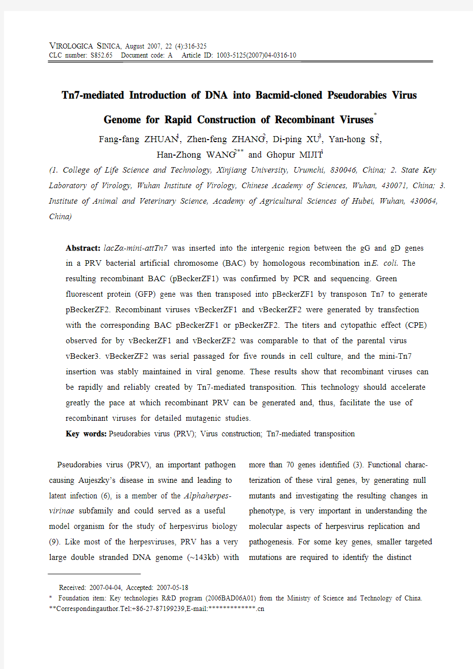
Fig. 1. Insertion of lacZα-mini-attTn7 region into pBecker3 and construction of recombinant bacmids by Tn7-mediated transposition are illustrated. A, Schematic diagram of the PRV-Becker genome depicting the unique long (UL), unique short (US), internal repeats (IR), and terminal repeats (TR) regions. A portion is expanded to show the gG and gD genes. For clarity, only this region of all subsequent viral bacmids is illustrated. B, Construction of pBeckerZF1 and pBeckerZF2. Viral genes are represented as rectangles with an arrowhead denoting the transcription direction. lacZα is represented as light gray boxes and attTn7 is represented as hatched boxes.
into viral bacmids and has proved to be a powerful tool in studies on herpesvirus genetics (2). In this study, to test whether this technology works well in PRV, a recombinant BAC pBeckerZF1 including lacZα-mini-attTn7 was constructed using the E. coli recombination system, which was inserted into the intergenic sequence between the putative polyadeny- lation signal of the gG gene and the putative TATA signal of the gD gene (3) without loss of any viral genes (Fig.1 and Fig.3).
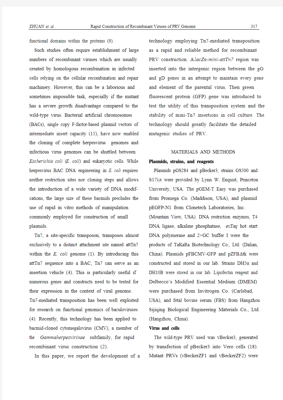
Insertion of lacZα-mini-attTn7 into the target site in pBecker3 is shown in Fig.2, evidenced by the different sizes of PCR products with pBeckerZF1 or pBecker3 as template. For PCR analysis of pBeckerZF1, two
Fig. 2. PCR analysis of pBeckerZF1. Two primers flanking the homology regions were used. M, DL3000 DNA marker; 1, pBecker3 as template; 2, pBeckerZF1 as template; 3, Double distilled H2O (ddH2O) as template.
ZHUAN et al. Rapid Construction of Recombinant Viruses of PRV Genome 321
primers flanking the homologous regions for allele exchange in E. coli were designed to exclude the possibility of contamination by transfer vector pZF5. The size of PCR product with the template pBec- kerZF1 is about 710 bp larger than that with pBecker3, which is corresponds exactly with the size of lacZ-mini-attTn7 region (Fig.2). Further sequencing of the PCR product with the template pBeckerZF1 established that the lacZ α-mini-attTn7 region was inserted 44bp downstream from the putative polya- denylation signal of gG and 23bp upstream from the putative TATA signal of gD (Fig.3.).
Transfection of pBeckerZF1 and characterization of vBeckerZF1 in cell culture
To test whether viruses vBeckerZF1 generated from
the pBeckerZF1 transfection included the lacZ α- mini-attTn7 region, PCR was performed with the same primers used for establishing pBeckZF1 was constructed correctly (see above). The size of PCR product with the vBeckerZF1 genome DNA as the template is about 710 bp larger than that obtained with vBecker3 (data not shown). Therefore, transfection of pBeckerZF1 resulted in the production of virus containing the lacZ α-mini-attTn7 region, vBeckerZF1, without the need for further plaque purification. To examine the growth properties of vBeckerZF1, PK 15 cells were transfected in parallell, with pBeckerZF1 or pBecker3, respectively. Viral titers of the resulted vBeckerZF1 were typically in the order of 108 to 109 TCID 50/mL, which is comparable to that
Fig. 3. Sequence analysis of lacZ-mini-attTn7 insertion into pBekcer3. lacZ-mini-attTn7 region is in bold and the attTn7 is framed. Homology regions for allele exchange are underlined with polyadenylation signal for gG and TATA signal for gD double underlined.
TATA signal
attTn7
polyadenylation signal
GCCCTCTGACATCTTCGTGACCCCCACCGGCAGCCCCGCCCTGCTCCTGGGCTTCCTGGGCAGCGCGCTCGCCTCGCGCCCCCTGCA CCTGACGGCCGGGGAGACGGCCCAGCACGTGCGCGAGGCCCAGCAGAAGAGCCGCCACATCCGCTCCCTCGGCGGCCTCCAGCTC TCGGTCGAGACCGAGACCACCAACACCACCACCACCCAGACGGGCCTGTCGGGCGACATCCGCACCTCGATCTACATCTGCGTCGC CCTCGCCGGCCTGGTCGTCGTGGGCATCGTCATCATGTGCCTCCATATGGCGATCATCAGGGCCCGGGCCCGGAACGACGGCTACCG
CCACGTGGCCTCCGCCTGACCCGGCCCCGCCCGACTCCCCCGCGATTCCCCCCCTCTCTCACCGGGTGTCCATCTTCAATAAAGTAT GTCTCAAACACCTAATTTGCGTACGGCCTTGCTTACGGGG AATTAAGTCTTCGAACCAATACGCAAACCGCCTCTCCCCGCGCG TTGGCCGATTCATTAATGCAGCTGGCACGACAGGTTTCCCGACTGGAAAGCGGGCAGTGAGCGCAACGCAATTAATGTGAG TTAGCTCACTCATTAGGCACCCCAGGCTTTACACTTTATGCTTCCGGCTCGTATGTTGTGTGGAATTGTGAGCGGATAACAAT TTCACACAGGAAACAGCTATGACCATGATTACGCCAAGCGCGCAATTAACCCTCACTAAAGGGAACAAAAGCTGGAGCTCC ACCGCGGTGGCGGCCGCTCTAGAACTAGTGGATCCCCCGGGCTGCAGGAATTCACATAACAGGAAGAAAAATGCCCCGCTT
ACGCAGGGCATCCATTTATTACTCAACCGTAACCGATTTTGCCAGGTTACGCGGCTGTCGACCTCGAGGGGGGGCCCGGTA CCCAATTCGCCCTATAGTGAGTCGTATTACGCGCGCTCACTGGCCGTCGTTTTACAACGTCGTGACTGGGAAAACCCTGGCG TTACCCAACTTAATCGCCTTGCAGCACATCCCCCTTTCGCCAGCTGGCGTAATAGCGAAGAGGCCCGCACCGATCGCCCTTC CCAACAGTTGCGCAGCCTGAATGGCGAATGGGACGCGCCCTGTAGCGGCGCATTAAGCGCGGCGGGTGTGGTGGTTACGC
GCTTCGAAGACGCTCGA CGTGCGATCCACGCCCAGCGGTCCATAAAATTGGGTTGGCGCCCCAGGTTCCCATACACTCACCCGCC AGCGCCATGCTGCTCGCAGCGCTATTGGCGGCGCTGGTCGCCCGGACGACGCTCGGTGCGGACGTGGACGCCGTGCCCGCGCCGAC CTTCCCCCCGCCCGCGTACCCGTACACCGAGTCGTGGCAGCTGACGCTGACGACGGTCCCCTCGCCCTTCGTCGGCCCCGCGGACG TCTACCACACGCGCCCGCTGGAGGACCCGTGCGGGGTGGTGGCGCTGATCTCCGACCCGCAGGTGGACCGGCTGCTGAACGAGGC GGTGGCCCACCGGCGGCCCACGTACCGCGCCCACGTGGCCTGGTACCGCATCGCGGACGGGTGCGCGCACCTGCTGTACTTTATCG AGTACGCCGACTGCGACCCCAGGCAGATCTTTGGGCGCTGCCGGCGCCGCACCACGCCGATGTGGTGGACCCCGTCCGCGGACTAC ATGTTCCCCACGGAGGACGAGCTGGGGCTGCTCATGGTGGCTCCGGGGCGGTTCAACGAGGGCCAGTACCGGCGCCTGGTGTCCGT CGACGGCGTGAACATCCTCACCGACTTCATGGTGGCGCTCCCCGAGGGGCAAGAGTGCCCGTTCGCCCGCGTGGACCAGCACCGC
322 V IROLOGICA S INICA V ol.22, No 4
obtained for vBecker3. The CPE observed for vBeckerZF1 was also indistinguishable from that observed for vBecker3. Together, these data indicated that the insertion of lacZ α-mini-attTn7 in the intergenic region between the gG and gD genes did not visibly affect the viral growth at recorded levels of viral titers and CPE.
Tn7-mediated transposition for introduction of sequences into PRV bacmid
Three components are necessary for Tn7-mediated transposition: an attachment site, attTn7, a shuttle plasmid (pFastBac Dual or its derivatives) including both the right and left arms of Tn7, and a third plasmid (pMON7124) encoding the necessary trans -acting factors required in transposition (4). In this study the lacZ α-mini-attTn7 region containing an attTn7 site in lacZ α gene without disrupting its function was used (4). To evaluate the utility of Tn7-mediated transposition for rapid construction of the recombinant virus, a derivative shuttle vector pFastBacCMV-G containing the GFP gene under P CMV was transformed into DH10B harboring both pBec- kerZF1 and pMON7124. Transformants were selected
on a plate containing Cm, X-Gal and IPTG . White colonies were further screened by PCR with the two primers flanking the attTn7 site. Integration of mini-Tn7 is shown in Fig.4. The PCR product with pBeckerZF2 as a template was 3.5 kb (lane 2) in length as expected, while that with pBeckerZF1 carrying the intact attTn7 as template was about 300 bp in length (lane 3). Transfection of pBeckerZF2 resulted in production of infectious recombinant virus
vBckerZF2 with GFP expression (Fig.5).
Fig. 4. PCR analysis of pBeckerZF2. M13 primers flanking attTn7 were used. M 1, λDNA/Eco R I+Bam H I+Hin d III; 1, pBeckerZF2 as template; 2, pBeckerZF1 as template. 3, ddH 2O as template; M 2
, DL2000 DNA marker.
Fig. 5. Recovery of infectious vBeckerZF2 by transfection. Pictures were taken 48 hours post transfection. Recovery of vBeckerZF2 was monitored by CPE and GFP expression. A : PK15 cells transfected with pBeckerZF2 exposed to ultraviolet light; B : PK15 cells transfected with pBeckerZF2 exposed to visible light; C : Normal PK15 cells exposed to ultraviolet light; D : Normal PK15 Cells exposed to visible light.
ZHUAN et al. Rapid Construction of Recombinant Viruses of PRV Genome 323
To test whether the mini-Tn7 insertion impaired viral growth, a strategy for examining the growth above). Viral titers and CPE developed by vBecker- ZF2 were indistinguishable from that developed by vBecker3. So insertion of at least 3.5 kb mini-Tn7 region into the attTn7 site in vBeckerZF1 did not visibly affect the viral growth at levels of viral titers and CPE.
Stability of mini-Tn7 insertions in vBeckerZF1
For Tn7-mediated transposition to be useful in herpesvirus genetics, the insertions must be stable in the recombinant virus resulting from transfection. In this study, the stability of mini-Tn7 was tested in cell culture. vBeckerZF2 harvested from transfection of pBeckZF2 was serial passaged in PK15 cells for five rounds at a low motility of infection (MOI) (~0.01 TCID50/cell). The virus harvested was applied to a plaque assay for GFP expression. Of 200 plaques counted, 100% expressed the GFP. This indicates that a 3.5 kb mini-Tn7 insertion by Tn7-mesitad trans- position in vBeckerZF2 is stable in cell culture.
DISCUSSION
Members of the Herpesviridae family are characterized with large (130~230kb) double stranded DNA genomes capable of encoding as many as 200 potential genes (12). The genetic analysis of these complex viruses has been a challenge to herpes- virologists. Successful techniques for gene- ration of recombinant herpsviruses have been developed by homologous recombination between the viral genomes and the plasmids containing the desired mutations in infected cells (8, 10). However, these techniques are limited by the fact that wild-type virus is invariably present during the selection process, and, therefore, extensive purification is required to isolate the recombinant virus (5). Mutations which confer a growth disadv- antage to the recombinant virus may make purifi- cation very problematic (5). BAC technology has undoubtedly opened new avenues for mutagenesis of these large virus genomes and enabled the systematic mining of the information stored in virus genomes. The resultant mutant bacmids are clonal and free of contaminating wild type sequences, facilitating rapid production of pure recombinant viruses (2). While this represents a significant advance, mutant construction of these large size bacmids is still time consuming es- pecially when detailed mutagenesis are needed for structure-function studies of viral proteins.
To address this problem, Gabriele Hahn et al adapted a technology employing Tn7-mediated site-specific transposition as a rapid and highly reliable method to create mutants of cytomegalovirus (2). In the present study, we engineered the lacZα-mini-attTn7 region into PRV BAC by homologous recombination in E. coli, and recombi- nant viruses could then be created by transposition of a mini-Tn7 region containing the desired DNA sequences into the attTn7 site. This technology should greatly facilitate detailed mutagenesis of PRV since most of the effort of recombinant virus production will now be located at the point of plasmid construction of the desired mutations.
Since our primary interest lies in functional genomics of PRV, the emphasis of our design was to maintain every gene and element of the parental virus. Thus the lacZ-mini-attTn7 region should be positioned in the viral genome, so as not to interfere
322V IROLOGICA S INICA V ol.22, No 4
with expression of viral genes or function of cis elements. This is not necessarily a straightforward task since the herpesvirus genomes are very compact with few regions that are not transcribed (13). After consideration we therefore chose to place the lacZα- mini-attTn7 region in a relatively large intergenic space between the gG and the gD genes (3). After transfection, the recombinant virus vBeckerZF1 (carrying the lacZα-mini-attTn7 region) and the vBeckerZF2 virus (carrying the 3.5 kb mini-Tn7 insertion region) were recovered and their growth properties were found to be indistinguishable from that of the wild-type virus vBecker3 at the examined levels of viral titers and CPE. Never- theless, further studies are required to determine the growth properties of vBeckerZF1 and vBecker- ZF2 in tissue cultures and their virulence in animals.
Stability of the insertions in recombinant viruses generated by BAC technology should be taken into consideration. In studies reported by Smith and Enquist, F-plasmid sequences inserted in the gG ORF of PRV were unstable following passage, however, the lacZ insertion at the same locus or the F-plasmid insertion at another locus showed no signs of instability (17, 18). More information is needed to understand these differences. We tested the stability of mini-Tn7 insertions in the intergenic region between the gG and gD genes and results showed that insertion of a 3.5 kb mini-Tn7 into vBecker1 by transposition was stable in cell culture. Future work is needed to make clear whether larger insertions or insertions of different sequences into vBeckerZF1 are still stable. The size of insertions should also be taken into consideration. It is yet unclear how many hetero- logous genes may be packaged into the BAC- derived viruses before size constraints become problematic. Although HSV can package up to 30 kb in excess of its genome, the packaging constraints of other herpesviruses are yet unknown. Initial studies with MCMV BACs did report stability problems once more than 5 kb of extra DNA was inserted into the viral genome (5). In this study, to minimize the size of insertion, the lacZα-mini-attTn7 region was inserted alone but not together with an antibiotic resistance gene (which is standard for homologous recombination in E. coli). Although more complicated procedures were needed to get the clonal recombinant bacmid, this did permit a capacity for accommodating larger insertions.
In summary, a technology employing Tn7- mediated site-specific transposition as a rapid method to introduce novel sequences into bacmid- cloned PRV genome was developed and which allowed recombinant viruses to be rapidly and reliably created. This technology should accelerate greatly the pace at which recombinant viruses can be created and, thus, facilitate the use of recombinant viruses for detailed mutagenic studies.
Acknowledgements
The work is supported by key technologies R&D program (2006BAD06A01) from the Ministry of Science and Technology, People’s Republic of china. We are especially grateful to Prof. Lynn Enquist for providing pBecker3, pGS284, GS500 and S17λπ. Additionally, we are grateful to Dr. Marlies Eld- ridge (Princeton University, USA) and Dr. Greg Smith (Northwestern University, USA) for their
ZHUAN et al. Rapid Construction of Recombinant Viruses of PRV Genome 323
technical assistance.
References
1.Craig N L. 1996. Transposon Tn7 . Curr Top Microbiol
Immunol, 204 (1): 27-48.
2.Hahn G, Jarosch M, Wang J B, et al. 200
3. Tn7-mediated
introduction of DNA sequences into bacmid-cloned cytomegalovirus genomes for rapid recombinant virus construction. J Virol Methods, 107: 185-194.
3.Klupp B G, Hengartner C J, Mettenleiter T C, et al.
2004. Complete, annotated sequence of the pseudorabies virus genome. J Virol, 78 (1): 424-440.
4.Luckow V A, Lee S C, Barry, G F, et al. 1993. Efficient
generation of infectious recombinant baculovirus genome propagate in Escherichia coli. J Virol, 67 (8): 4566-4579. 5.MacGregor A, Schleiss M R. 2001. Recent advances in
herpesvirus genetics using bacterial artificial chrom- osomes . Mol Genet Metab, 72 (1): 8-14.
6.Mettenleiter T C. 2000. Aujeszky’s disease (pseudorabies)
virus: the virus and molecular pathogenesis-state of the art, June 1999. Vet Res, 31 (1): 99-115.
https://www.doczj.com/doc/173407734.html,ler V L, Mekalanos J J. 1988. A novel suicide vector
and its use in construction of insertion mutations: osmoregulation of outer membrane proteins and virulence determinants in Vibrio cholerae requires toxR. J Bacteriol, 170 (6): 2575-2583.
8.Mocarski E S, Post L E, Roizman B. 1980. Molecular
engineering of the herpes simplex virus genome: insertion of a second L-S junction into the genome causes additional genome inversions. Cell, 22 (1): 243- 255.
9.Pomeranz L E, Reynolds A E, Hengartner C J. 2005.
Molecular biology of pseudorabies virus: impact on neurovirology and veterinary medicine. Microbiol Mol Bio Rev, 69 (3): 462-500.
10.Post L E, Roizman B. 1981. A generalized technique for
deletion of specific genes in large genomes: alpha gene 22
of herpes simplex virus 1 is not essential for growth . Cell,
25 (1): 227-232.
11.Ried J L, Collmer A. 1987. An nptI-sacB-sacR cartridge
for constructing directed, unmarked mutations in gram- negative bacteria by marker exchange-eviction muta- genesis. Gene, 57 (2-3): 239-246.
12.Roizman B. 1993. The herpesviridea, a brief introduction.
In: Roizman B, Whitley R J, Lopez C. (eds), The human herpesviruses . Raven Press, New York, p 1-9.
13.Roizman B, Sears E. 1996. In: Fields B N, Knipe D M,
Howley PM. (eds), Fundamental virology. Lippincott- Raven, Philadelphia, pp. 1043-1107.
14.Schwartz J A, Brittle E E, Reynolds A E, et al. 2006.
UL54-null pseudorabies virus is attenuated in mice productively infects cells in culture . J Virol, 80 (2): 769-784.
15.Shizuya H, Birren B, Kim U J, et al. 1992. Cloning and
stable maintenance of 300-kilobase-pair fragments of human DNA in Escherichia coli using an F-factor-based vector. Proc Natl Acad Sci USA, 89 (18): 8794-8797.
16.Simon R, Priefer U, Pühler A. 1983. A broad host range
mobilization system for in vivo genetics engineering: transposon mutagenesis in gram negative bacteria.
Biotechnology 1, 784-791.
17.Smith G A, Enquist L W. 1999. Construction and
transposon mutagenesis in Escherichia coli of a full-length infectious clone of pseudorabies virus, an alphaher- pesvirus . J Virol, 73(8): 6405-6414.
18.Smith G A, Enquist L W. 2000. A self-recombining
bacterial artificial chromosome and its application for analysis of herpesvirus pathogenesis. Proc Natl Acad Sci USA, 97 (9): 4873-4878.
19.Wang H Z, Deng F, Pijlman G P, et al. 2003. Cloning of
biological active genomes from a Helicoverpa armigera single-nucleocapsid nucleopolyhedrovirus isolate by using
a bacterial artificial chromosome. Virus Res, 97: 57-63.
伪狂犬病毒上海株糖蛋白G (gG )基因的克隆及序列分析 黄伟坚1,2,姜焱1,陈德胜1,芦银华1,张雪莲1,陈溥言13 (11南京农业大学农业部动物疫病诊断和免疫重点开放实验室,江苏南京210095; 21广西大学动物科学技术学院,广西南宁530005) 摘要:应用PCR 方法从PRV SH (上海株)病毒培养物扩增了gG 基因,克隆入pcDNA3质粒,并进行了序列测定。结果表明,扩增的gG 基因片段长1719bp ,包含一个1491 bp 的开放阅读框(ORF ),在起始密码子上游有TA TAAAA 盒,在终止密码子下游有AA TAAA 的poly (A )尾,编码由496个氨基酸组成的蛋白质。与Rice 株相应的gG 片段相比,核苷酸序列同源性达9711%。但推导的氨基酸序列同源性仅有8716%,主要是在编码区内插入和缺失核苷酸,出现了突变和移码,导致氨基酸序列同源性降低。gG 具有膜蛋白的结构特点。 关键词:伪狂犬病毒;gG 基因;PCR 扩增;核苷酸序列;氨基酸序列 中图分类号:S852165+911 文献标识码:A 文章编号:10002030(2003)01010704 Cloning ,sequence analysis of the gG gene of Pseudorabies virus SH strain HUAN G Wei 2jian 1,2,J IAN G Yan 1,CHEN De 2sheng 1,L U Y in 2hua 1, ZHAN G Xue 2lian 1,CHEN Pu 2yan 13 (1.Key Laboratory of Animal Disease Diagnosis and Immunology ,Ministry of Agriculture , Nanjing Agric Univ ,Nanjing 210095,China ; 2.College of Animal Science and Technology ,Guangxi Univ ,Nanning 530005,China ) Abstract :The gG gene of PRV SH strain was am plified by PCR technique and cloned into pcDNA3,moreover ,it was sequenced by Sanger ′s sequencing technique 1The amplified DNA fragment was 1719bp included an ORF which encoded a protein of 496amino acids with a characteristics of TA TAAAA box in u pstream and AA TAAA poly (A )in downstream of codon 1Compared with gG gene of Rice strain ,the homology of nucleotides sequence was 9711%,however ,because of inserts or deletion in the nu 2cleotides sequence of ORF ,the homology of the deduced amino acids between PRV SH strain and Rice strain was onl y 8716%1The gG was one of membrane proteins 1 K ey w ords :Pseudorabies virus ;gG gene ;PCR amplification ;nucleotide sequence ;amino acids sequence 伪狂犬病毒(PRV )又称猪疱疹病毒Ⅰ型,引起多种家畜及野生动物的伪狂犬病,对猪的危害最为严重。PRV 基因组为长150kb 的线性双链DNA 分子,可编码70~100种蛋白质。其中最受关注的是位于病毒表面的糖蛋白,其不仅与病毒的吸附、复制、释放有关,而且与病毒的毒力、诱发机体产生免疫有密切关系[1]。糖蛋白G (gG 旧称gX )是伪狂犬病毒在体外复制的非必需蛋白,是惟一分泌到病毒外的糖蛋白[2],可刺激机体产生抗体。gG 与病毒的毒力无关,其缺失不会导致毒力下降,且缺失gG 对病毒免疫原性的影响比缺失gE 要小[3]。但对gG 在PRV 的生命活动过程中的作用了解甚少。在有些α2疱疹病毒中,如HSV 21中已发现g G 在病毒侵入细胞过程中有重要作用[4]。在BHV 1中影响病毒在宿主细胞内有效增殖、细胞间扩散及细胞凋亡[5]。g G 具有很强的抗原性[6],且在自然界没有发现g G 缺 收稿日期:20020203 基金项目:上海市农委重点攻关课题(998057) 作者简介:黄伟坚(1963),副教授,在职博士生,从事动物分子病毒学研究。3通讯作者Corresponding author ,E 2mail :aid @ njau 1edu 1cn 南京农业大学学报 2003,26(1):107~110Journal of N anjing A gricultural U niversity
V IROLOGICA S INICA, August 2007, 22 (4):316-325 Received: 2007-04-04, Accepted: 2007-05-18 * Foundation item: Key technologies R&D program (2006BAD06A01) from the Ministry of Science and Technology of China. ** Corresponding author. Tel: +86-27-87199239, E-mail: wanghz@https://www.doczj.com/doc/173407734.html,
ZHUAN et al. Rapid Construction of Recombinant Viruses of PRV Genome 317 functional domains within the proteins (9). Such studies often require establishment of large numbers of recombinant viruses which are usually created by homologous recombination in infected cells relying on the cellular recombination and repair machinery. However, this can be a laborious and sometimes impossible task, especially if the mutant has a severe growth disadvantage compared to the wild-type virus. Bacterial artificial chromosomes (BACs), single copy F-factor-based plasmid vectors of intermediate insert capacity (15), have now enabled the cloning of complete herpesvirus genomes and infectious virus genomes can be shuttled between Escherichia coli (E. coli) and eukaryotic cells. While herpesvirus BAC DNA engineering in E. coli requires neither restriction sites nor cloning steps and allows the introduction of a wide variety of DNA modif- cations, the large size of these bacmids precludes the use of rapid in vitro methods of manipulation commonly employed for construction of small plasmids. Tn7, a site-specific transposon, transposes almost exclusively to a distinct attachment site named attTn7 within the E. coli genome (1). By introducing this attTn7 sequence into a BAC, Tn7 can serve as an insertion vehicle (4). This is particularly useful if numerous genes and constructs need to be tested for their expression in the context of viral genome. Tn7-mediated transposition has been well exploited for research on functional genomics of baculoviruses (4). Recently, this technology has been applied to bacmid-cloned cytomegalovirus (CMV), a member of the Gammaherpesvirinae subfamily, for rapid recombinant virus construction (2). In this paper, we report the development of a technology employing Tn7-mediated transposition as a rapid and reliable method for recombinant PRV construction. A lacZα-mini-attTn7 region was inserted into the intergenic region between the gG and gD genes in an attempt to maintain every gene and element of the parental virus. Then green fluorescent protein (GFP) gene was introduced to test the utility of this transposition system and the stability of mini-Tn7 insertions in cell culture. The technology should greatly facilitate the detailed mutagenic studies of PRV. MATERIALS AND METHODS Plasmids, strains, and reagents Plasmids pGS284 and pBecker3, strains GS500 and S17λπ were provided by Lynn W. Enquist, Princeton University, USA. The pGEM-T Easy was purchased from Promega Co. (Maddison, USA), and plasmid pEGFP-N1 from Clonetech Laboratories, Inc. (Mountain View, USA). DNA restriction enzymes, T4 DNA ligase, alkaline phosphatase, exTaq hot start DNA polymerase and 2×GC buffer I were the products of TaKaRa Biotechnology Co., Ltd. (Dalian, China). Plasmids pFBCMV-GFP and pZFBΔtk were constructed and stored in our lab. Strains DH5α and DH10B were stored in our lab. Lipofectin reagent and Delbecco’s Modified Essential Medium (DMEM) were purchased from Invitrogen Co. (Carlsbad, USA), and fetal bovine serum (FBS) from Hangzhou Sijiqing Biological Engineering Materials Co., Ltd. (Hangzhou, China). Virus and cells The wild-type PRV used was vBecker3, generated by transfection of pBecker3 into Vero cells (18). Mutant PRVs (vBeckerZF1 and vBeckerZF2) were
实验三伪狂犬病的诊断 一.实验目的: ⒈熟悉伪狂犬病的临诊特点。 ⒉熟悉伪狂犬病的诊断方法。 二.实验原理: 利用分离的伪狂犬病毒注射实验兔,病毒在动物机体繁殖一段时间后,会影响实验动物的神经系统,实验兔会出现强烈的痒觉,根据这些症状来判断是否含有伪狂犬病毒。 三.内容及方法 1. 伪狂犬病的临床特点 本病发生于牛、绵羊,犬、猫、鼠及猪。野生动物亦可发生。牛、绵羊、犬及猫感染本病后症状很特殊而明显。主要表现为某部皮肤的强烈痒觉。常使劲地于墙柱上摩擦,直到皮肤撕碎,仍不断摩擦,病畜像疯狂一样,用力制止亦无效果。体温可达40℃以上。常发病后48小时内死亡。成猪一般为隐性感染,怀孕母猪可发生流产、死胎、木乃伊胎儿。仔猪,尤于新生仔猪病情极严重,常可发生大批死亡。主要侵害神经系统,表现为神经症状。 2. 实验室诊断 2.1 病料的采集与处理 分离病毒的材料于发热期最好采取中脑、桥脑及延脑,或采取病总部之水肿液、侵入部神经干及脊髓。病料用培养液制成1:10组织悬浮液。 2.2 兔体接种试验上述悬液经2000r/min离心1Omin,取上清液1~2ml经腹侧皮下或肌肉接种家兔,通常在36-48h后注射部位出现剧痒,病兔啃咬注射部位皮肤,皮肤脱毛、破皮和出血,继之四肢麻痹,体温下降,卧地不起,最后角弓反张,抽搐死亡。但这种症状只维持几小时,一般常于夜间死亡,可见死兔口内有接种部位咬下的被毛。亦可脑内接种小鼠,症状可维持12小时,但其敏感性不如兔。亦可用细胞培养来分离病毒。许多种哺乳动物细胞均能繁殖本病毒,但最常用的是猪肾传代细胞。 2.3 兔、猪及牛肾原代细胞培养。病料接种细胞后最早经18小时出现病变(病毒量大),一般经48小时,病毒量很低时要到96小时。其典型的病变是出现巨细
详解猪伪狂犬的病理过程及其防治 核心提示:本文详细阐述猪伪狂犬病的临床表现、发病过程及诊断要点。 猪伪狂犬病病(PPV)是由疱疹病毒属的伪狂犬病毒引起的疾病。在猪群中该病毒能够通过妊娠从上一代传递到下一代的垂直传播,还能够通过猪只之间互相接吻以及蚊蝇叮咬而水平传播,属于乙型传播疫病。主要经呼吸道传播,病毒感染后首先在鼻咽部、扁桃体中增殖,再经神经侵袭脑、脊髓的同时,也可由鼻腔粘膜经呼吸道侵入肺泡。这是临床初生仔猪无其他明显症状,但若发生抽搐后迅速死亡的主要原因。 一、临床表现 感染早期体重25kg以上的商品猪,病毒侵袭导致猪的肝脏的胆汁分泌机能亢进,突然增多的大量胆汁刺激十二指肠和幽门壶腹部,病猪表现胃肠痉挛和粪便发黑,阵发性腹痛,频频排粪,采食量下降;体温39-41℃。进入4-10天的感染中期,由于十二指肠和幽门壶腹的持续痉挛,幽门突红肿,溃疡,形成胆汁排泄障碍和向胃部的逆流;同时,由于持续增值的病毒对外周淋巴的侵袭引起的全身发热,水排泄的增强,继发胆汁粘稠,胆囊内壁充血、出血和胆囊壁增厚等炎症变化。与此同时,大量胆汁进入胃部,打破胃内容物的酸碱平衡,使pH值上升,直接引发病猪发生呕吐。pH值偏高的胃液长时间的浸润,一方面使得胃神经反射机能麻痹,呕吐很快停止,病猪出现采食量下降,这是饲养密度过大或观察不及时,个体发病后不易被发现的主要原因;另一方面,贴近胃壁的高pH值胃液直接损伤胃底粘膜,引起胃底充血、出血和穿孔、胃痛,形成病猪有食欲,但采食量下降或呈间断性采食(即添加饲料时猪反映明显,但采食数口后停顿下来,稍后继续采食,并再次发生采食停顿)、粪便发黑的现象。到了10-15天(部分单一性病例可达20天)的感染中后期,由于胃底损伤严重,病猪采食量下降至正常采食量的1/5-1/8,甚至
狂犬病防治手册 1.为何要停止生产含氢氧化铝佐剂的狂犬病疫苗? 研究发现,含氢氧化铝佐剂的狂犬病疫苗较无佐剂狂犬病疫苗免疫人体后中和抗体的产生晚7天左右。狂犬病疫苗的暴露后免疫是一种应急使用,抗体的产生越早越好。因此,氢氧化铝佐剂对狂犬病的暴露后治疗十分不利。另据报道,使用了丹麦Statens血清研究所生产的氢氧化铝吸附的百白破疫苗,导致546例注射部位出现顽固性硬结性瘙痒的严重不良反应。其中77%的不良反应病例经皮肤试验确认为对氢氧化铝过敏。狂犬病疫苗中的的氢氧化铝佐剂同样可以导致不良反应增多。因此,2004年12月的人用狂犬病疫苗质量工作会议上,国家食品药品管理局已要求各生产企业在2005年6月30日前停止氢氧化铝佐剂人用狂犬病疫苗的生产。 2.为什么人用纯化狂犬病疫苗禁止臂部肌肉注射? 因为臂部脂肪较多,疫苗注射后不易扩散,可能会影响免疫效果。因此,要求成人在上臂三角肌注射,儿童最好选择大腿前外侧区肌肉注射。 3.正在接种其它疫苗,是否可以注射狂犬病疫苗? 正在接种其它疫苗,仍可注射狂犬病疫苗,但接种部位应远离前一种疫苗的接种部位。 4.使用疫苗的同时使用抗生素,会影响疫苗的效果? 二者同时应用不会影响疫苗的效果。 5.年幼儿童注射疫苗为什么选择大腿前外侧? 因为臂部肌肉脂肪较多,疫苗注射后不易扩散。上臂三角肌不发达,会影响疫苗的吸收。大腿前内侧因有大血管和神经经过,在此接种易发生危险。所以,年幼儿童应在大腿前外侧区肌肉注射,这里肌肉丰厚,易接种。
6.狂犬病病毒从感染至发病有哪些步骤? 狂犬病病毒从感染到发病的步骤为:①病毒感染;②病毒在肌肉内复制;③病毒在神经肌肉结合处,与乙酰胆碱受体结合;④病毒通过快速轴突传递方式在周围神经的轴突内传播;⑤在脊髓的神经元与局部的周围感觉(背根)神经节内复制并快速上行到脑;⑥脑部神经元感染伴发神经功能障碍;⑦沿神经离心扩散到唾液腺、皮肤、角膜以及其他器官。 7.狂犬病病毒是如何与神经细胞相互作用的? 狂犬病病毒与神经细胞的相互作用分四个阶段:①吸附:狂犬病病毒吸附于健康的神经细胞;②侵入:病毒被细胞吸入,进入细胞内;③复制:在细胞内,病毒迅速繁殖;④出芽:新的狂犬病病毒离开宿主细胞,吸附于其他神经细胞。然后,病毒从脑通过神经扩散到身体的其他器官。 8.狂犬病病毒的特性有哪些? 狂犬病病毒是嗜神经性病毒,对神经组织有特殊亲和力。病毒不能穿透健康皮肤,主要通过损伤皮肤和粘膜入侵,少数由呼吸道吸入感染。病毒侵入后,沿传入神经到达中枢神经,侵害中枢神经细胞,然后再由中枢沿传出神经侵入各脏器组织,如唾液腺、眼、舌、皮肤、心脏等。因唾液腺最适狂犬病病毒繁殖,故唾液中含病毒最多,早在症状出现前14天即有病毒出现。因此唾液为主要传染源,既可通过舔咬感染人和畜,又可通过流涎污染环境,引起吸入性感染。 9.狂犬病的病原体是什么? 狂犬病的病原体是弹状病毒科狂犬病病毒属的狂犬病病毒。整个病毒由最外层的脂质双分子层外膜、结构蛋白外壳和负载遗传信息的RNA分子构成。 10.曾经注射过狂犬疫苗的人又被犬咬伤还用再打针吗? 全程接种符合效价标准的疫苗后1年内再次被动物致伤者,应于0和3天各接种一剂疫苗;在1-3年内再次被动物致伤,且已进行过上述处置者,应于0、3、7天各接种一剂疫苗;超过3年者应接种全程疫苗。此外,对暴露前后所用的疫苗效价无法证实者及免疫回忆应答无法确认者仍应进行全程免疫。
十一、伪狂犬病检测方法 伪狂犬病病毒分离鉴定 1 材料准备 DMEM培养基、BHK-21细胞、新生犊牛血清、青霉素、链霉素溶液、0.22ul 微孔滤膜、细胞培养瓶、CO2培养箱、倒置显微镜。溶液配制见附录A(标准的附录) 2 操作步骤 2.1病料的采集对于刚死亡或活体送检并处死的动物,无菌采取肝、脾、肺、肾及其脑组织,尤其是三叉神经节、嗅球,4℃送实验室检测。 2.2样品处理待检组织在灭菌乳钵内剪碎,加入灭菌玻璃砂研磨,用灭菌生理盐水或DME培养基制成1:5乳剂,—70℃反复冻融后,经3000rpm离心30分钟后,取上清液经0.22μm微孔滤膜过滤后,加入青链霉素溶液至最终浓度为100U/mL,—70℃保存作为接种材料。 2.3 病料接种将病料滤液接种已长成单层的BHK-21细胞,接种量为培养基量的10%,37℃恒温箱中吸附1小时后,加入含10%新生犊牛血清(经过56℃水浴灭活30分钟,过滤除菌,无支原体)的DMEM培养基,置37℃温箱中培养。 2.4观察结果接种后24—48小时,BHK-21细胞应出现典型的细胞病变效应(Cyto pathogenic effect, CPE),表现为细胞变圆,脱落。如第一次接种不出现CPE,应将细胞培养物冻融后盲传三代,如仍无CPE,则判为伪狂犬病病毒阴性。 2.5病毒的鉴定将出现CPE的细胞培养物反复冻融后,用聚合酶链式反应、荧光抗体试验等两种方法中任一方法作进一步鉴定。 伪狂犬病聚合酶链式反应 1 材料准备:待检组织、组织匀浆器、蛋白酶K,十二烷基磺酸钠(SDS),苯酚、氯仿,异戊醇(分析纯)、TEN缓冲液。溶液配制见附录C(标准的附录) 引物:扩增伪狂犬病病毒基因中434—651bp之间217bp基因片段,由上海生物工程公司合成。 序列为,上游引物P1:5’-CAGGAGGACGAGCTGGGGCT-3’, 下游引物P2:5’-GTCCACGCCC-CGCTTGAAGCT-3 仪器设备有:凝胶电泳紫外线检测仪,PCR扩增仪,电泳仪 2操作步骤: 2.1 样品的采集:对于病死或扑杀动物,取脑组织;对于待检活猪,用已灭菌的棉签,伸入猪鼻腔中,采取鼻粘液,即为鼻拭子,冷藏条件送实验室检测。2.2样品处理所采病料经组织研磨器充分研磨,按1:5用TEN缓冲液悬浮收集于离心管内,-70℃反复冻融3次,7000r/min离心5min,如样品为鼻试子,则加入2ml TEN缓冲液,充分挤压,取出棉签,7000rpm,离心5分钟,取上清液。取上清液472.5μl,加入25μl 10%SDS和2.5μl的20mg/ml 蛋白酶K,50℃水浴摇床上放置2h后加入等量的饱和酚500μl,涡旋20s。离心取上清液,加等量的酚:氯仿:异戍醇(25:24:1)抽提一次,再用氯仿:异戍醇(24:1)抽提一次,最后用乙醇沉淀,真空抽干后加入20μl双蒸水溶解,-20℃贮存备用。
(19)中华人民共和国国家知识产权局 (12)发明专利申请 (10)申请公布号 (43)申请公布日 (21)申请号 201910244928.7 (22)申请日 2019.03.28 (71)申请人 中国科学院武汉物理与数学研究所 地址 430071 湖北省武汉市武昌区小洪山 西30号 (72)发明人 贾凡 徐富强 李莉 缪欢 吕培 施祥玮 (74)专利代理机构 武汉宇晨专利事务所 42001 代理人 王敏锋 (51)Int.Cl. C12N 7/01(2006.01) C12N 15/85(2006.01) A61K 49/00(2006.01) A61K 35/76(2015.01) C12R 1/93(2006.01) (54)发明名称 一种高亮表达红色荧光蛋白的逆向神经环 路示踪的重组伪狂犬病毒的制备方法和应用 (57)摘要 本发明公开了一种高亮表达红色荧光蛋白 的逆向神经环路示踪的重组伪狂犬病毒的制备 方法和应用,包括(1)制备表达红色荧光蛋白的 重组伪狂犬病毒;(2)在标记神经环路中的应用, 该平台成功制备高亮表达红色荧光蛋白的重组 伪狂犬病毒。本发明成功获得高亮表达红色荧光 蛋白的逆向神经环路示踪的重组伪狂犬病毒,在 神经环路标记、药物筛选平台的建立、药物抑制 病毒作用机制、病毒疫苗和诊断试剂的研发、动 物模型的建立、病毒复制和致病机制的分析等方 面具有广泛的应用价值。 权利要求书1页 说明书5页序列表8页 附图2页CN 109943539 A 2019.06.28 C N 109943539 A
权 利 要 求 书1/1页CN 109943539 A 1.一种高亮表达红色荧光蛋白的逆向神经环路示踪的重组伪狂犬病毒的制备方法,包括下述步骤: 1)具备高亮表达红色荧光蛋白能力的克隆:以pCDNA3.1(+)为载体,采用同源重组的方式将SEQ ID NO.2、SEQ ID NO.3、SEQ ID NO.1、SEQ ID NO.4和SEQ ID NO.5顺次插入到载体,获得克隆命名为pCDNA3.1(+)-CAG-兔β-globin-mRuby3-F2A-mRuby3-T2A-mRuby3-WPRE-BGHpA; 2)构建高亮表达红色荧光蛋白的重组伪狂犬病毒:将SEQ ID NO.16插入到经过MluI和ClaI双切的pCDNA3.1(+)-CAG-兔β-globin-mRuby3-F2A-mRuby3-T2A-mRuby3-WPRE-BGHpA,从而获得质粒pCDNA3.1(+)-left arm-CAG-兔β-globin-mRuby3-F2A-mRuby3-T2A-mRuby3-WPRE-BGHpA;将SEQ ID NO.19插入到经过AscI单切的pCDNA3.1(+)-left arm-CAG-兔β-globin-mRuby3-F2A-mRuby3-T2A-mRuby3-WPRE-BGHpA,从而获得质粒pCDNA3.1(+)-left arm-CAG-兔β-globin-mRuby3-F2A-mRuby3-T2A-mRuby3-WPRE-BGHpA-right arm;将所得的质粒转染BHK21细胞后感染伪狂犬病毒Bartha株,收集细胞培养基上清;纯化后即获得高亮表达红色荧光蛋白的重组伪狂犬病毒。 2.权利要求1所述制备方法制得的重组伪狂犬病毒在建立伪狂犬病毒的药物筛选平台中的应用。 3.权利要求1所述制备方法制得的重组伪狂犬病毒在研究药物抑制伪狂犬病毒作用机制中的应用。 4.权利要求1所述制备方法制得的重组伪狂犬病毒在伪狂犬病毒感染的动物模型的建立中的应用。 5.权利要求1所述制备方法制得的重组伪狂犬病毒在伪狂犬病毒病毒复制和致病机制的分析中的应用。 6.权利要求1所述制备方法制得的重组伪狂犬病毒在对哺乳动物的脑神经环路进行示踪中的应用。 2
狂犬病病毒实验室检测方法 摘要:狂犬病(Rabies)是一种重要的人兽共患传染病,由狂犬病病毒(Rabies virus,RV)感染温血动物和人后引起,近年来又有感染上升的趋势。一种准确、灵敏、快速的实验室检测诊断方法就显得极为重要。现就酶联免疫吸附试验(ELISA)、荧光抗体方法(FAT)、快速荧光抑制灶技术(RFFIT)、反转录-聚合酶链反应(RT-PCR)、荧光定量 RT-PCR,基因芯片技术和恒温扩增技术等狂犬病病毒实验室诊断方法做一综述。 关键词:狂犬病狂犬病病毒检测方法 狂犬病(Rabies)是一种重要的人兽共患传染病,由狂犬病病毒(Rabies virus,RV)感染温血动物和人后引起,以恐水、畏光、吞咽困难、狂躁、急性致死性脑脊髓炎,进行性麻痹和最终死亡为主要临床特征。RV可感染多种温血动物引起死亡,表现为高度嗜神经性。脑组织感染RV后遭到破坏,使得狂犬病感染的病死率几乎 100%。 据WHO数据显示狂犬病在全世界150个国家和地区出现过狂犬病病例。尽管狂犬病可以通过疫苗免疫进行预防,全世界每年仍有超过 5.5 万人死于该病,主要集中在亚、非、拉等发展中国家[1]。中国狂犬病疫情较严重,居世界第2位[2~3],近年来,狂犬病疫情呈现回升的趋势[4]。检测狂犬病抗原抗体、分析狂犬病的流行特点,并建立高效、快速、可靠的实验室检测方法可以有效控制此病的流行。下面主要针对RV的形态特征和分子结构及主要的检测技术进行概述。 1、狂犬病病毒形态特征 RV属于弹状病毒科(Rhabdoviridae)狂犬病病毒属(Lyssavirus)血清/基因 1 型,单股负链RNA病毒。电镜下观察病毒粒子直径为70~80nm,长160~240nm,一端钝圆,另一端平凹,整体呈子弹状[5]。病毒有双层脂质外膜,其外面镶嵌有1072-1900个8-10nm长的纤突(spike),为糖蛋白,每个糖蛋白呈同源三聚体形式,电镜还显示了糖蛋白具有“头”和“茎”结构。病毒双层脂质包膜的内侧主要是膜蛋白
猪伪狂犬病的预防与用药 -----本文由深圳安多福整理 最近,河南的一养殖户,使用成都天邦的疫苗,却死了3000多头母猪。是成都天邦的伪狂犬疫苗有问题,还是母猪已经感染了强毒或是母猪在注射后被蓝耳病和其他病致死的?相信不久,真相就会揭晓。 那么猪伪狂犬是怎么样的一种病,发病有哪些症状,怎么样去预防和治疗呢? (一)综述 猪伪狂犬病是由伪狂犬病毒引起家畜和野生动物的一种急性传染病。特征为成年猪呈隐性感染或有上呼吸道卡他性症状;妊娠母猪发生流产死胎;哺乳仔猪出现脑脊髓炎(神经症状)和败血症状(发热),最后死亡。没有明显的季节性,但以寒冷的冬季发病较多。 本病主要通过与病猪和带毒猪接触,经呼吸道、消化道、损伤的皮肤感染,也可通过配种、哺乳感染,妊娠母猪感染后,可感染胎儿。(二)症状 本病潜伏期3~11天。临床症状随猪年龄不同而有很大差异。妊娠母猪常发生流产,产出的弱胎通常在3~4天死亡,流产率可达50%;适龄母猪表现为不育症,返情率高,但屡配不孕。成年猪一般为隐性感染,即使有症状也是轻微的,只表现为一般性发热,精神沉郁,有的有呕吐、咳嗽、一般4~8天恢复;可引起新生仔猪大量死
亡,主要表现为刚生下的仔猪第一天无异常,常从第二天开始发病,3~5天内达到死亡高峰,表现明显的神经症状,病猪昏睡、鸣叫、呕吐、拉稀、流涎、发抖、痉挛,有时不自主地前冲、后退或转圈运动;随着病情的发展,发现四肢麻痹,倒地侧卧,头向后仰,四肢乱动或划水样运动,最后昏迷死亡;可引起断奶仔猪发病死亡,发病率在20%~40%,死亡率在40%~60%,主要症状表现为神经症状,拉稀,呕吐等。 (三)病理变化 剖检主要表现为脑膜充血,水肿、出血,脑脊液增多,淋巴结肿大,胃肠黏膜可见卡他性炎症,胃底部有明显出血区,上呼吸道黏膜及扁桃体出血,水肿,并有纤维素性坏死性伪膜覆盖。有的肾脏布满针尖样出血点,有的出现肺水肿。肝肾有特征性坏死灶,中央灰白色,外周有红色晕圈,具有诊断意义。流产胎儿的肝、脾及胎盘绒毛膜有凝固性坏死。 (四)防治 1、种猪每6个月背颈皮下注射伪狂犬病基因缺失油佐剂苗3毫升,母猪在产前1个月左右加强免疫一次;种用仔猪28~35日龄注射一次1.5毫升,4~6周重复注射一次,育肥仔猪30日龄注射1.5毫升。 2、严格执行消毒措施。猪舍地面、墙壁、设施及用具等每周定期消毒1次,粪便放发酵地或沼气池处理。发生疫情时则2~3天消毒1次,消毒液可用安多福万金水按1:500稀释。
中国畜牧兽医 2017,44(4):1175- 1181China Animal Husbandry &Veterinary Medicinedoi:10.16431/j .cnki.1671-7236.2017.04.033猪伪狂犬病病毒gB蛋白表达及免疫原性分析 贾 刚1, 樊梅娜2*,谷 巍1(1. 山东宝来利来生物工程股份有限公司研究院,泰安271000;2.泰安大凡神农制药有限公司研究院,泰安271000)摘 要:本试验旨在研究猪伪狂犬病病毒(pseudorabies virus,PRV)gB蛋白的表达并分析其免疫原性,以PRV病毒液为模板,设计特异性引物,扩增大小为612bp的保守片段并测序,将其克隆到表达载体pET-28a中,转化表达菌BL21(DE3),经诱导表达、纯化得到目的蛋白,进行Western blotting分析验证并分析免疫原性。结果表明,表达的gB蛋白大小为30ku,主要以包涵体形式存在,复性后浓度为106μg/mL,且具有良好的反应原性。应用市售试剂盒检测到样品中含13份PRV阳性血清和16份阴性血清,利用检出的阳性血清,初步可建立以PRV gB蛋白为包被抗原的PRV抗体ELISA检测方法。 关键词:猪伪狂犬病病毒;g B蛋白;原核表达;免疫原性中图分类号:S852.65 文献标识码:A 文章编号:1671-7236(2017)04-1175- 07收稿日期:2016-07- 06基金项目:国家863计划专项基金(新型抗感染功能型微生态制剂的研制与应用)(2013AA102806)作者简介:贾 刚(1988-),男,山东泰安人,硕士,研究方向:动物微生态学,E-mail:gang 0712@163.com*通信作者: 樊梅娜(1987-),女,河南南阳人,硕士,研究方向:动物微生态学,E-mail:xuemei412.com@163.comExpression and Immunogenicity Analysis of Porcine Pseudorabies Virus gB ProteinJIA Gang1, FAN Mei-na2*,GU Wei 1 (1.Institute of Shandong Baolai-leelai Bio-industrial Group,T ai’an271000,China;2.Institute of Taian DaFanShenNong Pharmaceutical Co.,Ltd.,Tai’an271000,China)Abstract:In order to study the expression and immunogenicity of porcine pseudorabies virus(PRV)gB protein,the specific primers were designed with the template of PRV preserved in thelaboratory,and the 612bp conserved gene fragments were amplified and sequenced,then it wascloned into the expression vector pET-28aand transformed into E.coli BL21(DE3),the targetprotein was obtained after induced expression and purification.Western blotting was performed toanalyse its immunogenicity.The results showed that gB protein was 30ku,which mainly ex-pressed in the form of inclusion body,and the concentration of the protein was 106μg/mL,withwell reactogenicity.13PRV positive serum and 16negative serum in the samp les were detectedusing ELISA Kit on sale,using positive serum,the PRV antibody detection method was initiallyestablished with the PRV gB protein as antigen packag e.Key words:porcine pseudorabies virus;gB protein;prokaryotic expression;immunogenicity 伪狂犬病,又称Aujeszky’ s病,是一类由疱疹病毒科α疱疹病毒亚科伪狂犬病病毒(p seudorabiesvirus,PRV) 引起的危害家畜及多种野生动物的急性传染病[1] ,主要感染猪等动物,可造成怀孕母猪流 产或死胎,新生仔猪发热,出现神经症状等,极大地 影响了中国养殖业的健康发展[2- 4]。该病传播快、传 播范围广、传播途径多、死亡率高,已成为危害养猪 业最为严重的疾病之一,需要加以重视[ 5] 。PRV作为一种猪疱疹病毒, 基因组为双链线性DNA,其病毒粒子脂质囊膜由多达16种糖蛋白组成[6] 。其中,gB蛋白是PRV最主要的保护性抗原蛋白,是PRV复制、感染必不可少的囊膜组分,不仅能够诱导宿主产生免疫应答,而且也是病毒入侵 增殖和在细胞间传播的必需组分[7- 9]。gB基因位于
龙源期刊网 https://www.doczj.com/doc/173407734.html, 伪狂犬病概述 作者:潘余乐 来源:《国外畜牧学·猪与禽》2015年第08期 伪狂犬病是一种急性、高致死性疾病,广泛分布在世界各地,主要感染猪和其他家畜,偶尔会感染野生动物。自20世纪60年代以来,伪狂犬病毒在美国已经成为一个重要的病原体,可能是因为在分娩猪舍的发病增加,或因为有毒力更强的毒株出现。猪以外动物的临床症状类似于狂犬病的症状,因此得名“疯痒”(但猪不表现出这种症状)。伪狂犬病是一种需向政府相关部门报告的疾病,并已成功地从美国绝大多数地区根除。 1 病因 伪狂犬病毒是一类DNA疱疹病毒。猪是该病毒的唯一宿主,但病毒可以感染牛、羊、猫、狗和山羊,以及野生动物,包括浣熊、负鼠、臭鼬和啮齿动物。在灵长类动物上进行的实验研究表明,恒河猴和狨猴易感染,但黑猩猩不易感染。人感染伪狂犬病毒的报告很有限,且基于血清转化而不是病毒分离得出的结果。马感染狂犬病毒的病例很罕见。目前伪狂犬病毒只有一个血清型,但使用单克隆抗体制备物、限制性内切酶测定法以及胰蛋白酶灭活标记法可以鉴别出毒株的差异。 2 流行病学 伪狂犬病毒可通过鼻-鼻或粪-口途径传播。间接传染一般通过吸入雾化的病毒后发生传播。传染性病毒可在相对湿度≥55 %的空气中存活长达7 h。来自英国的研究数据表明,在特定的气候条件下病毒可以通过气溶剂传播到2 km以外的地方。其他研究证明,该病毒可在无氯井水中生存长达7 h;在厌氧的湖泊水、青草、土壤、粪便和去皮的玉米中生存2 d,在塑料瓶中的鼻腔洗涤液和猪颗粒饲料中存活3 d,在稻草类垫料中存活 4 d。伪狂犬病毒外有包膜,因此干燥、阳光和高温[≥37 ℃(98.6 ℉)]可使其失活。濒死宿主,如狗、猫、野生动物,可以在农场之间传播病毒,但这些动物在感染后只能存活2 d~3 d。昆虫作为病毒传播载体的潜在作用正在调查中。鸟类在病毒的传播中似乎并无作用。 3 临床症状和发病机理 猪的临床症状取决于受感染猪的年龄。幼龄的猪高度易感,且日龄低于7 d的仔猪死亡会达到100 %。一般情况下,幼龄猪感染后中枢神经系统症状(例如颤动和摇晃)很容易出现。如果断奶仔猪受到感染,呼吸系统疾病是主要的临床症状,特别是并发感染致病性细菌后症状将复杂化。据报道,伪狂犬病毒能够抑制肺泡巨噬细胞的功能,因此病毒会降低这些细胞处理和破坏病毒的能力。全身性发热[41 ℃~42 ℃(105.8 ℉~107.6 ℉)]、厌食和体重减轻可见于各年龄段受感染的猪上。生长和肥育猪的死亡率非常低(1 %~2 %),但保育猪的死亡率 可能达到50 %。打喷嚏和呼吸困难很常见,偶尔有报道可见到中枢神经系统疾病。在非猪的
狂犬病题目及答案标准化管理处编码[BBX968T-XBB8968-NNJ668-MM9N]
狂犬病暴露处置技术培训试题 姓名:单位:分数: 一.填空题(每题4分,共20分) 1.狂犬病是由感染人体引起的一种传染病,发病后病死率达。 2.狂犬病病毒是一种侵犯中枢神经系统,引起____和____狂犬病的病原体. 3.人被犬、猫等宿主动物咬、抓伤后,凡不能确定伤人动物为健康动物的,应立即进行受伤部位的彻底清洗和消毒处理。用或彻底冲洗伤口至少分钟。彻底冲洗后用涂擦伤口。 4.狂犬疫苗接种程序为:、、、、天各注射一支狂犬疫苗,成人、儿童用量。婴幼儿可在大腿前外侧肌肉内注射。禁止注射。对于Ⅲ类暴露及免疫功能低下者Ⅱ类以上的暴露,接种疫苗的同时要在伤口周围浸润注射或。二.单选题(每题5分,共50分) 1.关于狂犬病病毒不正确的描述是() A.狂犬病毒为弹状病毒科 B.狂犬病毒是非嗜神经性病毒 C.不会引起化脓性脑炎 D.在中枢神经细胞胞浆内形成内基小体(NegriBodies) E.病毒对外界抵抗力不强,56℃30分钟即可杀灭 2.Ⅲ类暴露及免疫功能低下者Ⅱ类以上的暴露,最正确的处理措施是()
A.注射狂犬病毒免疫血清+抗病毒药物 B.注射大剂量丙种球蛋白+抗病毒药物 C.清创+抗生素 D.清创+注射狂犬病被动免疫制剂+接种疫苗 E.清创+注射狂犬病毒免疫血清 3.狂犬病标本采集叙述正确的是() A.从事标本采集和运送的工作人员均要进行暴露前免疫 B.在狂犬病病人入院后,尽可能早期采集标本 C.用于病原学检测的标本,以脑组织阳性率最高 D.A+B+C E.B+C. 4.狂犬病病毒最不可能感染的动物是() A.狗 B.猫 C.蝙蝠 D.家禽 5.暴露前的免疫程序是() A. 0、7、21 天各 1 剂的程序 B. 0、3、7、14 和28天各接种 1 剂 C. 0天两剂,7、21 天各1剂的程序 D.直接注射免疫球蛋白 6.狂犬病临床表现有:() A.有怕水、怕光、怕声的“三怕”症状
猪伪狂犬病净化方案 (见农业部《伪狂犬病防治技术规范》) 我国种猪场伪狂犬病净化方案内容由6个不同阶段组成,即:调查阶段、强化免疫控制、检疫淘汰、全部或部分清群、监测认证阶段和维持阶段。种猪场根据本场实际情况,分阶段逐步实施。 一、材料准备 疫苗:使用国家批准的基因缺失疫苗。 抗体检测试剂盒:检测伪狂犬病全病毒抗体或gB抗体试剂盒、区分免疫猪和感染猪的抗体鉴别检测试剂盒。试剂盒必须获得国家相关批准文号。 待净化猪群的确定:应选择健康、生产成绩稳定的种猪群开展伪狂犬病的净化。在初次开展本病净化的大型种猪场中,可先选定500-1000头母猪群开展净化,逐步推进。 二、净化方案 1、调查阶段 如果种猪群母猪大于500头,按照种母猪群数量的10%、种公猪全群采血。分离血清,用gE-ELISA检测,根据野毒抗体阳性率高低,确定种猪群(种母猪和种公猪)伪狂犬病野毒感染状态。如果母猪数量小于500头,可一次采集全部种猪的样品,检测gE抗体,根据检测结果,确定进入何种净化阶段。 1.1如果所抽检样品全部为gE抗体阴性,则种猪群全群逐头检测,如所有血清样品均为阴性,可确认种猪群为伪狂犬病野毒抗体阴性,直接进入“监测与认证”阶段; 1.2如果血清样品的gE抗体阳性率低于10%,可进入“检测淘汰”—“监测与认证维持”的净化方案; 1.3如果gE抗体阳性率在10%-20%,可实行“强化免疫控制”—“检测淘汰”—“监测与认证维持”的净化方案; 1.4如果gE抗体阳性率大于30%,则实行“全群或部分清群”措施,暂不考
虑实施净化; 1.5在全部种猪群gE抗体阴性时,可直接进入“监测与认证维持”阶段。 2.强化免疫控制阶段 使用伪狂犬病gE基因缺失疫苗免疫该猪群,同时采取生物安全和灭鼠等综合措施,达到控制的目的。 2.1免疫程序的制定 2.1.1种母猪(种公猪) 每年普免3-4次。或者母猪配种前和产前4周分别免疫一次。普免时,避免在分娩前7天内接种疫苗,以减少接种应激对分娩的影响,但可于母猪分娩后补免。 公猪每年3次免疫,采用集中普免的方式。 2.1.2仔猪 分别测定仔猪在50日龄、60日龄和70日龄时的伪狂犬病抗体(gB抗体或全病毒抗体),每个阶段抽样30份,样品来自10窝猪,每窝3头,确保采样的均衡性。根据抗体水平确定仔猪的免疫日龄。猪场可根据产房小猪和保育猪是否出现伪狂犬病(如腹泻、神经症状等),使用出生猪滴鼻免疫的方式,克服母源抗体干扰,并以提高局部的黏膜免疫力。 2.2实验室评估 仔猪免疫后3周,检测30份血清gB或全病毒抗体和gE抗体。当gE抗体阴性时,如果85%的样品为gB抗体或全病毒抗体阳性,即可认为群体免疫合格。 2.3 临床评估 主要考察猪群的生长成绩。母猪无繁殖障碍,哺乳仔猪无尖叫、腹泻和转圈,最后死亡等现象;保育猪无神经症状,育肥猪无伪狂犬病病毒引起的呼吸道症状。 2.4配套的生物安全措施 2.4.1如需引种,只能引进伪狂犬病gE抗体阴性的后备种猪; 2.4.2对进出猪场的车辆和外来人员进行消毒; 2.4.3无害化处理病死猪和流产死胎等; 2.4.4定期灭鼠。 3、检测淘汰阶段