大鼠血管平滑肌细胞的讲义培养-课件(PPT·精·选)
- 格式:ppt
- 大小:1.28 MB
- 文档页数:33

大鼠胸主动脉血管平滑肌细胞的培养与鉴定徐索文肖平喜陈健文乐康沈晓燕黄河清刘培庆【摘要】目的建立大鼠胸主动脉血管平滑肌细胞的培养模型,为体外研究动脉粥样硬化的发病机制提供重要的实验手段。
方法采用改良的植块贴壁法,成功建立大鼠胸主动脉血管平滑肌细胞的培养模型;分别用倒置显微镜和SABC试剂盒对培养细胞进行形态学观察、免疫组织化学和免疫荧光鉴定(平滑肌细胞特异性的α- SM - actin)。
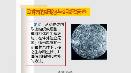
结果原代培养的血管平滑肌细胞在接种后3天开始从组织块周围游出,光镜下培养细胞呈梭形,放射状生长和典型的“峰谷”状生长;免疫组化和荧光染色显示胞浆内α平滑肌肌动蛋白阳性表达,证实培养的细胞为血管平滑肌细胞。
结论本法简便、可靠、经济、短期内可获大量高纯度、功能良好的血管平滑肌细胞,可作为研究动脉粥样硬化和移植血管再狭窄机制的有效模型。
【关键词】大鼠;胸主动脉;植块法;血管平滑肌细胞[Abstract]ObjectiveTo establish the culture model of rat aorta vascular smooth muscle cells (VSMCs) to provide important experimental method for the pathogenesis research of atherosclerosis in vitro. MethodsEmploying the explants technique, the primary and sub-culture were completed bymodified tissue-piece inoculation and trypsin digestion respectively. The cultured cells were identified by phase contrast microscopy and immunohistochemical and immunofluorescent staining.ResultsAfter 3 days of inoculation of tissue-pieces, VSMCs migrated from the vessel pieces. The cultured cells showed the typical “hills and valleys”morphological features under the microscope. Immunohistochemical and immunofluorescent staining with monoclonal antibody against mouse α-SM- actin demonstrated these cells were positive. ConculsionIt is a simple and reliable method for obtaining highly purified and satisfactory VSMCs in short term, providing a favorable experimental platform for research into the mechanism of vascular proliferous diseases such as atherosclerosis and restenosis.[Key words]rat;thoracic aorta;explants method;vascular smooth muscle cells血管平滑肌细胞(vascular smooth muscle cells, VSMCs)的异常增殖、迁移、泡沫化及凋亡与冠状动脉旁路移植术(CABG)和经皮冠脉腔内血管成形术(PTCA)术后血管再狭窄、颈动脉球囊损伤术致新生内膜形成(neointima formation)及动脉粥样硬化(atherosclerosis)密切相关[1~4]。

大鼠血管壁中膜平滑肌细胞的原代培养及鉴定敖锋;张自力;宋健【摘要】Objective To investigate the primary culture and identification of the smooth muscle cel s (SMC)from the medial of vascu-lar wal inrats.Methods SMC from the medial of vascular wal were cultured by modified tissue culture.The cel ular morphology was observed under inverted phase contrast microscope.SMC were isolated and identified with immunofluorescence.Results SMC were isolated successful y and cultured.The cel s had a typical peak val ey like growth,and showed fusiform.The positive rate of cel s i-dentified with immunofluorescence were more than 95%.Conclusion The modified tissue culture can isolateand cultivate SMC with high purity and good activity under in vitro conditions with simple,reliable method.%目的:探讨一种方便、快捷、大量原代培养大鼠血管壁中膜平滑肌细胞的方法,为心血管疾病的体外研究提供可靠的实验材料。
方法利用改良的组织贴块法进行血管壁中膜平滑肌细胞原代培养,使用倒置相差显微镜观察细胞形态,并用免疫荧光染色鉴定分离培养的细胞。
SD大鼠平滑肌的分离
1、300g左右大鼠戊巴比妥钠1ml注射麻醉(用20 mg/L的戊巴比妥钠按照50 mg/kg腹腔注射麻醉)。
10分钟后固定于解剖板上,酒精消毒,剪开腹部,挑开内脏,寻找腹主动脉,用静脉留置针尽量抽血。
2、抽干血液,剪开胸腔,翻开肺脏,寻找与心腔相连的胸主动脉(连接处有3个开口),沿着脊柱一直向下延伸(注意与食道区别,食道与胃连接),钝性分离周围脂肪组织,剪断后放入生理盐水中,冲洗干净血液,用镊子夹住一段,另一镊子从上至下用力刮脱,分离掉血管周围的脂肪组织。
3、转入超净台中,用PBS(或生理盐水)清洗,滴入少许胎牛血清至血管,在血清中剪碎血管组织,置入培养箱中2h。
4、随后加入20%胎牛血清低糖DMEM培液(最好加抗生素),继续培养。
