神经内科全英文疑难病例讨论 - 神经内科_20151022_133621
- 格式:doc
- 大小:20.50 KB
- 文档页数:4

神经内科病例讨论及诊疗思路神经内科作为医学领域的一个重要分支,主要研究和治疗与神经系统相关的疾病。
本文将通过讨论一个典型的神经内科病例,探讨其诊断和治疗的思路,以期为临床医生提供一定的参考和指导。
病例一:患者,男性,45岁,主诉头痛、恶心、呕吐已持续1周。
病史回顾:患者过去无相关疾病史。
最近半年来,患者的工作压力较大,睡眠质量欠佳。
体格检查:患者清醒,神志正常,颈部无僵硬。
血压为140/90 mmHg,脉搏为80次/分。
神经系统检查:患者的瞳孔对光反射正常;脑神经检查无异常;肢体肌力正常;腱反射生理正常。
辅助检查:头颅CT示无明显异常。
根据患者的主诉、病史回顾和体格检查结果,我们初步怀疑患者可能患有偏头痛。
偏头痛是一种常见的神经内科病症,表现为反复发作的头痛,伴随恶心、呕吐等症状。
针对这一病例,我们提出以下诊疗思路:1. 确诊偏头痛的标准:根据国际头痛学会(IHS)的标准,偏头痛的诊断包括典型的头痛发作、特定的头痛特征、头痛的持续时间等。
在确定是否为偏头痛之前,我们需要排除其他导致头痛的病因。
2. 详细了解病史:通过与患者充分沟通,了解其头痛发作的特点、频率、持续时间、伴随症状等,以帮助进一步确认诊断。
3. 辅助检查:除了头颅CT,我们还可以考虑进行其他辅助检查,如颅脑核磁共振成像(MRI)、脑电图(EEG)等,以排除其他潜在病因,并评估脑部结构和功能的异常情况。
4. 诱发因素的评估:在偏头痛患者中,一些特定的诱发因素可能导致头痛发作。
我们需要询问患者在头痛发作前是否有暴露于明显的诱发因素,如视觉刺激、气候变化、饮食因素等。
5. 头痛的急性治疗和预防治疗:对于急性头痛发作,我们可以使用非甾体类抗炎药、三叉神经阻滞剂等进行缓解。
对于频繁发作的患者,预防性治疗可能是必要的,包括口服药物、生物反馈治疗、针灸等。
6. 生活方式干预:我们可以建议患者调整生活方式,如规律作息、避免暴露于诱发因素、加强体育锻炼等,以减少头痛的发作频率和严重程度。


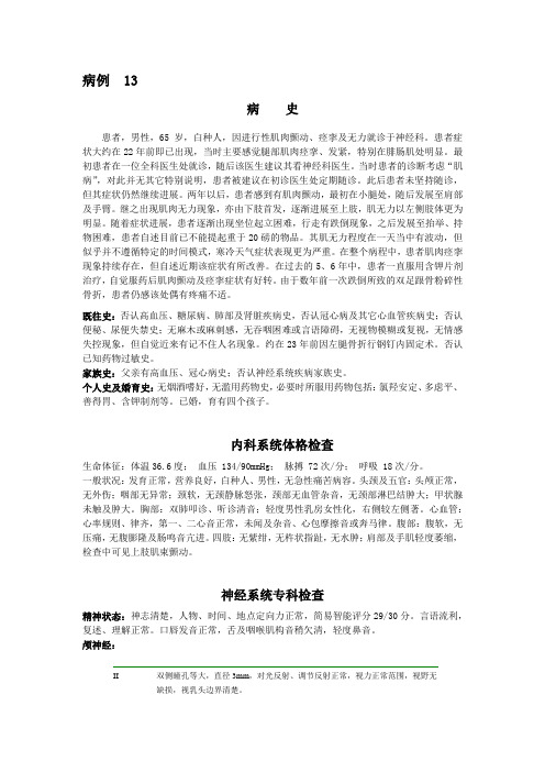
病例 13病史患者,男性,65岁,白种人,因进行性肌肉颤动、痉挛及无力就诊于神经科。
患者症状大约在22年前即已出现,当时主要感觉腿部肌肉痉挛、发紧,特别在腓肠肌处明显。
最初患者在一位全科医生处就诊,随后该医生建议其看神经科医生。
当时患者的诊断考虑“肌病”,对此并无其它特别说明,患者被建议在初诊医生处定期随诊。
此后患者未坚持随诊,但其症状仍然继续进展。
两年以后,患者感到有肌肉颤动,最初在小腿处,随后发展至肩部及手臂。
继之出现肌肉无力现象,亦由下肢首发,逐渐进展至上肢,肌无力以左侧肢体更为明显。
随着症状进展,患者逐渐出现坐位起立困难,行走有跌倒现象,之后发展至抬举、持物困难,患者自述目前已不能提起重于20磅的物品。
其肌无力程度在一天当中有波动,但似乎并不遵循特定的时间模式,寒冷天气症状表现更为严重。
在整个病程中,患者肌肉痉挛现象持续存在,但自述近期该症状有所改善。
在过去的5、6年中,患者一直服用含钾片剂治疗,自觉服药后肌肉颤动及痉挛症状有好转。
由于数年前一次跌倒所致的双足跟骨粉碎性骨折,患者仍感该处偶有疼痛不适。
既往史:否认高血压、糖尿病、肺部及肾脏疾病史,否认冠心病及其它心血管疾病史;否认便秘、尿便失禁史;无麻木或麻刺感,无吞咽困难或言语障碍,无视物模糊或复视,无情感失控现象,但自觉近来有记不住人名现象。
约在23年前因左腿骨折行钢钉内固定术。
否认已知药物过敏史。
家族史:父亲有高血压、冠心病史;否认神经系统疾病家族史。
个人史及婚育史:无烟酒嗜好,无滥用药物史,必要时所服用药物包括:氯羟安定、多虑平、善得胃、含钾制剂等。
已婚,育有四个孩子。
内科系统体格检查生命体征:体温36.6度;血压 134/90mmHg;脉搏 72次/分;呼吸 18次/分。
一般状况:发育正常,营养良好,白种人、男性,无急性痛苦病容。
头颈及五官:头颅正常,无外伤;咽部无异常;颈软,无颈静脉怒张,颈部无血管杂音,无颈部淋巴结肿大;甲状腺未触及肿大。


神经内科疑难病例讨论:多灶性运动神经病一例作者:郝以姝袁宝玉施咏梅张志珺多灶性运动神经病(Multifocal Motor Neuropathy, MMN)是一种罕见的、累及多数单神经的纯运动神经病;发病率约为0.3-3/10万,男女发病比例为2.7:1,多于50岁前发病,儿童发病少见;临床上主要表现为慢性进行性或阶梯样、非对称性肢体乏力伴萎缩,一般上肢症状较重,通常不伴有感觉缺失;神经电生理检查特点是持续多发的局灶性神经传导阻滞;为自身免疫介导相关性疾病,可有血清抗神经节苷脂GM1抗体滴度增高;丙种球蛋白治疗有效[1]。
该病容易与颈椎病、腰椎病、运动神经元病、慢性炎性脱髓鞘性多发神经病及脊髓性肌萎缩等混淆,尤其在基层医院,肌电图等检查不全面,神经肌肉活检等细胞分子病理学检查及基因检测未开展,导致患者未能得到及时诊断,延误治疗,造成病情加重,甚至出现残疾,严重影响生活质量。
该文报道一例反复就诊于多地高校附属医院和三甲医院,确诊为多灶性运动神经病的患者。
临床资料:患者男,38岁,货车司机,因“进行性四肢乏力伴肌萎缩五年余”于2015年7月16日入院。
患者五年前出现右下肢乏力,表现为踩刹车时力弱,无肢体麻木及疼痛,无腰痛,无肉跳,在当地医院就诊时发现右侧小腿肌肉萎缩,诊断“腰椎病”,予营养神经等药物治疗症状未见好转。
三年前患者出现左下肢无力,行走时左下肢拖拽,易绊倒,有时伴双下肢麻木及肉跳,无饮水呛咳及吞咽困难,当地医院仍按“腰椎病”治疗,症状无好转,两年前患者自觉左手不灵活,系扣子困难,并发现虎口处肌肉萎缩,症状逐渐加重,半年前自觉右手不灵活,遂到南京某医院就诊,查颈椎、腰椎MRI示:颈椎退变,颈椎盘变性,C5-T1椎间盘轻度突出,腰椎退变,L3-L5椎间盘变性伴突出。
查肌电图示:多灶性运动神经损害伴传导阻滞,血清肌酸激酶:295U/L,腰穿检查未见异常,抗核抗体系列及ANCA均未见异常,肿瘤标志物未见异常,叶酸、维生素B12检查未见异常,诊断“运动神经元病”,未予特殊治疗,遂到上海某医院查血清抗GM1(IgM,IgG)抗体,血清抗GQ1b(IgM,IgG)抗体均未见异常;复查肌电图示:多数单神经损害,累及部分运动神经,髓鞘损害伴轴索改变,MMN可考虑,门诊考虑:多灶性运动神经病,建议丙种球蛋白治疗,患者拒绝,后给予“美卓乐8mg qd”治疗五周,病情无好转,遂来我院就诊。
神经内科全英文疑难病例讨论 - 神经内科_20151022_133621History A 55-year-old left hand dominant man is evaluated at the hospital for muscle pain and weakness. He was in his usual state of health until four months prior to his initial clinic visit when he first noted stiffness and pain in his calves, the back of the knees and lower thighs. The pain was intermittent, lasting 10 to 15 seconds with spontaneous resolution, and was most prominent in the evenings. Over the next two months, the patient began to have increasing weakness in his arms and legs in addition to “aching pain” in his muscles. He had difficulty lifting objects, opening jars, and typing. He also had increasing difficulty climbing staircase and standing for prolonged periods. His symptoms were present throughout the day and would wax and wane with no identifiable provocative or alleviating factors. The muscle pain and stiffness gradually became more diffuse and spread to involve his trunk. It was exacerbated by physical activity and improved with prolonged rest. His muscles were tender with palpation and he even experienced mild chest discomfort when lying on his chest. One month prior to his clinic visit, he began to have increased swelling of his limbs especially in his feet, calves, hands and forearms. He gained over 15 pounds over two weeks prior to his clinic visit. Of note, the patient was evaluated by a community physician who performed serologic studies, including serum CPK, which were all normal. His electrodiagnosticstudies were felt to be suggestive of “inflammatory myositis” and he was referred to the Neuromuscular Clinic for furtherevaluation of possible inflammatory myopathy. He denied having fever, chills, night sweats, joint pain or rash. He did not have diplopia, dysarthria, dysphagia, neck weakness, loss of muscle bulk, numbness, paresthesia, incoordination, or bowel or bladder incontinence. He did have a burning sensation on his forearms, right more than left, most noticeably in the evenings. Review of System: He denied having other respiratory, cardiovascular, gastrointestinal or urinary symptoms. Past Medical History: He had no prior medical problems Past Surgical History: Renal calculi removal. Traumatic amputation of the left index finger requiring surgical repair. Allergies: No known drug allergies Medications: Multi-vitamin one per day, oralvitamin B12 supplement once daily, Flaxifish supplement. He did not take other health supplements or vitamins. Social History: He was a former smoker but quit 5 years ago. He drank alcohol occasionally. He denied illicit drug use. He was employed as a technology director. There was no history of toxic exposures. His only recent travel was to Hawaii with his wife two months after the onset of his symptoms. Family History: His father was 62 year old and his mother was 60 years old, both had a history of lung cancer. He had 2 sisters who were healthy. He wasmarried and had a daughter who was healthy. His paternal grandmother had type 2 diabetes mellitus. There was no family history of a similar condition or neuromuscular disorders. Physical Exam Vital signs: Hispulse was 66 beats per minute, respiration rate of 18 per minute and blood pressure was 102/68 mm Hg. His weight was 196 pounds. General appearance: Well built man, appears comfortable HEENT: Sclera anicteric, moist mucus membranes Neck: Supple, no thyromegaly, no lymphadenopathy Cardiovascular: S1, S2 regular rate and rhythm Chest: Clear to auscultation bilaterally with good air entry Abdomen: Normal bowel sounds, soft, non tender Extremities: Marked non-pitting edema up to the mid to proximal forearm and proximal calves (note photograph). No clubbing, cyanosis or rash. Range of motion is full. leg-edma Neurological Examination Mental status: He was awake, alert, orientedand was able to provide a detailed and comprehensive medical history.His speech was fluent without dysarthria. Cranial nerves: II. Pupils 3 mm bilaterally briskly reactive to light and accommodation. III, IV, and VI. Extraocular movements were full and intact. There was no nystagmus, ptosis, or diplopia. V. Sensation intact to pinprick, light touch, and temperature. Muscles of mastication showed good bulk and normal strength. VII. There was no facial asymmetry and no weakness the orbicularis oris or oculi. VIII. Hearing was intact to finger rub bilaterally. IX, X. Uvula and palate elevated in the midline. There was no dysphonia. XI. Sternocleidomastoid muscles were 5/5. XII. Tongue protruded in the midline. Atrophy or fibrillations were not noted. Motor examination: There was normal bulk and tone. There was tenderness with palpation of limb muscles. No myotonia, myokymia or fasciculations were present. MRC manual muscle testing were graded as 5/5 in all muscle groups includingthe neck flexors and extensor except for hip flexors, which were 4/5 and deltoids, triceps, wrist extensors, quadriceps, and hamstring muscles, which were 4+/5. Sensory examination: Intact to pinprick, temperature, light touch, vibration and proprioception in the extremities bilaterally. Romberg testing was negative. Deep tendon reflexes: 2+ and symmetric. No pathological reflexes were present. Coordination: Finger-to-nose andheel-to-shin maneuvers were slow but intact. Gait: Normal base and swing. Toe walking, heel walking, and tandem gait were normal. There was nogait ataxia.。