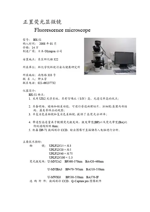
欣谕染色体分析系统显微镜BX
- 格式:doc
- 大小:83.00 KB
- 文档页数:3

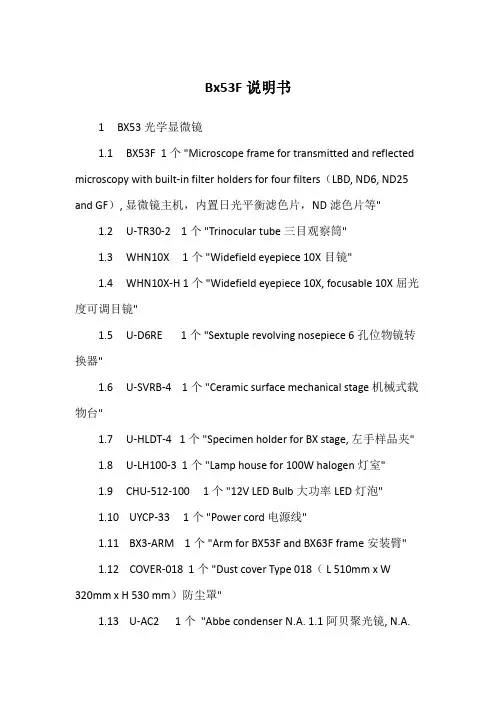
Bx53F说明书1BX53光学显微镜1.1BX53F1个"Microscope frame for transmitted and reflected microscopy with built-in filter holders for four filters(LBD, ND6, ND25 and GF),显微镜主机,内置日光平衡滤色片,ND滤色片等"1.2U-TR30-21个"Trinocular tube三目观察筒"1.3WHN10X1个"Widefield eyepiece 10X目镜"1.4WHN10X-H1个"Widefield eyepiece 10X, focusable 10X屈光度可调目镜"1.5U-D6RE1个"Sextuple revolving nosepiece 6孔位物镜转换器"1.6U-SVRB-41个"Ceramic surface mechanical stage机械式载物台"1.7U-HLDT-41个"Specimen holder for BX stage,左手样品夹"1.8U-LH100-31个"Lamp house for 100W halogen灯室"1.9CHU-512-1001个"12V LED Bulb大功率LED灯泡"1.10UYCP-331个"Power cord电源线"1.11BX3-ARM1个"Arm for BX53F and BX63F frame安装臂"1.12COVER-0181个"Dust cover Type 018(L 510mm x W320mm x H 530 mm)防尘罩"1.13U-AC21个"Abbe condenser N.A. 1.1阿贝聚光镜, N.A.1.1"1.14PLN4X1个"Plan achromat objective 4X/0.1, WD 18.5 4X 平场消色差物镜"1.15PLN10X1个"Plan achromat objective 10X/0.25, WD 10.6 10X平场消色差物镜"1.16PLN20X1个"Plan achromat objective 20X/0.4, WD 1.2 20X平场消色差物镜"1.17PLN40X1个"Plan achromat objective 40X/0.65, WD 0.6 40X平场消色差物镜"1.18PLN100XO1个"Plan achromat objective 100X/1.25, WD0.15 100X平场消色差油镜"1.19IMMOIL-8CC1个"Immersion oil 8cc镜油"2显微成像系统2.1D20001个"OPLENIC 20MP Digital camera USB3摄像头"2.2U-TV1X-21个"C-mount adapter holder接口适配器"2.3U-CMAD31个"1X C-mount adapter C型摄像头接口"2.4TS-M71个"Stage micrometer软件测量定标板"2.5USB320-ATB1个"USB3.0 cable A male to B male上等USB3数据线"3图像采集软件3.1静态图像采集3.2图片序列采集3.3延时摄影3.4视频录制3.5视频定标3.62D平面几何测量3.7多自由度图片及视频添加标尺3.8虚拟3D地形图3.9视频降噪3.10视频锐化3.11HDR / WDR宽动态范围图像获取3.12LIVE实时拼图/全景图像拼接工具3.13LIVE实时景深扩展EDoF/大景深图像合成3.14FISH多通道荧光叠加(含可编辑荧光染料库)3.15全黑背景图像降噪(用于拍摄荧光图片)3.16高阶拉普拉斯图像锐化3.17高阶色彩矫正3.18背景灰度校正3.19图文报告支持3.20分屏对比功能3.21预设参数加载、编辑、删除及导出导入3.22测量定标参数导出导入3.23多国语言包支持4图像工作站4.1处理器:Intel i3-81004.2主板:支持原生USB3高速传输芯片4.3硬盘:固态硬盘256GB4.4内存:8GB4.5操作系统:Windows 10 64-bit4.6显示器:23”4.7数据接口:USB3.04.8无线功能:蓝牙3.0、Wi-Fi4.9键盘鼠标:2.4G无线。
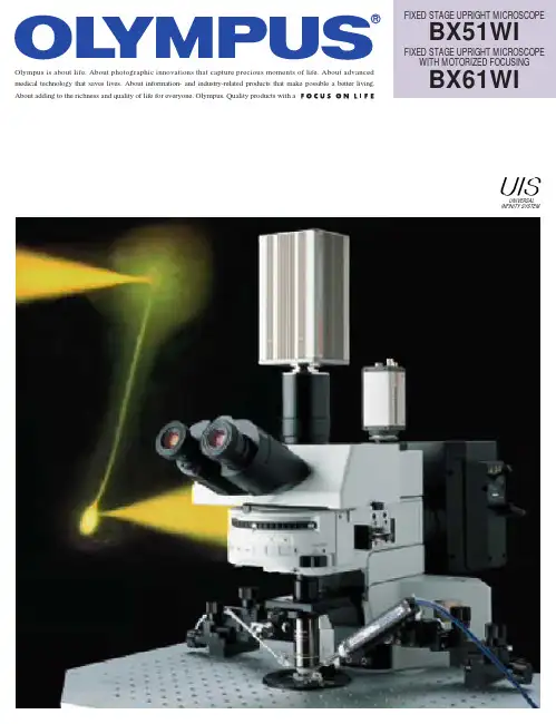
Olympus is about life. About photographic innovations that capture precious moments of life. About advanced medical technology that saves lives. About information- and industry-related products that make possible a better living. About adding to the richness and quality of life for everyone. Olympus. Quality products with a FIXED STAGE UPRIGHT MICROSCOPE BX51WIFIXED STAGE UPRIGHT MICROSCOPE WITH MOTORIZED FOCUSING BX61WIUNIVERSALINFINITY SYSTEMBX51WI with Luigs & Neumann Accessories.Combined with WI-DPMC.BX51WI with Burleigh AccessoriesA dual commitment:Preventing vibration andprotecting living cell specimensOne design theme was central to the development ofthe new fixed stage microscopes from Olympus — achieve an evenhigher standard of stability and reliability in electro-physiological applications.The result is a wide range of advanced new features to avoid and prevent vibration. These innovations include the introduction of a new observation method along withdetailed analysis of operability and further refinements in image clarity. These improvements work together to make patch clamp operations smoother and more efficient than ever before. Combined with the traditional excellence of UIS optics, the new Olympus fixed stage microscopes define new levels of quality in both performance and ease of use.Fluorescence macro objectives for membrane potential observation Full-system physiological confocal microscope,BX61WI with Z-axis motor is LSM ready7×Intermediate magnification 0.35 x 80×Intermediate magnification4 xN .A .0.95X L UM P L F L20x W Interchanging low and high magnifications without changing objectives.A new concept in vibration-free design.A major concern for researchers conducting electro-physiology experiments is the vibration which occurswhen switching objectives and the resulting interference this can cause to the specimens and adjacent equipment. To solve this problem, Olympus introduces a new concept —the provision of an intermediate magnification changer in combination withthe new High N.A. long working distance 20x objective that allows the user to switch between low and high magnifications without the need to switch objectives.New 20x objective (XLUMPLFL20XW) N.A. 0.95; W.D.: 2.0mm The new 20x water immersion objective makes high-resolutionobservation possible with a wide range of intermediate magnification lenses. Since exchanges between low and high magnification are performed through the intermediate magnification changer, vibration is reduced to a minimum and the usual concern about collisions between objectives and patch clamp electrodes is eliminated.CondenserFilter turretIR-DIC port IR-DICXLUMPLFL20xW N.A. 0.95Video port FL/DICLight path exchange leverIntermediate magnification change lever IR filter(not to use with visible light DIC observations)Observation port FL/DICDual port WI-DPMCMirror unit exchange turret 0.35x(0.25x) lensTube lensMirror unitDIC PrismAnalyzerC mountDichroic mirror 4x lensPolarizer1/4 wave plateDIC elementExcitation light Fluorescence light IR-DICTransmitted lightSimultaneous fluorescence and IR-DIC observations With the included 690nm dichromatic mirror in the WI-DPMC,fluorescence light is sent to the front port, and IR-DIC light is sent to the back port allowing two cameras to image simultaneously with no vibration introduced by light path selection.Variable magnification dual port (WI-DPMC)The WI-DPMC rear camera port includes a 2 position intermediate magnification selector. A high magnification 4x intermediate lens is included and a (0.25x, or 0.35x) low magnification lens is optional.High or low magnification selection is via a single lever with no click-stops or detents allowing a specimen to be scanned and measured with minimal disturbance from vibration. 775nm and 900nm IR-DIC compatible.*Available for 0.5x, 1x and 2x intermediate magnification lenses by special order.Variable Click-stopsAll click-stops, as when selecting between camera and observation modes, can be adjusted to the point of no click and thus no vibration.q Vibration-free shutterThe fluorescence shutter slides horizontally with no detents and no vibration.y A waterproofing sheetA waterproofing sheet, attached by the supplied magnets, provides protection against liquid overflow and spills. The sheet is large enough to protect the frame, condenser and focusing mechanisms .e Ample space around the condenserFrame designed for ample space around the condenser, making it easy to adjust Nomarski DIC contrast, exchange filters,adjust the condenser's aperture stop and to easily switch between visible light,Nomarski DIC or IR-DIC.w Mirror unit turret with adjustable click releaseThe click-stop on the 6 position turret can be released with a precision screwdriver.r Front focus knobs close to the operator's handFine focus control is located at the front on both sides of the microscope body. The knob on the right integrates both coarse and fine focus control.t Coarse focus lock leverWhen engaged at the desired position, the objective can be raised with the coarse focus knob and then returned precisely to its original position.Front operation with no shock and no noise.A new concept in experimental operation.rewqThe new front operation system prevents interference in patch clamping work. The design concept is simple and allows frequently performed operations like focusing or filter exchange to be done easily at the front of the unit.Ample space is provided on both sides of the microscope frame and condenser, so the necessary manipulation equipment can be positioned close to themicroscope.tu Remote power supply and hand switchThe remote TH4 power supply for transmitted light is designed with no cooling fan to minimize electrical noise.Features on/off and intensity controls. Can also be used with the optional TH4-HS hand switch providing light intensity and on/off control a maximal distance away from the Faraday cage.uyRaising the objective and lowering the stage to enable small animal experimentsThe arm height raising kit (WI-ARMAD) provides an additional 40mm of clearance and is mounted between the microscope frame and the reflected light illuminator. Small animal experiments usually do not require transmitted light thus allowing the removal of the substage condenser assembly. After removal, the stage may be lowered an additional 50mm, providing a total clearance increase of 90mm.A variety of convenient units toadd light sources and control the lightLamphouse adapter U-LHADThis adapter allows the mounting of the dual port (U-DP) between the microscope frame and lamp housing.Rectangular field stop BX-RFSSDesigned for use with CCD cameras, prevents photobleaching of the specimen outside of the imaging area.Pinhole unit BX-RFSPOTLighting the cell via a pinhole allows experimentation on reaction to light. Optional MELLES GRIOT's ø16pinhole is used.New functionality and solutions to meet a wide variety of needs.A powerful new concept.(photo shown is BX-RFA)Experimenting with small animalsPhotoactivationNormal configuration40mm more clearance via WI-ARMAD18mmW.D.18mm 40mmW.D.BX-RFSSBX Stage and adapter for injection experimentsThe stage adapter WI-STAD is designed to allow the attachment ofa traditional microscope right or left hand stage to the WI frame.The compact design of the BX2 stage (U-SVRB-4, or U-SVLB-4)reduces the distance between the specimen and the manipulatorand creates a stable platform for injections.MicroinjectionConfocal Microscope SystemBX61WI — Built in Z-axis focus motorThe BX61WI frame incorporates a precise Z-axis focus motor with 0.01µm step size. Designed to incorporate the Olympus Fluoview scan unit and software, the BX61WI is ready for confocal z-stacks. Microscope frame includes programmable buttons for a wide variety of applications.Convenient, optional focusing hand switch U-FH forremote operationThe remote hand switch allows the user to control the microscope remotely via a 2 meter connection cable. Hand switch allows the selection of coarse and fine focus movement, and nosepiece escape/ return. Hand switch buttons can also be custom programmed for individual needs.Optional Olympus Fluoview Confocal System FV500/FV300 With the scanning unit set at the back of the microscope body, compact layout in the cage is possible.Moving the microscope and scanning unit together(mover available by special order)Allows X and Y movement of both the microscope and scan unit together while the stage and specimen are fixed.Additional lasers and accessoriesAn assortment of lasers can easily be attached to satisfy a wide variety of applications.Setting example: BX61WI+FV300 U-FHUltimate image clarity for electro-physiological experim A new concept in live cell observation.Senarmont compensation for Nomarski DIC observation When using a Senarmont equipped condenser, all contrast adjustments are performed with the 1/4 wave plate below the condenser, thus eliminating the risk of bumping the stage, specimen, manipulators or nosepiece.IR-DIC/ Nomarski DIC observationOblique illumination observationOblique observation optimizes contrast by changing the direction of the specimen shadowOlympus has developed an oblique condenser (WI-OBCD) whose long working distance enables the angles of shadow to be altered through 360 degrees without moving the specimen. Requiring no additional accessories, oblique illumination is easy to set up and control. Plastic dishes (normally unsuitable for all types of DIC) are easy to image with oblique illumination. The oblique illumination slit aperture is variable in size and on a slider allowing quick changeover.IR-DIC Optimized Optics:Designed for observations at 775nm to 900nmThanks to the precisely aberration-compensated IR-DIC optics covering from visible to near infrared light of 775nm/900nmwavelength, the clarity of images observed under near infrared light has been improved still further, allowing clear observation of even deep sections of brain slice.• Visible light DICAllows operator high-resolution observation of the tissue surface.• 775nm IR-DICIn combination with an IR camera allows observation within the tissue slice. Optics are corrected for visible and IR wavelengths allowing fast switching between wavelengths with minimal refocusing.• 900nm Nomarski DICAllows observation deeper into the tissue (requires special polarizer and analyzer optimized for 900nm).Analyzer DIC prismObjectiveDIC prism 1/4 wave platePolarizerIR filter(not to use with visiblelight DIC observation)Nucleus of solitary tract from slice of rat medulla oblongata (thickness: 400µm )Prof. Fusao KatoSchool of Medicine Physiology Dept.,Jikei UniversityKato & Shigetomi, J. Physiol.(2001), 530: 469-486Universal condenser with DIC for improved contrastSuitable for use in visible and 775nm/900nm near-infrared light,the U-UCD8 universal condenser is a high N.A., short working distance condenser offering improved contrast in nerve cell observations, for example.AdjustableU-UCD8WI-TP137WI-DICTWI-OBCDRotatablements.Transverse cryostat section through the hippocampus of a mouseat postnatal day 10 was stained with a mouse monoclonal anti-neurofilament-L (Chemicon, MAB1615) .An FITC-conjugated anti-mouse antibody was used for detection of NF-L.Objective: XLFLUOR4x/340Masaharu Ogawa,Ph.DLaboratory for Cell Culture Development,Brain Science Institute, RikenObserving changes in membrane potentialFluorescence macro observationMeasuring changes inmembrane electric potential by using the XLUMPLFL20xW objective with N.A. 0.95The XLUMPLFL20xW objective, with its high N.A., and 2.0mm of working distance allows the measurement of cell membrane electric potential (as seen right). Also, the 4x macro objective (XLFLUOR4x/340) can be used to measure membrane potential at the tissue level. A water immersion cap (XL-CAP) can be attached to the macro 2x or 4x objectives to eliminate disturbances caused by water ripples.2x and 4x Macro lenses with high numerical apertures provide fluorescence imagesDesigned for GFP imaging of large cells such as neurons 2x and 4x low magnification fluorescence objectives and a special GFP observation mirror unit are available. The objectives have a long working distance for maximum flexibility. An optional waterimmersion cap (XL-CAP)is also available toremove imageaberrations caused by ripples on water surfaceof immersed specimens.Imaging of neuronal activity with voltage sensitive dyeSpread of neural activity in area CA1 of acute rat hippocampal slice (400µm thick) in response to a single stimulation applied to Schaffer collateral pathway imaged (at frame rate of 0.7 ms/frame) with a fluorescent voltage sensitive dye (VSD; Di-4-ANEPPS). The fluorescent image (90x60 pixels) captured by a digital high-speed CCD camera (MiCAM01, Brain Vision Inc.; with 20x objective and 0.5x adapter) is superimposed on the illustration of a hippocampal slice (upper left panel). The image is enlarged and shown on the illustration of pyramidal cells (solid line) (lower left panel). Each laminar of CA1 is shown as follows: SO-A, Stratum oriens-alveus; SP, Stratum pyramidal; SR, Stradum radiatum. The individual somas of cells were visible (indicated by dotted circle on the image) and were found along the stratum pyramidal. The changes in the fluorescence of VSD (optical signal) in accordance with the membrane potential change upon a stimulation (Stim) onto Schaffer collateral (Sch) were pseudo-color encoded and shown as consecutive images (upper right panel; number in each image shows time from the stimulation (ms)). The depolarizing signal (red) spread along Schaffer collateral,which was followed by a hyperpolarizing signal (blue) originated in stratum pyramidal. The time courses of optical signals in representative pixels are shown in lower right traces.Takashi Tominaga Ph.D, Brain-Operative Device Lab., Brainway Group, Brain Science Institute, RikenU-SLRE XLFLUOR2x/340XLFLUOR4x/340U-MF/XL U-MGFPA/XL U-MGFP/XLIntermediate magnification changer U-ECA, U-CAThe U-ECA, which includes a 2x intermediate magnification position,allows quick magnification changes to a camera or observer without the need to change objectives. The U-CA includes a 4 position turret that allows rapid switching between a 1x, 1.25x, 1.6x and 2x positions. Both changers accept standard Olympus adapters for attaching a wide range of cameras.* U-ECA and U-CA are notrecommended for IR observation with the U-TR30 trinocular observation head.AccessoriesMulti double port tube U-DPTSThe U-DPTS accepts an optional dichroic mirror allowing theincoming to be split between visible and infrared and be observed simultaneously using two cameras.* A fluorescence mirror unit is required.UIS ObjectivesU-CMDPTSU-PMDPTSU-DPTSU-ECAU-CAU-TVCACBX51WI/BX61WI specificationsSan-Ei building, 22-2, Nishi Shinjuku 1-chome, Shinjuku-ku, Tokyo, JapanPostfach 10 49 08, 20034, Hamburg, Germany2 Corporate Center Drive, Melville, NY 11747-3157, U.S.A.491B River Valley Road, #12-01/04 Valley Point Office Tower, Singapore 2483732-8 Honduras Street, London EC1Y OTX, United Kingdom.104 Ferntree Gully Road, Oakleigh, Victoria, 3166, AustraliaSpecifications are subject to change without any obligation on the part of the manufacturer.ISO9001Certificate No.69372CertificationDesign and productionadheres to ISO9001international quality standard.ISO 9001ISO14001Design and production at the OlympusOptical Co. Ltd. Ina Plant conforms withISO14001 specifications forenvironmental management systems.CertificationCertificate No. 70933ISO 14001U K A S*All brands are trademarks or registered trademarks of their respective owners.Web site address: BX51+WI-DPMC dimensions (unit: mm)。

本规程适用于OLYMPUS BX43型成像显微镜操作。
2依据文件OLYMPUS BX43型成像显微镜操作说明书。
3 操作过程3.1接通电源:接通成像系统所属显微镜、主机、显示器、打印机等设备电源。
3.2开启系统设备:依次开启显微镜、显示器、打印机及主机开关。
3.3调节光路:转动物镜转换盘,于低倍镜下观察、调节光路至视野中央。
3.4寻求目标物:将制好的标本放置于载物台上,移动载物台,调节光亮明暗度,调焦距,从低倍镜至高倍镜依次观察,直至目标物在视野中央且观察清晰,定位。
3.5数据传输:将显微镜的“拉杆”拉至中间(或全开),让定位好的目标物图像数据传输给主机。
3.6进入成像系统:鼠标左键双击电脑桌面“cellSens Entry”软件图标,进入成像系统软件操作界面。
3.7实时观察图像:点击软件操作界面“实时观察”按钮,向下依次点选曝光、区域、灵敏度、分辨率等按钮,旋转显微镜底座亮度调节轮调光,直至所选图像清晰。
3.8拍摄图像:点击操作界面“拍照”按钮,即刻将目标物像拍摄;若需重拍,则再点击“实时观察”按钮,再点“拍照”。
3.9保存图像:点击“文件”菜单,选“保存”,将已摄图像保存在指定或新建文件夹中。
3.10续摄图像:移动载物台,变换物镜,寻找下一个目标物,重复步骤6、7、8操作过程,摄像,保存。
3.11编辑图像:打开存有已摄图像的文件夹,新建一个“word”空白文档并打开,将已摄图像拖入该文档,进行编辑、放大、缩小、输入文本等,直至达到要求。
3.12打印图像:点击“文件”菜单,选“打印”,将编辑成形的图像文档进行打印。
3.13显微镜归位:工作完毕,退出操作软件,关闭显微镜电源,降下载物台,取下标本,镜头归位,清洁载物台,推回拉杆,使显微镜回到初始状态。
3.14关机:依次关闭主机、显示器、打印机,并切断电源。
3.15记录:每次用完应用时登记相关操作信息。
3.16注意事项:本系统电脑为专用(内装图像采集卡),禁止乱插U盘或他用;本系统显微镜只须在摄像时使用,不作一般显微观察用;每次编辑图像时最好先关掉显微镜电源、归位。

注:后附染色体配套设备表前言染色体是遗传物质的载体,近40-50年来,对染色体的研究发展迅速。
目前由染色体畸变引起的疾病发现的越来越多,应用染色体诊断疾病,探讨病因和发病机制,合理制定出治疗方案和合理用药,采取必要的预防措施及推算预后等都提供了科学的依据.因此对染色体的研究已成为临床医学中一个组成部分.染色体制备、分析日益广泛应用,如何正确识别人类染色体显得十分重要。
基于这种想法,编写这本材料,以供学习参考。
由于水平所限错误之处再所难免,敬请批评指正。
实验室的基本设备开展人类染色体研究的实验工作,应具有一定的条件、设备和器材才能顺利地开展工作。
一、实验室实验室的间数、大小应根据从事的研究的内容、工作人员和设备的多少,并根据各单位具体情况安排。
一般包括:1、准备室:为实验主要工作场所,除放置冰箱、烘烤箱、恒温箱、离心机、天平等仪器外,还备有箱厨和其它橱柜放置玻璃器皿、各种日常应用物品。
室中央设有操作台,以供实验操作用。
2、无菌室:为无菌操作室。
应设在实验室靠近最里面的位置,以利于防止空气流通引起污染。
在无菌室外要有缓冲室,室内应装空调、紫外线光源、清洁工作台等设备。
3、研究室:供电脑染色体分析用,并要有显微镜、整理研究资料等用。
二、大型器械1、电热恒温培养箱:最好用隔水式的,用于细胞培养。
2、电热恒温干燥箱:以中型较合适,因为干燥灭菌时的温度要达到160℃,故应购置能升温到300℃的较为安全适用。
3、高压蒸汽灭菌器:以电热恒温式较好。
4、普通冰箱:主要用于短期保存血清、培养液、生理盐水、消化液等各种溶液。
5、低温冰箱:作为培养基、血清等的长期保存,温度达到-40℃即可。
6、离心沉淀机:一般应购置0-4000转/分,最好12管,每管容量为10ml为水平式的。
若自制血清则购置0-4000转/分,4管,每管容量为250-300ml的水平式为好。
7、酸度计:又称为氢离子测定计或PH计,是测定各种溶液酸碱度的必须工具。

/axiovert-digital蔡司Axiovert 5数码款您的一体式细胞成像系统质臻至简,配备蔡司AI 技术100 µm从自动驾驶和智能家居,到为智能手机加密的面部识别系统,人工智能(AI )已为我们的日常生活提供了诸多便利。
是时候将人工智能也带入您的细胞实验室了。
Axiovert 5数码款采用人工智能和自动功能,助您轻松完成日常工作。
它能让您享受更高效的工作流程,并获得可重复性更高的结果。
即使面对繁多的工作任务,您也可以轻松应对。
Axiovert 5数码款的人工智能经过预先训练,汲取了蔡司丰富的经验:我们已导入大量数据集,使其尤为可靠。
只需按下一个按钮,便可以获得实时结果。
您的一体式细胞成像系统单击此处观看本段视频› 简介› 优势› 应用› 系统› 技术参数› 售后服务更简单、更智能、更高度集成开箱即用畅想一体式显微镜系统的诸多优势。
从常规的科学工作到基础研究,从相差到多通道荧光成像,使用Axiovert 5数码款,即使是新手也能采集到出色的图像。
打开系统,设置和调整已然就绪,您仅需专注于样品,无需进行繁琐操作,便可立即投入工作。
您也不必担心细胞在密闭培养箱内的状态,可以随时关注它们的变化。
Axiovert 5 数码款将可重复性和数据质量提升至新水平。
您可以始终依靠仪器的出色性能,得到可供发表的图像。
简单易用Axiovert 5数码款的设计支持相应的系统操作,是您多用户环境的理想之选。
其一体式成像系统具有直观的操作理念,只需点击一下拍照按钮即可实现以下功能:• 多达5个通道的图像采集(包括多通道 成像)• AI 细胞计数和融合度工作流,采集并实时分析图像• 视频记录Axiovert 5数码款将可靠的光学质量和简单易用巧妙结合。
节省时间,让人工智能为您效力借助Axiovert 5数码款,轻松节省您的宝贵时间,而这些时间可能对细胞的活力至关重要。
无论是设置系统和采集参数、培训新同事、采集图像,还是从图像到产生结果,在各个环节上您都可以节省时间。
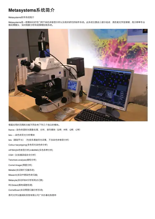
Metasystems系统简介
Metasystems软件系统简介
Metasystems是⼀款模块化的专门⽤于染⾊体核型分析以及相关研究的软件系统。
此系统主要由三部分组成:⾼性能光学显微镜;⾼分辨率专业数码摄像头;实时图象分析和显微镜控制系统。
根据应⽤的范围和功能不同含有下列⼏个独⽴的模块。
Ikaros---染⾊体透射光图象处理,分析,排列模块(G带,R带,Q带,C带)
Isis-----染⾊体荧光分析模块
Isis(基础平台)(包括多通道荧光采集,不含染⾊体核型分析)
Colour karyotyping(多⾊荧光染⾊体分析)
mFISH(24⾊核型分析)/mBAND(多⾊条带分析)
CGH(⽐较基因组杂交分析)
Telomere analysis(端粒分析)
Comet Imager(彗星分析)
Metafer(⾃动玻⽚扫描系统)
Msearch(⾃动中期染⾊体扫描)
Metacyte(⾃动FISH分析和斑点记数)
RCDetect(稀有细胞检测)
CometScan(⾃动彗星扫描分析系统)
蔡司光学仪器国际贸易有限公司⼴州办事处陈德林。

胚胎发育实验室设备采购在生命科学领域,胚胎发育研究是一项至关重要的工作。
而一个高效、精准且安全的胚胎发育实验室离不开先进、合适的设备支持。
本文将详细探讨胚胎发育实验室所需设备的采购要点,以帮助您打造一个功能完备的实验室。
一、显微镜系统显微镜是胚胎发育实验室的核心设备之一。
首先,倒置显微镜是必不可少的。
它能够提供清晰的细胞和胚胎图像,方便研究人员进行观察和操作。
在选择倒置显微镜时,要关注其光学分辨率、放大倍数范围以及照明系统的稳定性。
高分辨率可以更清晰地呈现胚胎的细微结构,而稳定的照明系统则能确保图像的质量不受光线波动的影响。
正置显微镜在某些实验中也能发挥重要作用,例如对组织切片的观察和分析。
此外,相差显微镜可以增强细胞和胚胎的对比度,使细微结构更加明显。
荧光显微镜则适用于特定蛋白质或基因标记的观察。
二、培养箱胚胎的培养需要一个稳定的环境,因此高质量的培养箱至关重要。
培养箱应能够精确控制温度、湿度和气体浓度。
温度的稳定性对于胚胎的正常发育至关重要,通常需要控制在37°C 左右,波动范围应极小。
湿度一般要保持在较高水平,以防止培养皿中的液体蒸发。
气体浓度的调节也非常关键,通常需要控制二氧化碳的浓度在 5%左右,以维持合适的酸碱度。
在选择培养箱时,要考虑其容量大小、内部结构是否易于清洁和消毒,以及是否具备报警系统,以在出现异常情况时及时通知实验人员。
三、体视显微镜体视显微镜在胚胎操作过程中有着广泛的应用。
它能够提供三维的视觉效果,方便实验人员进行胚胎的转移、注射等精细操作。
在选购体视显微镜时,要关注其景深、放大倍数和工作距离。
较大的景深可以使整个操作区域都保持清晰,合适的放大倍数和工作距离则能让实验人员操作更加舒适和准确。
四、离心机离心机用于分离和处理细胞和液体样本。
在胚胎发育实验室中,低速离心机常用于分离细胞和培养液,高速离心机则可用于提取核酸、蛋白质等生物大分子。
选择离心机时,要考虑其转速范围、离心容量、转头类型以及安全性等因素。

蔡司 Axiolab 5智能显微镜,让您的实验室日常工作更高效产品信息 版本 1.0Axiolab 5 适用于实验室中进行的常规显微镜检查工作。
其紧凑且符合人体工程学的设计可节省空间且易于操作。
Axiolab 5 是研究团队中的得力助手。
与 Axiocam 208 color 组合,充分发挥智能显微镜功能的优势——体验全新的显微数码成像方式。
只需聚焦样品并按下一个按钮,便可轻松获得清晰的真彩图像。
数字图像与您从目镜中观察的效果一致,所有细节和细微色差均清晰可辨。
另外,Axiolab 5 还会自动向图像添加正确的比例尺信息。
这一系列操作可单机完成,无需使用计算机或任何其他软件。
使用 Axiolab 5 既节省时间又节约成本,而且还可节约实验室的宝贵空间。
显微数码成像从未如此简单。
智能显微镜,让您的实验室日常工作更高效› 简介› 优势› 应用› 系统› 技术参数›售后服务更简单、 更智能、 更高度整合提升实验室日常工作的效率找到感兴趣区域后,只需按下显微镜主机两侧的拍照按钮,即可获得图像,操作简单便捷。
Axiolab 5 易于操作且符合人体工程学的设计理念,使其成为实验室日常工作的好帮手。
您甚至无需移动手的位置即可控制显微镜及其连接的相机。
智能显微镜系统将自动调节参数,以当时所显示的样品状况进行精确地记录,并获得包含细节信息的真彩图像。
同时也会自动添加正确的比例尺信息。
您无需再额外购买计算机或软件。
智能显微镜可以让您的工作更高效,始终专注于样品。
更经济、更可靠Axiolab 5 对您而言既节省成本又节能。
例如,启用 Eco 模式后,Axiolab 5 将在闲置 15 分钟后自动进入待机模式。
这一操作不仅节能,而且还延长了光源的使用寿命。
与传统照明系统相比,LED 的使用寿命更长。
在透射光下,全新的高性能白光 LED 让您能够观察到原色样品的图像。
即使是细微色差,仍清晰可辨。
在荧光应用中,具有不同波长的内置 LED 相比于传统汞灯使用更方便且更安全。
第1篇一、概述显微镜系统是一种用于观察微小物体的精密仪器,广泛应用于科研、教学、医疗等领域。
为确保显微镜系统的正常运行和使用效果,特制定本操作规程。
二、操作前的准备1. 确认显微镜系统状态:检查显微镜系统是否处于完好状态,电源线路、照明系统、物镜、目镜等部件是否完好。
2. 标本准备:将待观察的标本进行适当处理,确保标本的厚度适中,便于观察。
3. 清洁操作:确保操作台面、显微镜系统及其配件干净整洁。
三、操作步骤1. 开机:接通电源,开启显微镜系统电源开关。
2. 调节亮度:根据标本特性,调整内置照明灯光亮度及光圈大小,使视野亮度处于合适范围。
3. 固定装片:将处理好的标本放在载物台上,用标本片夹持器夹好,确保标本稳定。
4. 观察方法:a. 低倍物镜观察:转动转换器,使低倍物镜对准通光孔,转动粗准焦螺旋,使镜筒缓慢上移,直至视野清晰为止。
b. 高倍物镜观察:若需进一步观察,转动转换器,使高倍物镜对准样品,调节细准焦螺旋,使视野清晰。
c. 调整视野:若视野亮度不够,可通过调节内置照明灯光亮度或光圈大小进行调整;若视野不够清晰,可通过调节细准焦螺旋进行微调。
5. 观察操作:a. 旋转载物台:将需要观察的部位移至视野中央。
b. 调整瞳间距:左右目镜按需要推动两目镜筒向两旁分开或靠拢,确保两只目镜的机械筒长一致。
c. 调整视场:通过横向和纵向手轮,将需要观测的标本移至载物台的中央。
6. 关机:观察完毕后,关闭显微镜系统电源开关,取下载玻片,将物镜转成八“字形,转动粗调节螺旋使载物台下降到最低处,罩上防尘罩,将显微镜归位。
四、注意事项1. 操作过程中,避免用手触摸显微镜镜头,以免污染或损坏镜头。
2. 观察过程中,避免强烈震动显微镜系统,以免影响观察效果。
3. 定期清洁显微镜系统及其配件,确保显微镜系统的正常运行。
4. 严格按照操作规程进行操作,避免因操作不当导致显微镜系统损坏。
5. 使用完毕后,关闭电源,确保显微镜系统处于安全状态。
人类染色体的研究用于医学的目的,已有近半个世纪的历史,迄今已正式定名的染色体综合症已近70种,各种各样的染色体异常在500种以上,1970年以后,由于显带技术问世,可以确认各号染色体使染色体疾病的诊断和预防提供了有效方法。
“XY”染色体分析系统针对人的外周淋巴血细胞染色体的G带核型分析,研制了染色体核型自动分析系统。
该系统可以对G显带染色体细胞,进行分离和分割,然后自动进行识别排队,从而实现了G显带染色体核型分析。
同传统的人工核型分析相比,该系统大大减轻人工的繁锁劳动,可为临床遗传疾病的查检和对胎儿进行产前染色体疾病的诊断提供了一个有效的工具。
1.图像获取
2.预处理
染色体分割
3.染色体核型分析
执行核型分析程序后,程序自动对染色体进行配对处理,并将配对的结果保存于文件中。
4.核型图显示与调整
5.结果输出
6.分析系统的技术指标:
本系统的性能指标如下:分析速度:XY分析一个核型的最快时间2~3分钟;分析的准确性:>93%。
7.主要应用范围:
本系统可应用于以下几方面。
(1)对遗传疾病的诊治;
(2)开展优生优育的检查,以保证人口的质量;
(3)开展遗传病的普查工作;
(4)放射工作人员的剂量估算。
图像预处理:包括图像的获取、去噪、二值化;
图像的分割:对粘连或重叠染色体进行分割或分离,包括人工操作方法和计算机自动方法;
核型分析:对经过图像分割后的染色体,自动进行核型分析,按G带标准图分类排队;
核型图校正:通过人机交互方式,对排错的染色体进行纠正,包括:倒置、交换、移动;
结果打印:对正确的核型图,可以通过打印机打印,在打印核型图的同时,还可将检查与医生诊断结果一同打印,作为检查报告单;
资料管理:本系统还提供了数据管理功能,可将分析结果保存于数据库中,便于资料的维护、查寻。
本系统的性能指标:
核型分析的正确率:经过上千次的实验结果,对正常染色体标本的识别率达93%
分析速度:< 5分钟/每细胞(含对粘连和重叠染色体分割与分离的时间)
本系统的硬件包括:高清晰度彩色摄像机、真彩色图像获取板、高清晰数字图像采集卡、计算机和激光打印机。
染色体分析系统包含的配置
1.摄像机:日本JVC不低于9501配置
2.计算机:XY染色体软件专用品牌机:联想不低于M4350配置
3.显示器:液晶显示器:联想19宽屏
公司名称:上海欣谕仪器有限公司
网址:http://
联系人:宋杰
联系电话:
手机:
邮箱:
主营产品:冷冻水机,低温水浴槽,制冰机,冷冻干燥机,超低温冰箱,层析冷柜,气溶胶喷雾器,程序冷冻装置,染色体分析系统。