Clinical utility of total HCV core antigen quanti
- 格式:pdf
- 大小:140.10 KB
- 文档页数:8

胆固醇酯转运蛋白活性测定在心脑血管疾病监测中的意义林荔;李颂文
【期刊名称】《国际检验医学杂志》
【年(卷),期】2006(027)006
【摘要】目的探讨胆固醇酯转运蛋白(CETP)活性与心脑血管疾病的关系.方法应用体外合成14C标记的高密度脂蛋白(HDL)样颗粒,测定了45例冠心病患者、26例脑卒中患者和40例健康人CETP活性.结果冠心病患者、脑卒中患者CETP活性分别为(17.6±5.4)%、(15.2±3.8)%,而健康人为(12.7±2.0)%,两组患者与健康人比较差异均有统计学意义(P<0.01).结论 CETP与动脉粥样硬化的发生、发展密切相关.【总页数】2页(P493-494)
【作者】林荔;李颂文
【作者单位】511300,广东省增城市中医医院检验科;510010,广州军区总医院检验科
【正文语种】中文
【中图分类】R4
【相关文献】
1.测定血清胆固醇酯转运蛋白及血脂浓度对治疗糖尿病的意义 [J], 庄奕宏
2.人工合成高密度脂蛋白样颗粒测定血清胆固醇酯转运蛋白活性 [J], 楼正青;庄一义
3.2型糖尿病患者血清胆固醇酯转运蛋白浓度测定及其临床意义 [J], 杨文惠;张洪
4.心脑血管疾病患者胆固醇酯转运蛋白活性测定 [J], 楼正青;庄一义
5.14C标记合成脂蛋白样颗粒法测定血清中胆固醇酯转运蛋白活性 [J], 吉维民因版权原因,仅展示原文概要,查看原文内容请购买。

利用多层螺旋CT测量肝脏体积对肝硬化患者肝脏储备功能的评估价值杨卫【期刊名称】《医疗装备》【年(卷),期】2022(35)15【摘要】目的探讨利用多层螺旋CT测量肝脏体积对肝硬化患者肝脏储备功能的评估价值。
方法选取2018年6月至2021年8月在医院影像科接受多层螺旋CT 检查的87例肝硬化患者,将扫描图像传送至CT后台工作站测量肝脏体积,并利用肝脏理论体积公式计算肝脏理论体积;此外,依据Child-Pugh分级和终末期肝病模型(MELD)评分系统评估患者的肝脏储备功能,并根据上述标准将87例患者分为Child-A级(5~6分,32例)、Child-B级(7~9分,35例)和Child-C级(≥10分,20例)3个亚组和MELD评分<10分(29例)、10分≤MELD评分<20分(37例)、MELD评分≥20分(21例)3个亚组;最终比较肝硬化患者肝脏体积的CT测量值与理论值,以及不同肝脏储备功能肝硬化患者肝脏体积的CT测量值与理论值,分析肝硬化患者肝脏体积CT测量值与Child-Pugh分级的相关性。
结果87例患者肝脏体积的CT测量值为(975.38±137.40)cm^(3),小于理论值(1284.73±226.42)cm^(3),差异有统计学意义(P<0.05)。
随着Child-Pugh分级和MELD评分增加,肝脏体积的CT测量值呈依次下降的趋势(F=19.829、18.754,P<0.05),两两比较差异亦有统计学意义(P<0.05);且不同Child-Pugh分级和MELD评分肝硬化患者肝脏体积的CT测量值均小于理论值(P<0.05)。
Spearman等级相关分析显示,肝硬化患者肝脏体积CT测量值与Child-Pugh分级成负相关(r=-0.823,P<0.05)。
结论利用多层螺旋CT测量肝脏体积能够较好地反映肝硬化患者的肝脏损害程度,为临床量化评估肝脏储备功能提供依据。
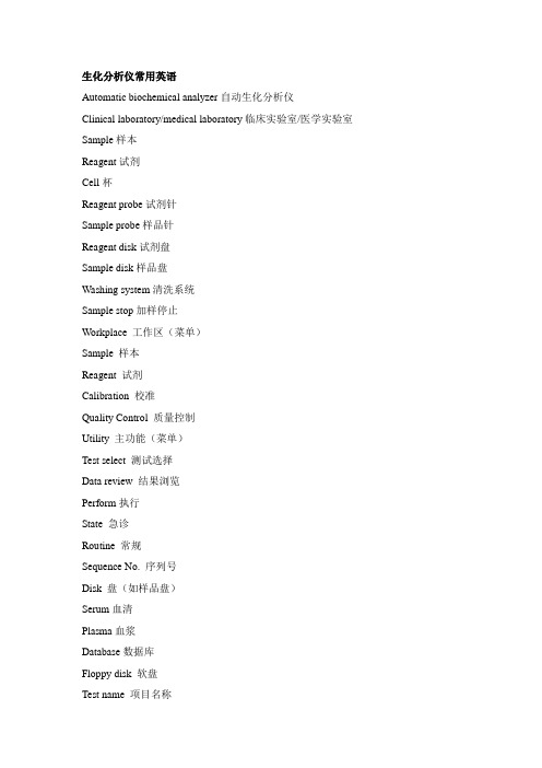
生化分析仪常用英语Automatic biochemical analyzer自动生化分析仪Clinical laboratory/medical laboratory临床实验室/医学实验室Sample样本Reagent试剂Cell杯Reagent probe试剂针Sample probe样品针Reagent disk试剂盘Sample disk样品盘Washing system清洗系统Sample stop加样停止Workplace 工作区(菜单)Sample 样本Reagent 试剂Calibration 校准Quality Control 质量控制Utility 主功能(菜单)Test select 测试选择Data review 结果浏览Perform执行State 急诊Routine 常规Sequence No. 序列号Disk 盘(如样品盘)Serum血清Plasma血浆Database数据库Floppy disk 软盘Test name 项目名称Reagent type 试剂类型Manual setting手工设置Barcode条形码Status 状态Start up calibration 起始校准Repeat calibration重复校准Installation设置Edit编辑QC table质控表QC chart质控图Realtime QC 实时质控Cumulative QC累积质控结果(日间质控)Installation安装、设置Maintenance维护Photometer check光度计检查Air purge排出空气Cell blank 杯空白Inc.(incubator) Water Exchange更换恒温水Analyze 测试/分析Normal正常量Decrease减量Increase增量Class类型(eg. Class 1::类型1)Abs.(absorbance)吸光度Range 范围Standard标准Delete删除Read parameter读参数Write parameter写参数Calibration type校准类型Expected range 参考值Default默认(eg. default age/sex)Instrument factor仪器因数Special wash特殊清洗Report format报告格式报警信息:Samp1 无法检知样品液面(样品量不足或是/忘记放置样品/液面传感器导线脱落)Reagn 试剂的液面无法检知/液体传感器在试剂流路检测到气泡ABS! 吸光度超限(样品浓度极高/试剂放错/试剂为准确配置)ABS! 吸光度超过3.3Abs(样品浓度极高、试剂放错等)Prozone 前带检查超过了设定的界限值(免疫项目样品浓度过高、界限值设定不当)Limt0/Limt1/Limt2 速率法中吸光度的值超过了设定的界限值(样品浓度过高、试剂配置不当或变质、分析参数“Abs. Limit”的“increase”或“decrease”设定不当)Lin./Lin.8 速率法的项目中反应的线性超过了设定的界限值(反应液中进入了脏物、样品高度浑浊、搅拌棒搅拌不良、光源灯老化、线性检查值设定不良)S1Abs? 第一标准液的吸光度不在设定的范围内(试剂变质、试剂放置错误、第一标准液吸光度范围设定不良)Dup 两次测定标准液吸光度的差不在设定范围内(因吸量器漏液而出现的重复不良、离散度的设定不良)Std?校准失败(试剂配制/放置错误;标准也的浓度/放置错误,其他)Sens 标准液的颜色变化比设定的吸光度小(第一标准液吸光度与最大浓度标准液吸光度之差不在应用画面的“灵敏度允许吸光度”的输入范围)(标准液、试剂放置错误、试剂变质、灵敏度界限设定不良)Calib K系数和上次的值比较,在±20%以上(测定新设定的分析项目、标准液/试剂放置错误、试剂变质、标准液浓缩、因吸量器连接部漏液而出现的重复性不良)Calibration factor error校准因素错误(两次校准结果之间的差值超过20%)Calibration error校准错误(校准失败)1、实验室管理常用英语CLIA’88 美国临床实验室修正法规(1988)ISO国际标准化组织IFCCCDC(centers for disease control and prevention)疾病控制与预防中心PT(proficiency testing)能力验证EQA(external quality assessment)室间质评Error误差NCCLS/CLSI(the National Committee for Clinical Laboratory Standards/Clinical and Laboratory Standards Institute)美国临床检验标准化委员会EP21-A 临床检验方法分析总误差的评估——批准指南TEa(total analytical error) 总误差Accuracy 准确度Assay CEDINGBias 偏倚Carry-over 携带污染Control material 质控品Inaccuracy 不准确度Measurement uncertainty 测量不确定度Measuring system 检测系统Specimen 标本Target value 靶值Validation 确认Precision 精密度Repeatability重复性Run 批Lot 批号Accuracy 准确度Trueness 正确度Systematic error系统误差Random error随机误差Total error总误差Measuring range 测量范围Blank 空白Limit of detection 检测限Sensitivity 灵敏度BLD(biologic limit of detection)生物检测限FS(functional sensitivity) 功能灵敏度CAP美国病历夹协会Interference干扰Analyte分析物Interfernce criteria干扰标准Matrix effect基质效应Assessment评价Pool标本Commutability互通性Traceability溯源性Outline离群值SD(standard deviation)标准差CV(coefficients of variation)变异系数Reference material参考物质SOP(standard operation procedure)标准操作程序Commutability互通性/互换性体外诊断试剂常用英语:IVD InVitroDiagnosticreagents体外诊断试剂HITERGENT:循环水添加剂、针清洗剂HIALKALI:碱性清洗液:去除有机物、纤维质、油脂等HIALKALI-D:碱性清洗液:去除有机物、纤维质、油脂等HICARRYNON:酸性清洗液:无机物、碳酸盐、铁化合物等HICHLOGENT-A:次氯酸盐清洗液:流路清洗HICHLOGENT-B:次氯酸盐清洗液:流路清洗ISE 清洗液(N):电极清洗、ISE流路清洗AKP/ALP alkaline phosphatase 碱性磷酸酶ACP acid phosphatase 酸性磷酸酶AST/GOT aspartate aminotransferase 天门冬氨酸氨基转移酶ALT/GPT alanine aminotransferase 丙氨酸氨基转移酶AML/AMS amylase 淀粉酶ALC/ALK alcohol 乙醇、酒精AIDS acquired immune deficidency syndrome 艾滋AL(ALB)albumin 白蛋白PAL(prealbumin)前白蛋白AFP alpha-fetoprotein 甲胎蛋白ADP adenosine diphospate 二磷酸腺苷AMP aednosine monophospate 一磷酸腺苷ATP aednosine triphospate 三磷酸腺苷APTT actived partial thrombolastin time 活化部分凝血酶时间AT-III antinyombin-III 抗凝血酶IIIA/G albumin-globulin ratio 白蛋白-球蛋白比值Bac bacteria 细菌Buffer 缓冲对BG blood group 血型BILI bilirubin 胆红素Blood 血液GLU glucose 葡萄糖BUN blood urea nitrogen 血液尿素氮BSA bovine serum albumin 牛血清白蛋白BUA blood urie acid 血尿酸C cell 细胞Ca calcium 钙C3 complement 3 补体C3Crea creatinine 肌酐CO/CHO cholesterol 胆固醇CHE cholinestetrase 胆碱脂酶CK/CPK creatine kinase/phosphokinase 肌酸(磷酸)激酶CV coefficient of vaviation 变异系数CEA carcinoembyonic antigen 癌胚抗原CRP C-reaction protien C- 反应蛋白CSF cerebral spinal fluid 脑脊液CBC complete blood count 全血细胞计数DC differential count of leucocyte 白细胞分类计数DIC disseminated intravascular coagulation 弥漫性血管内凝血DNA deoxyrinbonucleic acid 脱氧核糖核酸DD D-dimer D- 二聚体DB direct bilirubin 直接胆红素DW distilled water 蒸馏水E enzyme 酶ELISA enzyme linked immunosorbent assay 酶联免疫吸附试验FG fobrinogen 纤维蛋白原FDP fobrinogen degradation product 纤维蛋白原降解产物FFA free fatty acid 游离脂肪酸GGT gama glutamyltransferase γ-谷胺酰基转移酶GLU glucose 葡萄糖GN gram's negative 革兰氏阴性G6PD glucose-6-phosphate dehydrogenase 葡萄糖-6- 磷酸脱氢酶GOD glucose oxiddase 葡萄糖氧化酶GPB(GPC) gram's positive bacillus 革兰氏阳性杆菌HBDH hydroxybutyrate dehydrogenase 羟丁酸脱氢酶HDL-C high-density lipoprotein cholesterol 高密度脂蛋白胆固醇HB(HGB) hemoglobin 血红蛋白HP high power 高倍(显微镜用语)HPLC high gerformence liquid chromatography 高效液相色谱法HCT hematocrit 红细胞压积HC heavy chain 重链5-HT 5-hydroxy-tryptamince 5- 羟色胺HA V hepatitis A virus 甲型肝炎病毒HBV hepatitis B virus 乙型肝炎病毒HCV hepatitis C virus 丙型肝炎病毒HDV hepatitis D virus 丁型肝炎病毒HEV hepatitis E virus 戊型肝炎病毒HbsAg hepatitis B surface antigen 乙型肝炎表面抗原HbsAb hepatitis B surface antibody 乙型肝炎表面抗体HbeAb hepatitis B e antibody 乙型肝炎e 抗体HbeAg hepatitis B e antigen 乙型肝炎e 抗原HbcAg hepatitis B core antigen 乙型肝炎核心抗原HbcAb hepatitis B core antibody 乙型肝炎核心抗体HLA human leukocyte antigen 人类白细胞抗原HICN 氰化高铁血红蛋白HRP horseradish peroxidase 辣根过氧化物酶HSV herpes simple virus 单纯疱疹病毒HK hexokinase 己糖(磷酸)激酶Ig immunoglobulin 免疫球蛋白IgA immunoglobulin A 免疫球蛋白AIgG immunoglobulin G 免疫球蛋白GIgM immunoglobulin M 免疫球蛋白MIgE immunoglobulin E 免疫球蛋白EIgD immunoglobulin D 免疫球蛋白DIP inorganic phosphorus 无机磷IU international unit 国际单位IRMA immnnoradiometric assay 放射免疫试验IDL intermediate-density lipoprotein 中间密度脂蛋白IB indirect bilirubin 间接胆红素IBP iron binding protein 铁结合蛋白IC immune complex 免疫复合物IDA iron deficiency anemia 缺铁性贫血IE immune electrophoresis 免疫电泳IFA indirect fluorescent antibody 间接荧光抗体试验KET Ketone-bodies 酮体LDL-C low-density lipoprotein cholesterol 低密度脂蛋白胆固醇LYM lymphocyte 淋巴细胞β-LP β-lipoprotein β-脂蛋白LPA latex particles agglutination 郛胶凝集反应LEU leukocyte 白细胞M mol 摩尔Mb myoglobin 肌红蛋白β2-MG β2-microglobulin β2- 微球蛋白Mg magnesium 镁MSU mid-stream urine spceimen 中段尿标本MCV mean corpuscular volume 平均红细胞体积MCH mean corpuscular hemoglubin 平均红细胞血红蛋白量MCHC mean corpuscular hemoglubin concentration 平均红细胞血红蛋白浓度MPV mean platelet volume 平均血小板体积MAO micromine oxidase 单胺氧化酶Negative 阴性Normal range 正常范围NRBC nucleared red blood cell 有核红细胞NS normal serum 正常血清OSM osmol 渗透压(量)OD optical density 光密度P plasma 血浆P phosphorus 磷PCR polymerase chain reaction 聚合酶链式反应PR protein 蛋白质POS positive 阳性PLT platelet 血小板PLC platelet count 血小板计数PI isoelectric point 等电点POX peroxidase 过氧化物酶R reaction 反应RBC red blood cell 红细胞Routine 常规RNA ribonucleic acid 核糖核酸RET reticulockyte 网织红细胞RF rhermatoid factor 类风湿因子Sandard 标准Serum 血清Solution 溶液Specimen 标本SOD superoxide dismubase 超氧化物岐化酶SI international system unit 国际单位制Spectorphofluorometer 分光光度荧光计SI serum iron 血清铁SD standard deviation 标准差Test 试验、测定Time 时间TB tubercle bacillus 结核杆菌TP tatal protein 总蛋白TG triglyceride 甘油三酯TB(TBIL) total bilirubin 总胆红素TIBC total iron binging capacity 总铁结合力U unit 单位U urine 尿液UCR urine creatine 尿肌酐UCRE urine creatine 尿肌酸UCL urea clearance 尿素清除率UR urine routine 尿常规临床实验室URANAL urine analysis 尿分析Urine acid 尿酸V volume 体积VMA vanillyl mandelic acid 香草酸杏仁酸VWD von willebrand disease 血管性假血友病VLDL very low-density lipoprotein 极低密度脂蛋白WBC white blood cell 白细胞计数。

抗环瓜氨酸肽抗体在老年类风湿关节炎早期诊断中的意义陶蕾;李兴福【摘要】类风湿关节炎(RA)是常见的全身自身免疫性疾病,病情呈现进行性,导致身体残疾,严重损害老年人的健康,因此 RA 的早期诊断、早期治疗至关重要。
抗环瓜氨酸肽(CCP)抗体是 RA 的特异性自身抗体,可以在 RA 的早期被发现,并可以作为判断预后的重要标志物。
早期诊断 RA,远离关节畸形与功能丧失,提高老年人群的生活质量,需要高特异性、高敏感性、可在疾病早期出现的检测指标,抗 CCP 抗体是目前的首选。
【期刊名称】《临床荟萃》【年(卷),期】2014(000)011【总页数】3页(P1319-1321)【关键词】关节炎,类风湿;类风湿因子;瓜氨酸;老年人【作者】陶蕾;李兴福【作者单位】泰山医学院附属医院风湿免疫科,山东泰安 271000;山东大学齐鲁医院风湿免疫科,山东济南 250012【正文语种】中文【中图分类】R593.22类风湿关节炎(RA)是一种全身多关节的慢性滑膜炎症[1],导致关节破坏、畸形的系统性自身免疫性疾病。
在各个年龄段均可发病,高峰在30~50岁,女性多于男性。
该疾病有高度的致残性,70%的关节软骨及骨破坏发生在发病最初的2年内,3年内超过75%的患者会致残[2]。
因此,早期诊断,尽早达到缓解,减少该病的关节破坏,降低致残率是RA诊疗中的首要环节。
通常将≥60岁发病的RA称为老年RA[3]。
RA的发病率国内尚无精确的统计,估计在1.08%左右,北京、汕头等地的调查结果显示患病率在0.36%左右[4]。
RA在美国老年人群中的发病率达2%[5],占RA患者的10%~33%,可推断我国老年发病的RA是一个庞大的人群。
老年RA与中青年RA相比有其自身的临床特征[3,6]:①男性患者比例明显升高。
②急性发病多见。
③更容易首发累及肩、膝、髋等大关节,易误诊。
④症状不典型,类风湿因子(RF)特异性不高,老年人出现低滴度的RF时应结合临床做出判断。

QCT和MR在非酒精性脂肪肝诊断中的应用价值陈刚; 索方方; 陈少武; 陈翠云【期刊名称】《《中国CT和MRI杂志》》【年(卷),期】2018(016)008【总页数】4页(P93-95,103)【关键词】定量CT; 磁共振成像; 非酒精性脂肪肝; 肝脏脂肪含量【作者】陈刚; 索方方; 陈少武; 陈翠云【作者单位】郑州市第六人民医院放射科河南郑州450000; 洛阳市中心医院放射科河南洛阳471000; 平顶山市第一人民医院MR室河南平顶山467000【正文语种】中文【中图分类】R575.5; R445.2; R445.3非酒精性脂肪性肝病(nonalcoholic fatty liver disease,NAFLD)是指除外酒精和其他明确的损肝因素所致的、以肝细胞内脂肪过度沉积为主要特征的临床病理综合征[1]。
随着肥胖及其相关代谢综合征全球化的流行趋势,NAFLD患病率不断升高,西方国家15%~30%,我国约15%[2]。
研究发现[3],早期诊断和及时干预可阻止或延缓NAFLD进程,有助于提高患者的生存质量。
因此对肝脏脂肪变性进行早期风险评估,对阻断和延缓病情发展具有重要意义。
近年来如CT、MR等新技术逐渐应用于肝脏脂肪变性的定量评估中,并取得了较好的临床应用效果[4]。
本研究探讨定量CT(QCT)和磁共振成像(MR)在非酒精性脂肪肝(NAFLD)诊断中的应用价值,旨在为NAFLD早期诊断提供依据,现报道如下。
1 资料与方法1.1 一般资料选取2016年2月至2017年8月在我院治疗的NAFLD患者67例(NAFLD组),纳入标准:(1)诊断符合中华医学会肝脏病学分会制定的《非酒精性脂肪肝诊断标准》;(2)患者及家属知情同意。
排除标准:(1)因完全肠外营养等导致脂肪肝;(2)合并有恶性肿瘤、病毒性肝炎、心肺等重要脏器功能不全等;(3)有MRI检查禁忌症;(4)哺乳期或妊娠期妇女。
同时选取健康志愿者70例作为对照组,NAFLD组和对照组性别、年龄差异比较无统计学意义(P>0.05),见表1。

重症医学科患者血清降钙素原的检测意义
张剑宏;李国柱;陈军军
【期刊名称】《临床医药实践》
【年(卷),期】2018(027)008
【摘要】目的:探讨降钙素原(PCT)检测在重症医学科预防医院感染中的作用.方法:选择重症医学科收治的患者106例,将其分为感染组和非感染组.对两组患者的PCT、C反应蛋白(CRP)水平进行统计分析.结果:感染组PCT阳性率(96.82%)明显高于未感染组(16.28%),差异有统计学意义(P<0.05);感染组CRP阳性率(95.24%)明显高
于未感染组(18.60%),差异有统计学意义(P<0.05).结论:尽管完全依据PCT指导抗
生素治疗存在一定风险,但PCT和CRP的检测对重症医学科患者及时诊断感染有
重要的临床价值,可以有效避免抗生素滥用,减轻患者的经济负担.
【总页数】2页(P610-611)
【作者】张剑宏;李国柱;陈军军
【作者单位】山西医科大学第二医院,山西太原 030001;山西医科大学第二医院,山西太原 030001;山西医科大学第二医院,山西太原 030001
【正文语种】中文
【中图分类】R459.7
【相关文献】
1.血清降钙素原联合血培养对重症医学科血流感染患者病原菌的早期预测价值
2.血清降钙素原在重症医学科感染患者中的应用价值
3.血清降钙素原在重症感染临床
中检测意义4.血清降钙素原在重症感染临床中检测意义5.血清降钙素原、超敏-C 反应蛋白对新疆乌鲁木齐地区重症医学科患者鲍曼不动杆菌感染耐药预测分析
因版权原因,仅展示原文概要,查看原文内容请购买。
大鼠酒精性肝纤维化模型的构建周宁;厉有名;张宇;虞朝晖;陈韶华;季峰【期刊名称】《东南大学学报(医学版)》【年(卷),期】2003(022)002【摘要】目的:探索简便、经济的大鼠酒精性肝纤维化造模方法.方法:33只大鼠随机分为2组.模型组予酒精灌胃,于实验的第4、12、24周末分批采血,以测定生化指标,后行放血法处死.将取出的肝脏进行常规切片、HE染色及Masson三色胶原染色后行病理学观察.正常对照组以等量饮用水代替酒精灌胃,于实验第24周末处死,处理同模型组.结果:于实验第24周时观察到了除酒精性肝硬化外酒精性肝病的各种病理表现,并且随着酒精刺激时间的延长肝纤维化程度逐渐加重.结论:应用酒精灌胃法可成功复制大鼠酒精性肝纤维化模型,该方法简便、经济.【总页数】5页(P93-97)【作者】周宁;厉有名;张宇;虞朝晖;陈韶华;季峰【作者单位】浙江大学医学院附属第一医院,消化内科,浙江,杭州,310006;浙江大学医学院附属第一医院,消化内科,浙江,杭州,310006;浙江大学医学院附属第一医院,消化内科,浙江,杭州,310006;浙江大学医学院附属第一医院,消化内科,浙江,杭州,310006;浙江大学医学院附属第一医院,消化内科,浙江,杭州,310006;浙江大学医学院附属第一医院,消化内科,浙江,杭州,310006【正文语种】中文【中图分类】R332【相关文献】1.cAMP-PKA-CREB 信号通路在大鼠酒精性肝纤维化星状细胞模型中的作用 [J], 杨万枝;吕雄文;余世春;管文婕;翟丹丹;王和;李俊2.高脂饮食致大鼠非酒精性脂肪性肝炎肝纤维化模型的建立 [J], 孙林林;石军;郝菁华;任万华;阎春英;张捷;林晓燕;崔彬3.绞股蓝皂苷对2型糖尿病合并非酒精性脂肪性肝病模型大鼠肝纤维化影响 [J], 赵世印;邱华;贺琴;李德梅;谭华炳4.大鼠酒精性肝纤维化模型的构建 [J], 周宁;厉有名;等5.酒精性肝纤维化大鼠模型的建立及Smad7/TGF-β表达变化 [J], 何培元;侯志平;高淑梅;王明娟;马立新;李炳庆因版权原因,仅展示原文概要,查看原文内容请购买。
1前言甲胎蛋白(alpha-fetoprotein,AFP)是一种单链糖蛋白。
1970年Purves对肝癌患者血清作凝胶电泳时最先观察到AFP有不同的迁移率,因此提出甲胎蛋白异质体(alpha-fetoprotein variants)这一概念[1]。
随着生物化学及其相关分析技术的发展,研究人员发现不同来源的AFP,其糖链的组成和构型存在差异。
不同的植物凝集素能够特异性地识别一定的糖基,并与之结合。
根据与外源性凝集素亲和性的不同,可对AFP 的来源作出判断。
Taketa等[2]发现原发性肝癌(hepatocellular carcinoma,HCC)患者血清中AFP与小扁豆素(lens culinaris agglutinin,LCA)结合后,电泳分成三带,依次命名为AFP-L1、AFP-L2、AFP-L3,即LCA非结合型(AFP-L1、AFP-L2)和LCA结合型(AFP-L3)。
对比分析显示HCC患者AFP-L3比率比其它良性肝病患者明显升高。
现通常把与小扁豆素结合的AFP-L3称为AFP异质体,它是新一代的肿瘤标志物[3]。
2甲胎蛋白异质体(AFP-L3)检测的临床意义AFP属于胚胎性蛋白,是应用最广泛的肿瘤标志物。
大量研究证实,AFP分子的糖链异质性与其组织器官来源有关,不同生理病理状况可产生不同糖链结构,并且具有肿瘤特异性。
血清总AFP所含AFP-L1来自良性肝病,占其主要部分;AFP-L2来自孕妇;而AFP-L3则为肝癌细胞特有,AFP-L3>15%即可诊断为原发性肝癌。
AFP-L3诊断肝癌的敏感性为96.9%,特异性为92.0%,准确性为95.5%[4]。
AFP-L3值与总AFP 值无相关性,是独立于总AFP值的肝癌诊断因子[5],是目前公认的肝癌鉴别诊断和早期诊断的指标,具有非常重要的临床意义[6]。
3甲胎蛋白异质体的检测技术测定AFP-L3常用的方法,是根据AFP异质体对植物凝集素(如小扁豆凝集素LCA、刀豆素ConA或豌豆凝集素PSA)结合能力的不同进行分离,然后应用免疫学方法进行定量测定。
Potential utility of the Genedrive point-of-care test for HCV RNA detectionIn an article in Gut,1 Lemoine and Tillman reviewed our recent publication on the development and validation of a novel point-of-care (POC) HCV viral assay.2 They acknowledge that Genedrive is the only CE-In Vitro Diagnostic-qualified device for HCV detection, and that the on December 24, 2023 by guest. Protected by copyright./ Gut: first published as 10.1136/gutjnl-2018-317218 on 22 September 2018. Downloaded from19031904limit of detection (LOD: 1406–3203 IU/mL) permits identification of the vast majority of HCV chronic cases.3 However, they emphasise some limitations of the study and Genedrive. One limitation highlighted is the need for plasma which is still required for 100% of HCV clinical tests. Nevertheless, recent studies demon-strated microfluidic plasma separation without centrifugation 4 or with low-cost hand-powered paper centrifuges.5 It is feasible to foresee such devices over-coming the need for conventional centri-fuges and facilitating Genedrive testing.They also question whether 15µL of plasma can be obtained by fingerprick. Published studies report 20–25 µL perdrop of blood,6so 1 drop should be suffi-cient. Furthermore, devices to collect 100 µL of blood from a single fingerstick are now available.7 T echnology evolves rapidly to make new tools available which may further facilitate the use of the Gene-drive test. The Xpert technology (Cepheid) illustrates these constant improvements, with the latest version requiring only 100 µL blood,7 in contrast to the current 1 mL plasma. Whether Genedrive could be used for monitoring treatment responses was not addressed in our initial analysis but is part of ongoing studies. In reference to cartridges surviving extreme tempera-tures, while formal stability studies are ongoing, test reagents are certified at 2°C–28°C for 12 months, permitting cold chain-free storage. Regarding early sepa-ration of plasma being required to prevent HCV RNA degradation, it is stable in EDTA whole blood for 24 hours at room temperature and 4 days at 4°C 8. Although our study was not designed to test this, a large time between sample collection and testing was reported within African samples. For cases where high haemolysis was observed, there was no loss of perfor-mance, but additional studies are required.Lemoine and Tillmann highlight that 26/1055 samples required retesting with only 16 giving definitive results. If these ambiguous results are included, sensitivity (99.2%) and specificity (98.8%) are still very good. For the 10 samples without a result, 6 were HCV pos with viral loads close to LOD. Because a very small sample volume is used, and 2/3 replicates must be positive, in cases of low viral load, the absence of a viral particle ultimately affects the diagnostic accuracy . For the four HCV neg indeterminate samples, we can speculate on the possible effects of interfering substances. While a number were tested, we did not identify any that impacted Genedrive’s performance. Nonetheless, there are many compounds that could potentially affectassay function, as recently encountered for an IL28B single nucleotide polymorphism (SNP) POC assay .9While the authors are enthusiastic for GeneXpert, which has advanced the diagnostics options for HCV , they fail tomention one major limitation: the use of toxic cartridges requiring double cylinderhigh temperature (≥850°C) incineration, which is challenging to decentralise, in particular in resource-limited settings.10In contrast, Genedrive contains non-toxic materials, making waste management much easier.We agree that a real-life study in POCconditions is required, including a detailed cost analysis. However, as stated in our original discussion, we maintain that this study was a required step for devel-opment and validation of the Genedrive HCV test. It has helped to establish global distributions in Africa and Asia generatingreal-world data and will encourage newdevelopments to address this clinical need.It has resulted in a novel, easy-to-use, portable platform for decentralisation of HCV testing, which may have a positiveimpact on the continuum of care fromdiagnosis to treatment.Alba Llibre,1,2 Yusuke Shimakawa,3 Darragh Duffy 1,21Immunobiology of Dendritic Cells, Institut Pasteur, Paris, France 2Inserm U1223, Institut Pasteur, Paris, France 3Unité d’epidémiolotie des Maladies emergentes, Institut Pasteur, Paris, FranceCorrespondence to Darragh Duffy, Immunobiologie des cellules dendritiques, Institut Pasteur, Paris 75015, France;d arragh.d uffy@p asteur.f rContributors AL, Ys and DD wrote and edited the letter.Competing interests None declared.Patient consent Not required.Provenance and peer review Not commissioned;internally peer reviewed.Open access this is an open access article distributed in accordance with the Creative Commons Attribution Non Commercial (CC BY-NC 4.0) license, which permits others to distribute, remix, adapt, build upon this work non-commercially, and license their derivative works on different terms, provided the original work is properly cited, appropriate credit is given, any changes made indicated, and the use is non-commercial. see: http:// creativecommons.o rg/l icenses/b y-n c/4.0/.© Author(s) (or their employer(s)) 2019. re-usepermitted under CC BY-NC. No commercial re-use. seerights and permissions. Published by BMJ.To cite Llibre A, shimakawa Y , Duffy D. Gut 2019;68Received 20 July 2018revised 20 August 2018Accepted 1 september 2018Published Online First 22 september 2018Gut 2019;RefeRences1 Lemoine M, tillmann HL. What is required from HCVpoint-of-care tests to reduce the burden of hepatitis Cinfection? ’Development and clinical validation of thegenedrive point-of-care test for qualitative detection of hepatitis C virus ’. Gut 2018;67:1916–7.2 Llibre A, shimakawa Y , Mottez e, et al . Development and clinical validation of the Genedrive point-of-care test for qualitative detection of hepatitis C virus. Gut2018.3 WHO. Guidelines on hepatitis B and C testing. 2017.4 tripathi s, Kumar YV , Agrawal A, et al . Microdevicefor plasma separation from whole human blood using bio-physical and geometrical effects. Sci Rep2016;6:26749.5 Bhamla Ms, Benson B, Chai C, et al . Hand-poweredultralow-cost paper centrifuge. Nat Biomed Eng 2017;1:9. 6 Peck Hr, timko DM, Landmark JD, et al . A surveyof apparent blood volumes and sample geometriesamong filter paper bloodspot samples submitted for lead screening. Clin Chim Acta 2009;400:103–6.7 Grebely J, Lamoury FMJ, Hajarizadeh B, et al . evaluation of the Xpert HCV Viral load point-of-careassay from venepuncture-collected and finger-stickcapillary whole-blood samples: a cohort study. LancetGastroenterol Hepatol 2017;2:514–20.8 sener K, Yapar M, Bedir O, et al . stability of hepatitis Cvirus rNA in blood samples by taqMan real-time PCr. J Clin Lab Anal 2010;24:134–8.9 Duffy D, Mottez e, Ainsworth s, et al . An invitro diagnostic certified point of care singlenucleotide test for IL28B polymorphisms. PLoS One 2017;12:e0183084.10 WHO Xpert MtB/rIF . WHO Policy update and Implementation manual , 2017.7:1903–1904.68:1903–1904. doi:10.1136/gutjnl-2018-317218 on December 24, 2023 by guest. Protected by copyright./Gut: first published as 10.1136/gutjnl-2018-317218 on 22 September 2018. Downloaded from。
丙肝病毒核心抗原检测的临床应用价值[摘要] 目的研究丙肝病毒核心抗原检测在丙肝感染诊断中的应用价值。
方法通过对2009年12月-2010年12月,我院收集的288例丙肝抗体检测标本进行了研究,首先进行丙肝抗体(hcv-ab)检测,分为阳性和阴性两组,并对两组样本进行核心抗原(hcv-cag)及hcv-rna检测,分析比较两组的检测结果。
结果 184例hcv-ab 阴性的标本中,检出hcv-cag阳性2例,hcv-rna阳性1例;104例hcv-ab阳性的标本中,检出hcv-cag阳性54例,hcv-rna阳性58例,hcv-cag和hcv-rna符合率为93.10%(54/58)。
结论 hcv-cag 是丙肝病毒感染的早期标志物之一,检测hcv核心抗原有利于hcv 的早期诊断,为患者的早发现、早诊断、早治疗提供依据。
[关键词] 丙型肝炎核心抗原抗体临床应用[中图分类号] r512.6+3[文献标识码] a[文章编号] 1005-0515(2011)-08-314-01病毒性肝炎是一种危害人类健康的传染性疾病,最为常见的是三种病毒感染,即甲型病毒肝炎、乙型病毒肝炎和丙型病毒性肝炎,目前尚无特效治疗方法,甲型病毒肝炎和乙型病毒肝炎均有疫苗进行预防,但丙型肝炎并无疫苗。
丙型肝炎(hepatitis c virus,hcv)主要通过血液进行传播,我国丙肝的感染率为3.2%,大多数hcv感染都是没有症状的,并且慢性感染的比例很高,有较高的比例会发展成为肝纤维化、肝硬化,进而发展成为肝癌[1]。
因此,对丙型病毒肝炎进行及早的诊断,对于治疗及预后均有重要的意义。
1 样本与方法1.1 样本来源 2009年12月-2010年12月,我院收集的288例丙肝抗体检测标本,其中男性140例,女性148例,年龄在22岁-73岁之间,平均年龄44.2岁。
通过hcv-ab检测,将样本分为hcv-ab 阳性组和阴性组,其中,hcv-ab阳性有104例,hcv-ab阴性有184例,再分别对两组样本进行hcv-cag和hcv-rna检测。
Clinical Utility of Total HCV Core AntigenQuantification:A New Indirect Markerof HCV ReplicationMagali Bouvier-Alias,1Keyur Patel,2Harel Dahari,3St´e phanie Beaucourt,1Patrick Larderie,1Lawrence Blatt,2Christophe Hezode,1,4Gaston Picchio,5Daniel Dhumeaux,1,4Avidan U.Neumann,3John G.McHutchison,2and Jean-Michel Pawlotsky1Hepatitis C virus(HCV)RNA detection,viral load quantification,and HCV genotyping arewidely used in clinical practice.Recently,the availability of an anticore antigen(Ag)mono-clonal antibody allowed development of an enzyme-linked immunosorbent assay(ELISA)detecting and quantifying total HCV core Ag in peripheral blood of HCV-infected patients.The aims of the present study were to investigate the biologic significance of this new markerin HCV infection,to establish the intrinsic performance of the current assay,and to deter-mine its potential utility in the management of HCV-infected patients.A panel of infectedsera calibrated to the World Health Organization International Standard and657serumsamples from infected patients receiving antiviral treatment were studied.We showed thattotal HCV core Ag quantification is an accurate,precise,and specific indirect marker of HCVreplication.We estimated that1pg/mL of total HCV core Ag is equivalent to approximately8,000HCV RNA international units(IU)/mL,although minor between-patient differencesmay exist.In conclusion,total HCV core Ag quantification can be used in the variousindications of viral load monitoring,including the evaluation of baseline viral load beforetherapy,the assessment of the virologic response to antiviral treatment,and the study of earlyviral kinetics during therapy.Nevertheless,the total HCV core Ag assay cannot be used as amarker of viral replication for HCV RNA values below20,000IU/mL,limiting its use in themonitoring of late events during and after antiviral treatment.(H EPATOLOGY2002;36:211-218.) H epatitis C virus(HCV)is a single-strand RNAvirus belonging to the Flaviviridae family.Itsgenome is contained in an icosahedral capsid(or core),itself contained within the viral envelope.Thecapsid is formed by polymerization of the HCV core pro-tein,a structural viral protein encoded by the5Јend of theHCV open reading frame.1After translation,host signalpeptidases cleave the HCV core protein from the precur-sor polyprotein and remove the signal peptide located atits C-terminus.1The mature HCV core protein is a21-kdphosphoprotein made of the191first amino acids of thepolyprotein.In the cytoplasm of infected cells,it is locatedin close vicinity to the perinuclear membranes and theendoplasmic reticulum,where it polymerizes in the pres-ence of genomic RNA to form viral capsids.1The HCVcore protein is antigenic,interacts with numerous cellu-lar proteins,induces specific cellular and humoral re-sponses,2,3and through various pathways could play animportant role in the pathogenesis of HCV infection.4Virologic diagnosis and monitoring of HCV infectionare based on the use of serologic assays detecting specificanti-HCV antibodies(including anticore antibodies),and assays that can detect,quantify or characterize thecomponents of HCV viral particles.5In past years,clinicalhepatitis C studies have for the most part used molecularbiology–based HCV RNA techniques.Indeed,HCVRNA detection,viral load quantification,and HCV geno-typing are widely used in clinical practice.6Recently,theavailability of an anticore antigen(Ag)monoclonal anti-Abbreviations:HCV,hepatitis C virus;ELISA,enzyme-linked immunosorbentassay;OD,optical density;bDNA,branched DNA.From the1Departments of Virology(EA3489)and4Hepatology and Gastroen-terology,Hoˆpital Henri Mondor,Universite´Paris XII,Cre´teil,France;2the Divi-sion of Gastroenterology and Hepatology and the5Department of Immunology,Scripps Clinic,La Jolla,CA;and3Faculty of Life Sciences,Bar-Ilan University,Ramat-Gan,Israel.Received December19,2001;accepted April12,2002.Address reprint requests to:Jean-Michel Pawlotsky,M.D.,Ph.D.,Service deBacte´riologie-Virologie,Hoˆpital Henri Mondor,51avenue du Mare´chal de Lattrede Tassigny,94010Creteil,France.E-mail:jean-michel.pawlotsky@hmn.ap-hop-paris.fr;fax:(33)14-981-2839.Copyright©2002by the American Association for the Study of Liver Diseases.0270-9139/02/3601-0027$35.00/0doi:10.1053/jhep.2002.34130211body allowed development of an enzyme-linked immu-nosorbent assay(ELISA)that showed the presence of HCV core Ag in peripheral blood of HCV-infected pa-tients.7,8Results obtained in Japan with afirst-generation assay showed a significant relationship with the presence of HCV RNA and suggested that HCV core Ag detection, and quantification might be used to follow patients and predict the response to interferon alfa(IFN-␣)therapy.9 A standardized commercial assay using the same monoclonal antibody has been developed for the qualita-tive detection of HCV core Ag(Ortho Antibody to Hep-atitis C Core Antigen ELISA Test System;Ortho-Clinical Diagnostics,Raritan,NJ).This assay,conceived for the screening of blood donations,increases viral safety by sig-nificantly reducing the serologic window occurring before seroconversion during acute infection.Several studies have shown that core Ag can be detected,on average,1to 2days after HCV RNA during the preseroconversion period.10-13However,this screening assay exhibits de-creased sensitivity when analyzing HCV antibody–posi-tive samples.This is probably caused by the absence of a preliminary immune-complex dissociating step in the as-say’s procedure.In this regard,a new assay with potential application in the clinical setting,incorporating an im-mune-complex dissociating step and using the same monoclonal antibody,has been recently developed for detection and quantification of total HCV core Ag(Total HCV core Ag assay;Ortho-Clinical Diagnostics).The aims of the present study were(1)to investigate the biologic significance of this new marker in HCV in-fection,(2)to establish the intrinsic performance of the current assay,and(3)to determine its potential utility in the management of HCV-infected patients. Materials and MethodsMaterials.The following materials were used in this study:(1)A panel of infected sera calibrated to the World Health Organization International Standard,14the Nu-cleic Acid Panel(NAP)-HCV RNA(AcroMetrix Inc., San Francisco,CA),including the following panel mem-bers:50HCV RNA international units(IU)/mL,500 IU/mL,5,000IU/mL,50,000IU/mL,200,000IU/mL, 500,000IU/mL,and2,000,000IU/mL.15(2)A total of 392serum samples from44patients from Henri Mondor University Hospital,Cre´teil,France.These patients,in-fected with HCV genotype1b,received various schedules of IFN-␣with or without ribavirin.Blood samples were taken before treatment in all patients and frequently thereafter during thefirst weeks of treatment in20cases (i.e.,14days and2hours before the start of therapy,at the start of therapy,every4hours for24hours,every6hours for36hours,every12hours for48hours,at day14,and at day28of therapy).HCV RNA loads were measured with both the third-generation signal amplification-based branched DNA(bDNA3.0)assay Versant HCV RNA Quantitative Assay(Bayer Diagnostics,Emeryville,CA) and the reverse transcriptase-competitive polymerase chain reaction-based SuperQuant assay(National Genet-ics Institute,Los Angeles,CA).This collection of serum samples covered a wide range of viral loads representative of those routinely encountered in clinical practice.(3) Sixty-five serum samples from patients from Henri Mon-dor University Hospital infected with HCV genotype3a, taken before or at month6of IFN-␣treatment.HCV RNA loads were measured with bDNA3.0.(4)Two hun-dred samples from40patients infected with various HCV genotypes and receiving various schedules of IFN-␣or pegylated IFN-␣and ribavirin treatment at Scripps Clinic,La Jolla,CA.Blood samples were collected at the start of therapy,at weeks4and12of therapy,at the end of treatment,and at the end of follow-up6months later. HCV RNA loads were measured with the SuperQuant assay in35cases and with another reverse transcriptase polymerase chain reaction–based assay,the Amplicor HCV Monitor assay version2.0(Roche Molecular Sys-tems,Pleasanton,CA)in the remaining5cases.(5)A total of124sera from immunocompetent patients referred to Henri Mondor hospital for various medical conditions that tested negative for anti-HCV antibodies(ORTHO HCV3.0ELISA Test System with Enhanced SAVe;Or-tho-Clinical Diagnostics)and for HCV RNA in a quali-tative,nonquantitative polymerase chain reaction assay with a lower detection cutoff of50HCV RNA IU/mL (Amplicor HCV2.0;Roche Molecular Systems).(6)Se-rum samples from3HCV-infected patients containing different amounts of HCV RNA.(7)Serial one-third di-lutions of sera taken from7patients infected with differ-ent HCV genotypes,namely1a,1b,2b,3a,4a,5a,and6a, into a seronegative plasma.HCV Core Ag Testing.The NAP-HCV RNA panel was tested9times in5different runs to assess the dynamic range and linearity of quantification of the assay,as well as its precision and repeatability.To eliminate any interfer-ence,both the concentrated solution and the diluent used for preparing the panel also were tested.The392blood samples from Henri Mondor hospital were tested for total HCV core Ag and the results were compared with HCV RNA testing to assess the value of total HCV core Ag as a marker of viral replication,to determine the sensitivity of the core Ag assay relative to molecular biology techniques, to establish a relationship between total HCV core Ag and HCV RNA units,and to study the potential utility of total HCV core Ag quantification to monitor early HCV212BOUVIER-ALIAS ET AL.HEPATOLOGY,July2002replication kinetics during antiviral therapy.The200 samples from the Scripps Clinic were tested to assess the value of total HCV core Ag quantification as a routine marker of the virologic response to antiviral therapy.The sera from124anti-HCV antibody-negative,HCV RNA–negative individuals were tested to determine the specific-ity of the total core Ag assay.To determine the precision and reproducibility of HCV core Ag quantification,the2 positive controls provided in the kits were tested22times in22distinct runs and the sera from3patients containing different HCV RNA loads were tested4times in3dis-tinct runs.Finally the one-third dilutions of sera from7 patients infected with different genotypes were tested to verify that quantification was linear whatever the HCV genotype.HCV Core Ag Assay.Blood samples were assayed according to the manufacturer’s instructions.The kit lot number was DX-00001.One hundredL of samples and controls were mixed with50L of a pretreatment buffer containing detergents and pretreated for30minutes at56°C to dissociate HCV core antigen-antibody com-plexes.For the ELISA reaction,100L of pretreated samples and controls were incubated for60minutes at 25°C in the monoclonal antibody-coated wells of a mi-crotiter plate.The plates were washed and incubated for 30minutes at25°C with200L of conjugate,washed again,and incubated for30minutes at25°C with200L of substrate.The optical densities(ODs)were read in a spectrophotometer at490nm using a620nm reference. The samples and controls were tested in duplicate and the mean OD of each duplicate testing was used.The samples that exhibited more than25%variation between the two ODs were considered invalid and retested.As recom-mended by the manufacturer,the lower detection cutoff was established for each run and corresponded to the mean OD of the2negative controls plus0.041.A sample was considered positive only when the mean OD was higher than the cutoff OD of the corresponding run.The amount of total HCV core Ag in pg/mL was calculated by means of a standard curve established in each run by test-ing serial dilutions of a standard containing400HCV core Ag pg/mL.HCV RNA Detection and Quantification.The3 HCV RNA assays were performed according to the man-ufacturer’s instructions.The results were expressed in HCV RNA IU/mL.ResultsTotal HCV Core Ag Is a Marker of HCV Replica-tion.Figure1shows the relationship between total HCV core Ag and HCV RNA in392HCV genotype1b clinical samples covering a wide range of HCV RNA loads.HCV RNA was quantified in IU/mL with bDNA3.0,an accu-rate HCV RNA quantification assay.16In the293samples that were positive in both markers,total HCV core Ag and HCV RNA were significantly related(rϭ0.92;PϽ.0001).The relationship between HCV RNA in IU/mL and total HCV core Ag in pg/mL was given by the fol-lowing equation:HCV core Ag(log10pg/mL)ϭ0.9714ϫHCV RNA(log10IU/mL)Ϫ3.7461 Based on this equation,it was possible to calculate that 1pg/mL of total HCV core Ag is equivalent to7,688 IU/mL.Thus,total HCV core Ag is a reliable marker of HCV replication,and1core pg/mL is equivalent to ap-proximately8,000HCV RNA IU/mL in clinical samples from HCV-infected patients.The correlation between HCV RNA IU/mL and HCV core Ag pg/mL varied around this average ratio when individual samples were considered,with the majority of the ratios lying between 5,000and12,000HCV RNA IU/mL per core Ag pg/mL (Fig.2A).Because we studied several samples taken at different time points in the same patients,we were able to assess whether these differences were sample-or patient-related.As shown in Fig.2B,the correspondence between HCV RNA IU/mL and HCV core Ag pg/mL did not vary substantially within individual patients,but larger(al-though not dramatic)differences were seen between dif-ferent patients.To assess if the total HCV core Ag-HCV RNA rela-tionship was genotype-independent,we tested65geno-Fig.1.Relationship between HCV RNA(in log10IU/mL)and total HCV core Ag(in log10pg/mL)in293samples out of392tested that were positive for both markers.The2markers were significantly related(rϭ0.92;PϽ.0001).HEPATOLOGY,Vol.36,No.1,2002BOUVIER-ALIAS ET AL.213type 3a samples for both markers.Again,total HCV core Ag and HCV RNA were signi ficantly related (r ϭ0.90;P Ͻ.0001;data not shown).The relationship between HCV RNA and total HCV core Ag was given by the following equation:HCV core Ag (log 10pg/mL)ϭ0.9028ϫHCV RNA (log 10IU/mL)Ϫ3.2604One pg/mL of total HCV core Ag was equivalent to approximately 6,711IU/mL,a conversion factor of the same order as in genotype 1b.Performance of the Total HCV Core Ag Assay as a Marker of HCV Replication.Based on 22runs,the lower detection cutoff of the total HCV core Ag assay was on average 2.26Ϯ0.48pg/mL.Thus,based on the rela-tionship with HCV RNA mentioned above,the total core Ag assay did not detect HCV replication below 16,631Ϯ3,374HCV RNA IU/mL (i.e.,below approximately 20,000IU/mL).The clinical sensitivity of the assay was con firmed when testing the NAP –HCV RNA panel (Fig.3).Indeed,3panel members (200,000HCV RNA IU/mL,500,000IU/mL,and 2,000,000IU/mL,respec-tively)were always quanti fied in the core Ag assay.In contrast,the panel member containing 50,000HCV RNA IU/mL was found positive only once out of 9tests,with a value of 2.17pg/mL,just above the cutoff of the corresponding run.As shown in Fig.3,quanti fication was linear within the tested range of viral loads.Overall,the dynamic range of quanti fication of the total HCV core Ag assay was between its lower detection cutoff (approxi-mately 2pg/mL)to at least 250pg/mL.In addition,serial one-third dilutions of clinical samples from patients in-fected with various HCV genotypes (including 1a,1b,2b,3a,4a,5a,and 6a)showed that total HCV core Ag quan-ti fication was linear within its dynamic range of quanti fi-Fig.3.Total HCV core Ag detection and quantification in the NAP–HCV RNA panel,a panel calibrated to the HCV RNA IU/mL (AcroMetrix Inc.).Total HCV core Ag was constantly found in 3panel members (i.e.,200,000HCV RNA IU/mL,500,000IU/mL,and 2,000,000IU/mL,respectively),and its amount was significantly related to HCV RNA load in the corresponding range of values.The panel member containing 50,000HCV RNA IU/mL was found HCV core Ag-positive once out of 9experiments,with a core Ag value higher than the cutoff of the corre-sponding run,but lower than the mean of those in all of theexperiments.Fig. 2.Relationship between HCV RNA IU/mL and HCV core Ag pg/mL.On average,in the 293tested samples that were positive in both assays,the ratio was approximately 8,000HCV RNA IU/mL per HCV core Ag pg/mL.(A)HCV RNA IU/mL to HCV core Ag pg/mL ratio in 293individual samples testing positive in both assays.(B)Mean Ϯstandard deviation of the HCV RNA IU/mL to HCV core Ag pg/mL ratio of different samples taken at different dates in 20patients.214BOUVIER-ALIAS ET AL.HEPATOLOGY,July 2002cation irrespective of the HCV genotype(data not shown).One sample was found positive for total HCV core Ag among124samples from anti-HCV–negative,HCV RNA–negative patients,but was negative on repeat test-ing.The specificity of the assay was thus99.2%(95% confidence interval:94.3%-100.0%).The assay precision and reproducibility were assessed by testing the2positive controls provided in the kits22times in22distinct runs, and by testing sera from3patients containing different HCV RNA loads4times in4distinct runs.Overall,the within-run coefficients of variation ranged from7.5%to 33.2%,and the between-run coefficients of variation ranged from16.3%to26.7%.As recommended by the manufacturer,each sample was tested in duplicate,and the mean of the2determinations was used as thefinal result.Only8samples out of392(2%)had duplicate testing differing by more than25%and were retested in our study.The between-laboratory reproducibility was not evaluated.Clinical Use of Total HCV Core Ag Quantification to Monitor the Response to Therapy.Forty treated patients were tested before and during treatment,at the end of treatment,and at the end of follow-up for both HCV RNA and HCV core Ag,as detailed in the Materials and Methods section.They were classified into3catego-ries based on HCV RNA testing,including sustained vi-rologic responders(negative HCV RNA at the end of treatment and at the end of follow-up6months later); responder-relapsers(negative HCV RNA at the end of treatment,but positive HCV RNA detection at the end-of follow-up);nonresponders(positive HCV RNA detection throughout the entire follow-up period). Total HCV core Ag quantification allowed the same classification as the use of molecular biology testing in35 patients out of40(87%).The remaining5patients (13%)were misclassified with the core Ag assay.One patient with a baseline HCV RNA of265,000IU/mL was found to be HCV core Ag–negative at this time point,and throughout the entire follow-up period;he achieved sus-tained virologic responders after therapy.It is not yet clear whether this patient had no circulating HCV core Ag or the assay was not able to detect it.A nonresponder patient was HCV core Ag-negative at the end of treatment(viral load:10,300IU/mL)and6months later(viral load:150 IU/mL).Another nonresponder was found to be HCV core Ag–negative at day0(despite of a315,000IU/mL viral load)and was then classified as a responder-relapser with the core Ag assay(undetectable core Ag versus 10,000HCV RNA IU/mL at the end of treatment,59 core Ag pg/mL versus more than420,000HCV RNA IU/mL[undiluted sample]at the end of follow-up).Fi-nally,2responder-relapsers were HCV core Ag–negative at the end of follow-up(in spite of viral loads of1,900 IU/mL and2,450IU/mL,respectively).Overall,it ap-peared that most of the response misclassifications were related to the lower sensitivity of the core Ag assay relative to the HCV RNA assay.Clinical Use of Total HCV Core Ag Quantification to Study HCV Kinetics During Therapy.HCV repli-cation kinetics have been recently shown to be character-ized by a2-phase decrease during IFN-␣–based therapy.17,18Thefirst-phase decline occurs at day1,is rapid,and is likely related to direct inhibition of viral production or release.The second-phase decline starts at day2or later,is slower,and could be related to infected cell death in the context of efficient virus production blocking.17,18Our study of44patients receiving various IFN-␣–ribavirin schedules with frequent sampling showed that the changes from1time point to the next in HCV core Ag and in HCV RNA strongly correlated for all samples above20,000HCV RNA IU/mL(rϭ0.80, PϽ.001).The nonlinearfit of thefirst-phase of viral kinetics hada good agreement between HCV RNA and core Ag(Fig.4).This was attributable to the fact the former are deter-mined by early samples with HCV RNA titers generally higher than20,000IU/mL.Indeed,the mean effective-ness of3million units of IFN-␣to block HCV virion production was calculated to be89%Ϯ10%and91%Ϯ10%with core Ag and HCV RNA kinetics,respectively. The mean half-life of free virions was calculated to be 1.7Ϯ0.5and1.9Ϯ0.6hours with core Ag and HCV RNA kinetics,respectively.In contrast,the second-phase kinetics differed between the2assays.This was because of the fact that this phase is more dependent on latter samples that can fall below 20,000HCV RNA IU/mL(Fig.5).Indeed,the calcu-lated half-life of infected cells determining the second-phase slope differed on average between the2assays: 6.9Ϯ3.5and4.2Ϯ2.0days with the core Ag and HCV RNA kinetics,respectively.Overall,7out of10patients with a rapid viral response according to HCV RNA mea-surement(defined by a decrease of more than0.3logs per week during thefirst4weeks)also were identified as such with HCV core Ag measurement.In the remaining3 rapid responders,HCV core Ag kinetics apparently were slower because core Ag gave aflat second-phase slope, being below the limit of detection.DiscussionWe show in this study that total HCV core Ag is an accurate indirect marker of HCV replication that can be used in clinical practice.Indeed,HCV core Ag is signifi-HEPATOLOGY,Vol.36,No.1,2002BOUVIER-ALIAS ET AL.215cantly related to viral load measured by molecular biol-ogy –based techniques in clinical samples.However,in contrast with HCV RNA,HCV core Ag is not a product and direct marker of viral replication.We have established that 1HCV core Ag pg/mL is equivalent,on average,to approximately 8,000HCV RNA IU/mL,the standard-ized HCV RNA unit currently used in commercially available HCV RNA assays.Nevertheless,the core Ag to HCV RNA ratio can vary slightly from one HCV-in-fected patient to another,suggesting minor differences in the amount of core protein per HCV RNA molecule in the peripheral blood of different infected individuals.Be-cause the number of capsomers per virion is constant and each virion supposedly contains only 1genome molecule,our results are in keeping with the presence of core pro-teins not associated with viral genomes in peripheral blood,or vice versa.These discrepancies cannot be ex-plained by the recent report of nonenveloped viral nucleo-capsids in the serum of infected patients.19These various forms of the core protein in peripheral blood and their respective relationships to HCV replication remain to be determined.Despite these differences,our findings suggest that to-tal HCV core Ag quanti fication can be used as a marker of HCV replication in various clinical settings.Nevertheless,with the current version of the assay,assessment of viral replication below approximately 20,000HCV RNA IU/mL is not possible,reducing its utility in situations in which low viral loads are expected.In our hands,the performance of the total HCV core Ag assay was satisfactory.A qualitative assay using the same monoclonal antibody but lacking the pretreatment step failed to detect HCV core Ag in a signi ficant propor-tion of chronically infected patients,whereas it easily de-tected HCV core Ag at the preseroconversion phase,when no antibodies are present.10-13In contrast,the pre-treatment step aimed at dissociating HCV core Ag-anti-body complexes 7,8allowed the total HCV core Ag assay to detect and quantify HCV core Ag in all of the patients with suf ficiently high levels of replication.We found the assay to be speci fic and precise,reproducible,and accu-rate.This good reproducibility led the manufacturertoFig. 5.Two examples (A and B)showing discrepancies between second-phase viral kinetics estimated by HCV RNA testing (●)and total HCV core Ag testing (ᮀ),respectively,in patients receiving IFN-␣and ribavirin therapy.Despite similar results at the beginning of therapy,the curves diverged later on during therapy because of the lower sensitivity of total HCV core Ag assay at low replicationlevels.Fig. 4.Two examples (A and B)showing the close relationship between first-phase (days 1and 2)viral kinetics estimated by HCV RNA testing (●)and total HCV core Ag testing (ᮀ),respectively,in patients receiving IFN-␣and ribavirin therapy.216BOUVIER-ALIAS ET AL.HEPATOLOGY,July 2002recommend single determinations instead of duplicate ones in the currently available version.In addition,total HCV core Ag quantification appeared to be independent of the HCV genotype.We used total HCV core Ag quantification in various clinical settings where viral load assessment is indicated and compared the results with HCV RNA quantification by means of molecular biology assays.Our results show that total HCV core Ag quantification can be used in2 settings,namely before treatment and to monitor treat-ment efficacy.Before treatment,HCV core Ag quantifi-cation may help tailoring treatment dose and/or duration and serves as a reference for subsequent monitoring.Clin-ical trials are now needed to establish the HCV RNA and HCV core Ag thresholds that could be useful to tailor chronic hepatitis C treatment with the combination of pegylated IFN-␣and ribavirin(unpublished data).20To-tal HCV core Ag quantification also may be used during and after treatment to assess the response to therapy with the limitation of its lower detection cutoff,equivalent to approximately20,000HCV RNA IU/mL.The study of HCV RNA kinetics during treatment was recently shown to aid in understanding the mechanisms of action of an-tiviral drugs.17,18,21In addition,early viral kinetics have a good predictive value for the lack of sustained virological response(i.e.,ultimate treatment failure),and can be used to make early clinical decisions aimed at improving the overall results of therapy while reducing side effects(un-published observations).20,22,23HCV RNA kinetics dur-ing IFN-␣–ribavirin-based therapy have been shown to include2successive phases of viral decrease.Thefirst rapid phase occurs at day1and is likely caused by IFN-␣blocking of HCV production or release,whereas the sec-ond phase starts at day2or later,is slower,and could be related to infected cell death in the context of efficient blocking of viral production.17,18The latter leads to HCV RNA disappearance in the absence of breakthrough dur-ing therapy and to sustained virologic response in the absence of posttreatment relapse.A sustained response corresponds to a cure of the disease in the vast majority of cases,as a result of definitive infected cell clearance.24,25 Our study also shows that total HCV core Ag quantifica-tion can be reliably used to studyfirst-phase kinetics.This could prove to be particularly useful in research studies and in clinical practice to evaluate the antiviral effect of IFN-␣and make early predictions of response and appro-priate clinical decisions.Nevertheless,one should be more cautious when using total HCV core Ag quantification for monitoring the long-term response to therapy.Indeed, the assay does not correlate with HCV viral load below 20,000HCV RNA IU/mL,so that the assessment of sec-ond-phase kinetics,end-of-treatment response and sus-tained virologic response may differ between the2assays. In such indications,HCV RNA detection with a sensitive assay(lower detection cutoff of the order of50IU/mL) remains the standard.6In summary,total HCV core Ag quantification is an accurate and precise indirect marker of replication in HCV-infected patients.One total HCV core Ag pg/mL is equivalent to approximately8,000HCV RNA IU/mL. Total HCV core Ag quantification can be used in vari-ous settings in which there is a need to monitor viral load,although clinically relevant decision thresholds need to be established in prospective clinical trials.Nev-ertheless,the assay cannot be used as a marker of vi-ral replication for HCV RNA values below20,000IU/ mL.A new assay with better sensitivity is currently under development.Acknowledgment:The total HCV core Ag kits used in this study kindly were provided by Ortho-Clinical Diag-nostics.The authors thank David Hendricks and Fran-c¸oise Huisse(Bayer Diagnostics)for providing the bDNA 3.0kits,Paul Neuwald(AcroMetrix Inc.)for providing the NAP-HCV RNA panel,and Charles Tackney for helpful discussion.References1.Reed KE,Rice CM.Overview of hepatitis C virus genome structure,polyprotein processing,and protein properties.Curr Top Microbiol Im-munol2000;242:55-84.2.Nelson DR,Marousis CG,Davis GL,Rice CM,Wong J,Houghton M,Lau JY.The role of hepatitis C virus-specific cytotoxic T lymphocytes in chronic hepatitis C.J Immunol1997;158:1473-1481.3.Satoi J,Murata K,Lechmann M,Manickan E,Zhang Z,Wedemeyer H,etal.Genetic immunization of wild-type and hepatitis C virus transgenic mice reveals a hierarchy of cellular immune response and tolerance induc-tion against hepatitis C virus structural proteins.J Virol2001;75:12121-12127.i MMC,Ware CF.Hepatitis C Virus Core Protein:Possible Roles inViral Pathogenesis.In:Hagedorn CH,Rice CM,eds.The hepatitis C viruses.Berlin:Springer-Verlag;2000;117-134.5.Pawlotsky JM.Diagnostic tests for hepatitis C.J Hepatol1999;31(Suppl1):71-79.6.Pawlotsky JM.Molecular diagnosis of viral hepatitis.Gastroenterology2002(in press).7.Orito E,Mizokami M,Tanaka T,Lau JY,Suzuki K,Yamauchi M,et al.Quantification of serum hepatitis C virus core protein level in patients chronically infected with different hepatitis C virus genotypes.Gut1996;39:876-880.8.Aoyagi K,Ohue C,Iida K,Kimura T,Tanaka E,Kiyosawa K,Yagi S.Development of a simple and highly sensitive enzyme immunoassay for hepatitis C virus core antigen.J Clin Microbiol1999;37:1802-1808.9.Tanaka E,Ohue C,Aoyagi K,Yamaguchi K,Yagi S,Kiyosawa K,Alter HJ.Evaluation of a new enzyme immunoassay for hepatitis C virus(HCV) core antigen with clinical sensitivity approximating that of genomic am-plification of HCV RNA.H EPATOLOGY2000;32:388-393.10.Courouce AM,Le Marrec N,Bouchardeau F,Razer A,Maniez M,La-perche S,Simon N.Efficacy of HCV core antigen detection during the preseroconversion period.Transfusion2000;40:1198-1202.HEPATOLOGY,Vol.36,No.1,2002BOUVIER-ALIAS ET AL.217。