国外会议论文学术交流ppt万能模板
- 格式:pptx
- 大小:296.16 KB
- 文档页数:6


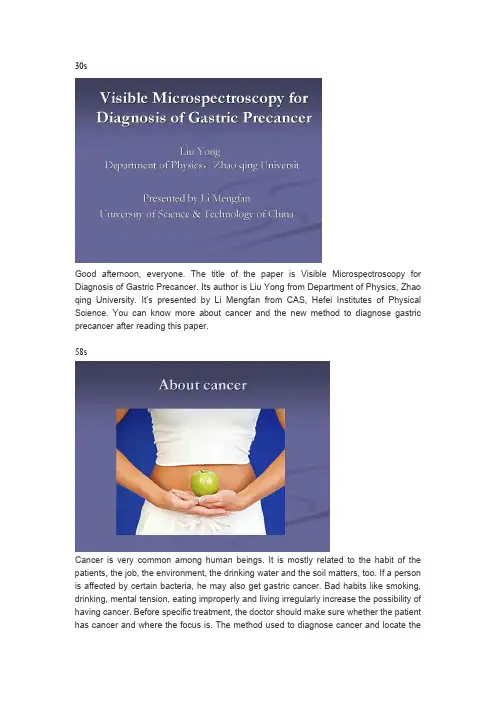
30sGood afternoon, everyone. The title of the paper is Visible Microspectroscopy for Diagnosis of Gastric Precancer. Its author is Liu Yong from Department of Physics, Zhao qing University. It’s presented by Li Mengfan from CAS, Hefei Institutes of Physical Science. You can know more about cancer and the new method to diagnose gastric precancer after reading this paper.58sCancer is very common among human beings. It is mostly related to the habit of the patients, the job, the environment, the drinking water and the soil matters, too. If a person is affected by certain bacteria, he may also get gastric cancer. Bad habits like smoking, drinking, mental tension, eating improperly and living irregularly increase the possibility of having cancer. Before specific treatment, the doctor should make sure whether the patient has cancer and where the focus is. The method used to diagnose cancer and locate thecancerous area is very important.42sBefore we design a method to diagnose cancer, we should know more about cancer like where it starts and what it causes.Firstly, about 90% of the human’s cancer start from the epithelial layer of skin, internal organs and tubular organs in the body. Secondly ,cancer usually accompanied by the changes of organization structure in the cell level.So, the changes which appear in the structure of organization and chemical composition provides important evidence for diagnosis of precancer.42sNormal method to diagnose gastric precancer is Reflectance spectroscopy. It’s very prospective in early diagnosis of cancer and is commonly used these years.One of the most common technology is diffuse reflectance visible spectroscopy, it can acquire the approximate information of the target tissue. This method is low cost, fast and non-invasive, it provides important means for diagnosis of malignant damage of organization of human organs. However, there are also some disadvantages of this method which need to be improved.55sThis is the system of diffuse reflectance spectroscopy, the light beam emitted from the tungsten halogen lamp is guided into the integral ball by the source fiber. The light come out of the integral ball irradiate the standard board, the reflected light of the standard board is treated as the background. Then take target tissue substitute for the standard board, and scan the reflected light of it. The reflected light spectra of the gastric tissue mucosal layer and the submucosa layer is guided into the spectrometer by the collector fiber. The spectrometer and the computer is connected by USB, spectrum can be processed using computer.36sFirst of all, only average visible spectrum of the target tissue can be acquired, it means that the docters cannot make out the location of the cancerous tissue precisely.What’s more, the device is easy to be affected by changes of optical path length induced by different set-up of prober and the variation of hemoglobin concentration due to the pressure between prober and tissue.50sTo avoid the problems of the commonly used techniques, diffuse reflectance visible spectroscopy, scientists do a lot of researches to find out a new way to diagnose precancer.The new method is Visible Microspectroscopy. The visible microspectroscopic measurement device is accomplished on the basis of fiber confocal microscope, its 3-d imaging principle devote to the optical tomography ability of the system.The visible microspectrum of cancerous and normal tissue can be analysed andcontrasted, so it’s more useful than the normal method.45sThis is the structure of the visible microspectroscopy device .The fiber coupler can separate the lighting beams from the signal beam. The two lens can focus the light on the tissue ,The 3-D electronic controlled platform move along the direction of optic axis, so that the device can acquire the visible microspectroscopy signal of the sample in different depth, precisely locate the boundary of the cancerous tissue.The signal measured from different position can be stored in the computer after analysed by the spectrometer.31sThis is the spectrum chart of the normal tissue and cancerous tissue, they are analyzed by the spectrometer before being transmitted to the computer. The upper chart is the visible spectrum of normal tissue, it is regular. Contrasting to this, in the nether chart, the spectrum of the cancerous area shows that the strength of reflected light grows as the wave length grows.49sIn comparison with the normal method, there are a lot of advantages of the new method.Firstly, the structure of visible microspectroscopy device is very simple. The main parts consist of source, fiber coupler, 3-D electronic controlled platform, spectrometer and the computer.Secondly, the 3-D space microspectrum can be acquired , which can help the doctors efficiently make out whether the gastric tissue is cancerous.What’s more, the optical tomography ability enabled the device to precisely locate the edge of the cancerous area.32si.In conclusion, the main goal of this research is to find a new way to diagnose thegastric precancer.ii.The spectrum of the normal and cancerous gastric tissues which can be measured by visible microspectroscopy in cell level is the central idea .iii.This new method can obtain the cell level information, and is not affected bychanges of optical path length and hemoglobin concentration.50si. The device is very simple, so it can have a small size, which can reduce the sufferings of patients.It can efficiently make out whether the tissue is canceration.ii. Besides, the 3-D space microspectrum and optical tomography ability can make the device accurately distinguish the cancerous tumor area from the normal tissue in cell level, thus the boundary of the cancerous tissue can be ascertained accurately.iii. The result of the research shows that this device may be used to non-invasively diagnosis of early gastric cancer.20sThis device is only researched in the lab, there are still a lot of works to do to make this device applied into practice. So, s pecial design of this device should be made which can be applied to the medical diagnosis.给大家推荐一个英语微信群Empty Your Cup英语微信群是目前学习英语最有效的方法,群里都是说英语,没有半个中文,而且规则非常严格,是一个超级不错的英语学习环境,群里有好多英语超好的超牛逼的人,还有鬼佬和外国美眉。