G 蛋白偶联受体介导的信号通路.ppt
- 格式:ppt
- 大小:3.53 MB
- 文档页数:34
细胞信号通路中的G蛋白偶联受体细胞是生命体系的基础单位,细胞内有着许多重要的分子,这些分子之间通过信号通路相互联系,从而调控细胞的各种生命活动。
在这些信号通路中,G蛋白偶联受体(GPCR)是最常见和最重要的一类受体,它们能感受到许多信号分子的刺激并在细胞内引起一系列反应。
一、GPCR的基本概念GPCR是一类七次跨膜受体,在细胞膜上具有多种不同的结构和功能。
GPCR的N端位于胞外,C端位于细胞内。
当受体受到激活物质的刺激时,N端的构象发生改变,从而使C端产生一系列反应。
GPCR激活后,它们能以特定的方式激活蛋白激酶、谷氨酸离子通道等下游蛋白,从而引发复杂的信号传导和调节细胞各项生理功能。
二、GPCR信号通路的分类根据GPCR的信号传导机制和调控功能的不同,可以将GPCR 信号通路分为四类:Gs、Gi、Gq和G12/13。
Gs和Gi通路都调节细胞内腺苷酸酰化酶的活性,前者可以激活腺苷酸酰化酶来降解环磷酸腺苷,后者则能抑制这一过程。
从而实现调节细胞内第二信使的含量和活性,对细胞的代谢、分化等生理功能都有影响。
Gq通路则能够激活蛋白激酶C、肌动蛋白的收缩等生理过程。
而G12/13通路则能够影响细胞的形态变化和迁移等生理功能。
三、GPCR的药物靶点和应用由于GPCR在细胞内信号通路的重要性,GPCR受体也成为了药物研发的重要靶点之一。
许多目前临床应用的药物都是通过干扰GPCR的功能和信号传导来发挥治疗作用。
例如,β-肾上腺素能受体拮抗剂可用于治疗高血压和心脏病,乙酰胆碱能受体激动剂可用于治疗哮喘和冠心病,5-羟色胺受体拮抗剂可用于治疗焦虑和抑郁症等等。
另外,GPCR在癌症的发生和发展中也扮演着重要角色。
许多癌细胞表达的GPCR与正常细胞不同,且它们的信号通路也有所变化。
因此,干扰特定GPCR的信号传导可以成为一种治疗癌症的手段。
例如,针对一些调节肿瘤细胞迁移的GPCR的抗体治疗已进入临床研究阶段。
四、GPCR的研究前景随着技术的不断进步,GPCR结构和功能的研究也迎来了新的契机。
《G蛋白偶联受体介导的信号通路共同点》近年来,G蛋白偶联受体(GPCR)介导的信号通路在细胞生物学和药物开发领域备受关注。
GPCR是一类跨膜受体蛋白,能感知细胞外的信号分子,从而引发细胞内的信号传导,对多种生理过程发挥重要作用。
在不同的细胞类型和组织中,不同种类的GPCR可能介导不同的信号通路,但同时也存在一些共同点。
本文将深入探讨G蛋白偶联受体介导的信号通路的共同点,并分析其对细胞生物学和临床治疗的意义。
共同点一:二级信号转导通路研究表明,GPCR介导的信号通路大多通过二级信号转导分子传递信号。
当细胞外的信号分子结合GPCR时,GPCR会激活其内在的G蛋白,并进而激活腺苷酸环化酶(AC)、磷脂酶C(PLC)、或细胞内钙离子等二级信号转导分子,最终引发细胞内信号传导级联反应。
这种二级信号转导通路是大多数GPCR共有的特点,为理解和干预GPCR介导的信号通路提供了重要线索。
共同点二:调控蛋白激活另一个共同点是,许多GPCR介导的信号通路均涉及调控蛋白的激活。
其中,蛋白激酶A(PKA)和蛋白激酶C(PKC)是最为典型的调控蛋白。
当GPCR被激活后,G蛋白会激活腺苷酸环化酶,进而使细胞内的cAMP水平升高,激活PKA。
另一些GPCR激活PLC,使细胞内的钙离子浓度升高,最终激活PKC。
这些调控蛋白激酶的激活对于细胞的功能和生理过程至关重要,因此成为了GPCR信号通路共同的重要环节。
共同点三:可能的信号转导交叉一些研究还发现,不同种类的GPCR介导的信号通路可能存在交叉。
某些GPCR激活PLC产生二酰甘油(DAG)和肌醇三磷酸(IP3),而IP3则可促使细胞内钙离子升高,从而激活PKC。
这种信号转导的交叉现象使得不同种类的GPCR之间产生相互影响和相互调节的可能性,增加了GPCR信号通路的复杂性和多样性。
总结与展望G蛋白偶联受体介导的信号通路具有一些共同的特点,如二级信号转导通路、调控蛋白激活和信号转导交叉等。

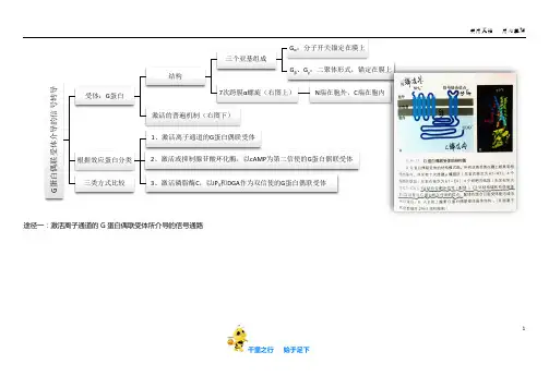
千里之行 始于足下1途径一:激活离子通道的G 蛋白偶联受体所介导的信号通路G 蛋白偶联受体介导的信号转导受体:G 蛋白结构三个亚基组成G α:分子开关锚定在膜上G β、G γ:二聚体形式,锚定在膜上7次跨膜α螺旋(右图上)N 端在胞外、C 端在胞内激活的普遍机制(右图下)根据效应蛋白分类1、激活离子通道的G 蛋白偶联受体2、激活或抑制腺苷酸环化酶,以cAMP 为第二信使的G 蛋白偶联受体3、激活磷脂酶C ,以IP 3和DGA 作为双信使的G 蛋白偶联受体三类方式比较千里之行 始于足下2图⑤ 图⑥典型例子心肌细胞M 乙酰胆碱受体激活G 蛋白开启K +通道附图p168(下图⑤)Gt 蛋白偶联的光敏感受体的活化诱发cGMP 门控阳离子通道的关闭附图p168(下图⑥)第二信使:cGMP千里之行 始于足下 3途径二:激活或抑制腺苷酸环化酶的G 蛋白偶联受体环化酶的G 蛋白偶联受体刺激AC 的物质肾上腺素、胰高血糖素、促肾上腺皮质激素受体:刺激性激素受体(Rs ),Gs α抑制AC 的物质前列腺素、腺苷受体:抑制性激素受体(Ri ),Gi αACAC 结构12次跨膜蛋白C 端与N 端均在细胞内胞质侧有两个大的相似的结构域,跨膜区有两个整合结构域AC 功能在Mg 2+或Mn 2+存在下,催化ATP 生成cAMP蛋白激酶A (PKA )未激活状态2个调节亚基与2个催化亚基结合激活状态激活物:cAMP调节亚基与催化亚基分开图⑦4 千里之行始于足下图⑧ 图⑨图115千里之行始于足下6 千里之行始于足下千里之行 始于足下7激活磷脂酶C 、以IP 3和DGA 作为双信使G 蛋白偶联受体介导的信号通路 图10第三条途径双信使(图10)来源磷脂酰肌醇(PI)代谢(图11)双信使介绍肌醇三磷酸(IP 3)机制与内质网上IP 3R 结合,开放Ca 2+通道功能引发贮存在内质网中的Ca 2+转移到细胞质基质中,使胞质中Ca 2+浓度升高二酰甘油(DAG)机制激活蛋白激酶C(PKC)降解DAG 激酶磷酸化后进入磷脂肌醇代谢DAG 脂酶水解成单酰甘油DAG 的维持原因细胞增殖、分化需要维持DAG 活性生成途径磷脂酶催化膜上磷脂酰胆碱断裂产生DAG蛋白激酶C(PKC)(图12)激活的信号分子与细胞分泌、肌肉收缩、细胞增殖、分化有关的信号分子作用途径一:磷酸化MAP 激酶途径二:磷酸化一种抑制蛋白8 千里之行始于足下千里之行 始于足下9激活离子通道的G 蛋白偶联受体激活/抑制腺苷酸环化酶的G 蛋白偶联受体 激活磷脂酶C 的G 蛋白偶联受体心肌细胞上K +通道的启闭 视杆细胞的光受体启闭效应蛋白 G 蛋白 PDE 腺苷酸环化酶(AC) 磷脂酶C(PLC)第二信使 无 cGMP cAMP IP 3、DAG生物学功能调节心肌细胞内外K +浓度,影响心肌收缩频率生物感光 调节肝细胞和肌细胞糖原代谢,对真核细胞基因表达调控 调节基因表达,与肌肉收缩、细胞增殖、分化有关图1210 千里之行始于足下。
g蛋白偶联受体(G protein-coupled receptors,简称GPCRs)是一类重要的跨膜蛋白,能够感知细胞外的信号并将其转导到细胞内部。
而g蛋白偶联受体介导的cAMP信号通路被认为是细胞内信号传导中最为重要的一种通路,对于细胞的正常功能起着至关重要的作用。
在人体生理学中,g蛋白偶联受体介导的cAMP信号通路涉及多种重要的生理过程,包括细胞分化、增殖、代谢调节、离子平衡以及神经递质释放等。
以下将详细介绍g蛋白偶联受体介导的cAMP信号通路在这些生理过程中的作用和意义。
【1】细胞分化g蛋白偶联受体介导的cAMP信号通路在细胞分化过程中发挥重要作用。
研究表明,特定的GPCRs激活cAMP信号通路后,能够促进干细胞向特定细胞系的分化,从而在组织发育和再生过程中发挥关键作用。
这一发现为干细胞治疗和再生医学领域的研究提供了重要的理论基础。
【2】细胞增殖cAMP信号通路对细胞增殖也具有调节作用。
在细胞周期调控中,cAMP信号通路能够通过激活蛋白激酶A(PKA)和调节蛋白激酶(Epac)等效应分子,调控细胞周期蛋白激酶的活性,影响细胞的增殖能力。
cAMP信号通路在肿瘤发生和发展过程中扮演着重要的角色,也成为肿瘤治疗的重要靶点。
【3】代谢调节在细胞能量代谢过程中,cAMP信号通路亦扮演着重要的调节作用。
通过激活PKA和Epac等效应分子,cAMP信号通路能够促进葡萄糖代谢、脂质代谢以及蛋白质合成等代谢过程,对维持细胞能量平衡起着至关重要的作用,并参与调节胰岛素分泌等内分泌功能。
【4】离子平衡cAMP信号通路对离子通道和转运蛋白的调节,也对细胞内离子平衡起着重要作用。
通过激活离子通道蛋白或转运蛋白,cAMP信号通路能够调节细胞内Ca2+、Na+、K+等离子的浓度,影响细胞内环境的稳态,参与细胞对外界环境的适应和响应。
【5】神经递质释放在神经系统中,cAMP信号通路对神经递质的释放及神经元兴奋性起着重要调节作用。
G蛋白耦联受体信号通路的研究进展近年来,G蛋白耦联受体(GPCR)信号通路在生命科学研究领域中引起了广泛关注。
GPCR是一个大家族,包括了多种不同类型的受体,并且广泛分布在人体各个组织器官中,对人体生理过程具有重要的调节作用。
细胞内信号传递的核心是受体激活导致的信号通路。
在GPCR信号通路中,最重要的是G蛋白的作用。
一.GPCR信号通路的基础原理1.1 GPCR简介GPCR是一类跨膜蛋白,它们通过细胞膜将信号从分子识别器至细胞内部转发。
GPCR的识别分子是非常丰富的,在协调细胞与环境间的交互过程中,GPCR扮演着至关重要的角色。
GPCR是一个世界上化学物质最多的跨膜受体家族,被估计为超过1,000种不同类型。
它们广泛分布于多种类型的细胞表面,参与不同的生理和病理过程,如视觉、味觉、嗅觉、免疫系统、内分泌调节、神经递质释放和心血管响应等。
1.2 GPCR信号通路基本原理组织和细胞通过GPCR自身特定的配体 - 受体相互作用进行相互沟通,并通过G蛋白激活,进而调节下游信号通路。
GPCR的特异性配体可以是小分子化合物、蛋白质、肽和胜肽等,为特异性的信号传递提供了必要的方式。
1.3 G蛋白激活的机制GPCR激活后,内生性媒介物如G蛋白介导固有和受体激动剂的活化状态。
G蛋白两个面向细胞内和细胞外的亚基,如串联蛋白一样将GPCR准确把握住,从而实现其与信号转导通路的连接。
这些G蛋白亚基可分为”Gα“和”Gβγ“两个亚基。
当GPCR受体活化时,它可以与G蛋白交互并激活G蛋白,导致Gα和Gβγ二者的分离,从而触发不同的下游信号通路。
二.G蛋白耦联受体信号通路的研究进展2.1 G蛋白G蛋白是GPCR信号通路的重要组成部分。
与GPCR的高度多样性相似,G蛋白也具有很高的多样性,包括多个亚型。
随着技术的发展和对G蛋白的研究,已经明确了G蛋白的具体分类及其在不同信号通路中的具体作用。
现在对G蛋白进行开发的技术,包括计算机模拟器、蛋白工程技术和生物化学方面的技术,相信将为细胞认识、疾病预防和治疗提供重要的启示。


细胞表面信号分子受体及G蛋白耦联受体介导的信号转导途径细胞通讯是指一个细胞发出的信息通过介质(又称配体)传递到另一个细胞并与靶细胞相应的受体相互作用,然后通过信号转导产生的胞内一系列生理生化变化,最终表现为细胞整体的生物学效应的过程。
1 细胞通讯的方式根据细胞分泌化学信号发挥作用距离的长短,可把细胞通讯分为:①内分泌,由内分泌细胞分泌信号分子到血液中,通过血液循环运送到体内各个部位,作用与靶细胞。
②旁分泌,细胞通过分泌局部化学介质到细胞外液中,经过局部扩散作用与临近靶细胞。
③自分泌,细胞对自身分泌的物质产生反应。
④通过化学突触传递神经信号,通过轴突及化学突触实现电信号---化学信号---电信号的转换。
⑤接触依赖性的通讯,细胞直接接触而无需信号分子的释放,代之以通过质膜上的信号分子与靶细胞质膜上的受体分子相互作用来介导细胞间的通讯。
2 信号分子与受体2.1信号分子信号分子是细胞的信息载体,根据其溶解性通常可以分为亲脂性和亲水性两大类①亲脂性信号分子,主要代表是甾类激素和甲状腺素,可穿过细胞质膜进入细胞,与细胞质或细胞核中受体结合形成激素---受体复合物,调节基因表达;②亲水性信号分子,包括神经递质、生长因子、局部化学递质和大多数激素,它们不能穿过靶细胞质膜的脂双分子层,只能通过与靶细胞表面受体结合,再经信号转换机制,在细胞内产生第二信使或激活蛋白激酶或蛋白磷酸酶的活性,引起细胞的应答。
此外,在20世纪80年代后期,发现和证实一氧化氮(nitric oxide,NO)这个氧自由基在生物体内是一种重要的信号分子和效应分子。
2.2受体受体是一种能够识别和选择性结合某种配基(信号分子)的大分子,当与配基(配体)结合后,通过信号转导作用将胞外信号转换为胞内化学或物理的信号,以启动一系列过程和生物学效应。
根据靶细胞上受体存在的部位,可将受体分为细胞内受体(intracellular receptor)和细胞表面受体(cell—surfacereceptor)。
G蛋白偶联受体信号传导通路的分子机制研究G蛋白偶联受体(G protein-coupled receptors,GPCRs)是人类体内最大的膜受体家族之一,广泛参与了生理和病理过程中的信号转导通路。
近年来,G蛋白偶联受体的研究变得非常火热,人们对其分子机制的研究也越来越深入。
本文将从G 蛋白偶联受体信号传导通路的分子机制、调控机制和应用前景三个方面进行讨论。
一、G蛋白偶联受体信号传导通路的分子机制G蛋白偶联受体最重要的功能是通过其三个亚基(α、β和γ)与G蛋白结合,从而触发下游的信号转导通路。
G蛋白偶联受体的典型结构包括七个跨膜区域和一个C端区域,其中N端朝向胞外,C端朝向胞内,七个跨膜区域之间的环状区域是GPCRs的关键结构域。
GPCRs与内源性配体结合后,促使G蛋白中α亚基与GDP结合状态下的GTP 交换,α亚基独立于β和γ亚基并与下游分子直接相互作用,从而影响线粒体、离子通道、酶等下游效应器的酶活性或电位水平。
G蛋白偶联受体信号传导通路在许多重要的生理和病理过程中都起着重要的作用。
二、G蛋白偶联受体信号传导通路的调控机制大量的实验证明,与GPCRs结合的配体分子可以调控G蛋白α亚基与GTP或GDP结合的平衡状态,进而引起了效应器的反馈调节。
此外,调控G蛋白偶联受体配体结合、转运和稳定性的工作也具有巨大的发展潜力。
一些潜在的药物分子也能够影响G蛋白偶联受体的稳定性和位置,从而具有重要的药物治疗作用。
三、G蛋白偶联受体信号传导通路的应用前景G蛋白偶联受体是当今的一个热门生物医学研究领域,已经被广泛应用于药物研究和发现、抗癌药物研究、病毒和细胞介导的疾病研究,以及治疗其他疾病的药物研究。
随着技术的不断进步,G蛋白偶联受体的应用前景变得越来越广泛,这将为人类健康的促进和医疗的进步带来更多的机会和挑战。
总之,G蛋白偶联受体信号转导通路的分子机制、调控机制和应用前景是当今生物医学研究的重要方向之一。
进一步的研究将有助于揭示G蛋白偶联受体信号转导通路在人类健康、疾病治疗等领域的应用潜力,从而更好地服务于人类的健康事业。
Theresa Filtz, PhDPhar 735, Winter 2005G protein-coupled Signal TransductionMain Objectives (the big chunks)•Describe in molecular detail the cascades of events in a generalized G protein-coupled signaling pathway•Describe the structure of a GPCR and how it relates to function•List the four major classes of G a subunits and the effects of toxins on their activity•List the major, proximal second messengers produced by adenylyl cyclase and PLC-b•Describe GPCR desensitization and supersensitization and discuss the consequences for drug action•Compare the effects of changing cyclic AMP and calcium levels in cardiac cells and in smooth muscle cellsG protein coupled receptors (GPCR)•FunctionThe receptor subtype mediating the actionsof hundreds to thousands ofendogenous and exogenous substancesCouple to a guanine nucleotide bindingprotein (G protein)G proteins activate effectors to initiatesignaling cascades•Examples of drug acting through GPCRsBronchodilators-albuterol (Ventolin®, etc) Stimulants- Ritalin®, ephedraDecongestants-Sudafed®Antipsychotics-haloperidol, Zyprexa®, Geodon®Antihistamines-Benadryl®, Claritin®, Allegra®Pain medication-morphine and opioid analgesics Labor inducing agents-pitocinAnti-hypertensives-b-blockersCardiac stimulants -atropine, dopamine, epinephrine, adenosine Epinephrine for severe allergic reaction•StructureOne of the most abundant proteinfamiliesAt least a thousand different genes for GPCR in the human genomePotentially a unique GPCR for everydectectable odorSeven transmembrane a helicesExternal NH 2 terminus and internal C tailBinding pocketInside the a helices for small molecules and at the NH 2 terminus for larger peptides Intracellular loopsBinding of activators to a helices affects configuration of the intracellular loops 2nd and 3rd loops and C tail interact with G proteinsGuanine nucleotide binding proteins (G Proteins)• Structure and FunctionTurn on effector enzymesHeterotrimer--composed of three protein subunits—G a , G b , and G gOther families of G proteins exist which serve other intracellular functions Bound by lipid chains to the intracellular surface of the plasma membrane G a subunitBinds GDP and associates with G bg in the resting state Binds GTP and dissociates from G bg in the active stateUpon activation by receptor, releases GDP and picks up GTP Dissociates from G bg upon binding GTPActivates effector enzymes when bound to GTP Intrinsic GTPase activitySelf-Hydrolyzes GTP back to GDPRejoins with G bg when bound to GDP to turn itself off G bg subunitExists mainly as a non-dissociable dimer Active when dissociated from G a subunitActivates different effector enzymes when separated from G a Inactivated by binding Ga•SubtypesG a subtypesFour major families, multiple subtypesG a sPredominantly activates adenylyl cyclaseConstitutively activated by cholera toxinG olf olfactory G a subunit, activates ion channels in nasal epitheliaG a iInhibited by pertussis toxinPredominantly inhibits adenylyl cyclaseG a t, tranducin-the retinal light transduction subunitactivates cyclic GMP phosphodiesterase in response to lightG a oinhibits some types of Ca++ channelsG a12,13Involvement with growth regulatory pathways still being experimentally confirmedG a qActivates phospholipase C-b enzymesG bg subtypesMultiple subtypes of each subunitActivate many enzymes including phospholipase C-b, cation channels, GPCR kinases,subtypes of adenylyl cyclase, among other signaling proteinsWhich combination of subtypes activate which effectors is still being investigatedEffector enzymes•Phospholipase C-bActivated by G a q subunits OR G bg subunits (depending on isoform)Hydrolyzes a low abundance membrane phospholipid, PIP2 (phosphatidylinositol 4,5-bisphosphate) into two products, IP3 (inositol trisphosphate) and DAG (diacylglycerol)IP3IP3 is water soluble and diffuses through the cytoplasm to bind to receptors on the endoplasmic reticulum (ER)IP3 receptor activation releases Ca++ from the ER into cell cytosolCells are exquisitely sensitive to changes in cytosolic Ca++ concentrationCa++ activates Ca++/calmodulin-dependent kinases (CaM kinases)CaM kinases activate multiple phosphorylation cascadesLithium carbonate inhibits inositol recycling back into PIP2, causing a build-up of IP3 anddepletion of PIP2DAGMembrane bound lipidActivates protein kinase C (PKC)PKC initiates multiple phosphorylation cascadesPhorbol esters are tumor promoters that mimic DAG and activate PKCActivation of PLC-b leads to many physiologic responses including smooth muscle contraction, learning and memory, and cell growth and differentiation•GPCR = G protein coupled receptor PKC = protein kinase CPLC-b = phospholipase C-bCaM = calmodulinAdenylyl CyclaseActivated by G a s subunits Inhibited by G a i subunits Transforms ATP into cyclic AMPcyclic AMPIncreases and decreases in cyclic AMP levels lead to different effects Activates cyclic AMP-dependent protein kinase (PKA)Hydrolyzed by phosphodiesterases (PDE)PDE is inhibited by theophylline (bronchodilator), caffeine, sildenafil (Viagra®), etc Involved in the classic glycogen metabolism pathway to liberate glucose for energyPKA phosphorylates glycogen synthetase to inhibits activity PKA activates phosphorylase kinasephosphorylase kinase converts glycogen phosphorylase b to glycogen phosphorylase a glycogen phosphorylase a promotes glycogen metabolism cyclic AMP also activates transcription factorscyclic AMP response element binding protein (CREB)G a sAdenylyl CyclaseG b gPKAGPCRP P PStimulatory Hormone GPCR = G protein coupled receptorPKA = cyclic AMP regulated protein kinase PDE = phosphodiesteraseG a iG b gGPCRInhibitory Hormone +-ATP cyclicAMPAMPPDERegulation of receptor responsiveness• DesensitizationUpon chronic application of agonists, cells will attempt to avoid overstimulation by blockingthe ability of the receptor to keep stimulating the signal transduction pathway Best understood for b 2 adrenergic receptors.Not all receptors desensitize, but many doExtent of desensitization seems to correlate with intrinsic activity of the agonist Short term desensitizationNegative feedback loop specific for the receptor that is bound by activator Occurs within seconds- minutes Sequelae of eventsActivation of b 2 receptors liberates G bg from G a s G bg activates a G protein receptor kinase (GR K)GRK phosphorylates the receptor at multiple sites in the C tailPhosphorylation of the C tail causes arrestin to bind to the receptor A rrestin binding inhibits further activation of G proteins by the receptorG a s GRKinaseG b gGPCRStimulatory Hormone GPCR = G protein coupled receptorGRKinase = G protein receptor regulated protein kinaseP PP PP PPPA r re s t i nXDownregulation and resensitizationDown-regulationLongterm negative feedback loopOccurs with continuous agonist administration over hours and days Receptors are removed from the membrane and degradedNo effect on agonist efficacy until all spare receptors are internalizedResensitizationActivator must be removedOccurs on same time scale as desensitization or downregulation Phosphatases can rapidly reverse simple desensitizationResensitization following downregulation requires hours to days for new protein synthesis• SupersensitizationLong term blockade (days to weeks) with antagonist leads to increased expression of receptorsat the membrane Up-regulation (increased levels) of receptors leads to supersensitivity upon removal of theantagonistAbrupt withdrawal of b -blockers could produce a myocardial infarction for up to 2 weeks following cessation of therapyP PPPPPPPPP ResensitizationDownregulation with internalizationDegradationCell membraneMore examples of G protein coupled signaling pathways•Cardiac contractionCyclic AMPIncreased or decreased by G a s or G a iPKA activation leads to increased phosphorylation of myofibrillary and sarcoplasmicproteins (troponin, phospholamban)Phosphorylation of myofibrillary proteins increases myocyte contractilityL-type Ca++ channelsActivated by G a s, inhibited by G a oOpening (activation) increases cardiomyocyte cytosolic Ca++ by stimulating release fromsarcoplasmic Ca++ storesCa++-activated troponin increases contractilityInward-rectifying K+ channelsActivated by G bg subunits released from G a i or G a oK+ channel opening leads to membrane hyperpolarizationImportant in slowing heart rate*Also important in decreasing neurotransmitter release from neurons*• Smooth muscle (e.g. blood vessel) contraction and relaxationIP3Increases lead to increased cytosolic Ca ++Increased Ca ++ leads to activation of Ca ++/CaM dependent myosin light chain kinase (MLCK)Phosphorylation of MLCK phosphorylates myosin P-myosin stimulates contractionCyclic AMPActivation of PKA leads to activation of membrane and sarcoplasmic Ca ++ pumps Activated pumps increase storage of Ca ++Decreased cytosolic calcium inhibits contraction Opposite of cardiac muscle effectsCyclic GMPSoluble guanylyl cyclase is stimulated by diffusible NO gas Cyclic GMP levels increase Smooth muscle relaxesMechanisms unclear Ca ++ levels decreaseCyclic GMP phosphodiesterase breaks down cyclic GMPInhibitors (sildenafil) increase smooth muscle relaxationb2-adrenergic receptor Endothelin receptorsM1 Muscarinic receptor Angiotensin receptor Histamine H1 receptorCysteinyl leukotriene receptor Oxytocin receptorP2Y purinergic receptors。