A spectral technique for correspondence problems using pairwise constraints
- 格式:pdf
- 大小:734.98 KB
- 文档页数:8

傅里叶红外光谱的英文傅里叶红外光谱的英文I. IntroductionInfrared spectroscopy is a common analytical method used for studying the chemical properties of a sample. Fourier transform infrared spectroscopy (FTIR), also known as Fourier transform infrared (FTIR) analysis, is a type of infrared spectroscopy that uses a Fourier transform to obtain the spectral information. In this article, we will discuss the English terminology used for FTIR.II. Basic Terminology1. Infrared spectrum: a representation of the absorption or transmission of infrared radiation as a function of wavelength or frequency2. Spectral range: the range of wavelengths or frequencies measured in the infrared spectrum3. Wavenumber: the reciprocal of wavelength, measured in cm-1 in the FTIR spectrum4. Absorbance: the logarithm of the ratio of the incident radiation to the transmitted radiation, measured in the FTIR spectrum5. Peak: a point on the FTIR spectrum that corresponds to a specific vibrational mode of the sample6. Baseline: the absorption background in the FTIR spectrumIII. Sample PreparationBefore performing FTIR analysis, the sample must be prepared in the formof a thin film or powder to ensure uniformity of the sample.IV. InstrumentationFTIR analysis requires a Fourier transform infrared spectrometer, which consists of a source, interferometer, and detector. The sample is placed in the path of the infrared beam generated by the source and the transmitted or absorbed radiation is measured by the detector. The interferometer is used to obtain the interferogram, which is then transformed into the FTIR spectrum.V. ApplicationsFTIR is used in various fields such as chemistry, pharmaceuticals, and material science. It is commonly used for the identification of unknown compounds, characterization of functional groups, and monitoring of chemical reactions.VI. ConclusionFTIR analysis is a powerful technique for studying the chemical properties of a sample. Understanding the basic terminology and instrumentation used in FTIR is essential for accurate interpretation of the spectral data.。
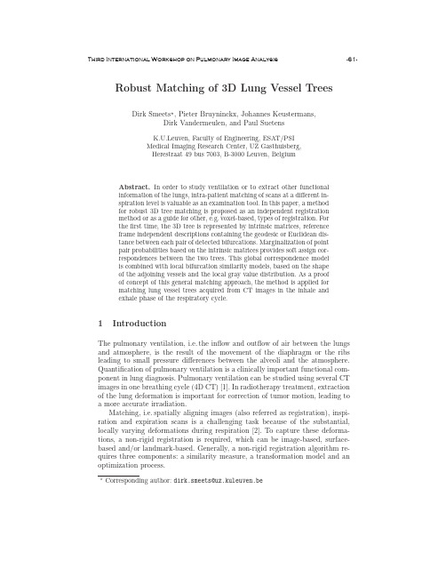
Robust Matching of3D Lung Vessel Trees Dirk Smeets ,Pieter Bruyninckx,Johannes Keustermans,Dirk Vandermeulen,and Paul SuetensK.U.Leuven,Faculty of Engineering,ESAT/PSIMedical Imaging Research Center,UZ Gasthuisberg,Herestraat49bus7003,B-3000Leuven,BelgiumAbstract.In order to study ventilation or to extract other functionalinformation of the lungs,intra-patient matching of scans at a different in-spiration level is valuable as an examination tool.In this paper,a methodfor robust3D tree matching is proposed as an independent registrationmethod or as a guide for other,e.g.voxel-based,types of registration.Forthefirst time,the3D tree is represented by intrinsic matrices,referenceframe independent descriptions containing the geodesic or Euclidean dis-tance between each pair of detected bifurcations.Marginalization of pointpair probabilities based on the intrinsic matrices provides soft assign cor-respondences between the two trees.This global correspondence modelis combined with local bifurcation similarity models,based on the shapeof the adjoining vessels and the local gray value distribution.As a proofof concept of this general matching approach,the method is applied formatching lung vessel trees acquired from CT images in the inhale andexhale phase of the respiratory cycle.1IntroductionThe pulmonary ventilation,i.e.the inflow and outflow of air between the lungs and atmosphere,is the result of the movement of the diaphragm or the ribs leading to small pressure differences between the alveoli and the atmosphere. Quantification of pulmonary ventilation is a clinically important functional com-ponent in lung diagnosis.Pulmonary ventilation can be studied using several CT images in one breathing cycle(4D CT)[1].In radiotherapy treatment,extraction of the lung deformation is important for correction of tumor motion,leading to a more accurate irradiation.Matching,i.e.spatially aligning images(also referred as registration),inspi-ration and expiration scans is a challenging task because of the substantial, locally varying deformations during respiration[2].To capture these deforma-tions,a non-rigid registration is required,which can be image-based,surface-based and/or landmark-based.Generally,a non-rigid registration algorithm re-quires three components:a similarity measure,a transformation model and an optimization process.Corresponding author:dirk.smeets@uz.kuleuven.beIn image registration,common voxel similarity measures are sum of square differences(SSD),correlation coefficient(CC)and mutual information(MI)[3, 4].To regularize the transformation,popular transformation models are elastic models,viscousfluid models,spline-based techniques,finite element models or opticalflow techniques[3].Voxel-similarity based techniques have been applied to lung matching in[5–7].They have the advantage that dense and generally accurate correspondences are obtained.Disadvantages are the sensitivity to the initialization and the computational demands.Surface-based registration meth-ods use a similarity measure that is a function of the distances between points on the two surfaces.Thin-plate splines are popular for regularization of the transfor-mation.A combination of an voxel-similarity based and surface based registra-tion for lung matching is presented in[8].Generally,landmark-based non-rigid registration algorithms allow large deformations.Because of their sparsity,they are very efficient.In[9]bifurcations of lung vessels arefirst detected after which these landmarks are matched by comparing the3D local shape context of each pair of bifurcations with theχ2-distance.In this paper,we present a robust registration method for matching vessel trees.After detecting candidate bifurcations(Sec.2.1),correspondences are es-tablished by combining a global and a local bifurcation similarity model.For the novel global model(Sec.2.3),the tree is represented by two reference frame inde-pendent,hence intrinsic,matrices:the Euclidean(rigid transformation invariant) and geodesic(isometric deformation invariant)distance matrix.Marginalization of point pair probabilities provides soft(not binary)correspondences between the detected bifurcations of the two trees.The local bifurcation similarity model reflects correspondences based on local intensities and is explained in Sec.2.3. The optimization to obtain hard correspondences is done using a spectral de-composition technique(Sec.2.4).The proof of concept of the general matching approach is shown in Sec.3.Finally we draw some conclusions.2MethodThe goal of the proposed matching framework is to establish correspondences between characteristic points in the image,independent of the reference frame (translation and rotation invariant)and,for characteristic points in structures that are assumed to deform isometrically,invariant for isometric deformations. Isometries are defined as distance-preserving isomorphisms between metric spaces, which generally means that structures only bend without stretching.The bifur-cations,the splittings of the vessels in two or more parts,are chosen as charac-teristic points,although their detection is not the main goal of this paper.We then assume the vessel tree to deform isometrically.This assumption of nearly constant vessel length has been made before in a.o.[10]and[11].2.1Bifurcation detectionBifurcations are locations where a blood vessel splits into two smaller vessels.A subset of the bifurcations that involves only major vessels can be detected in a robust way by analyzing the skeletonization of the segmented vessels.As preprocessing,the lungs are segmented by keeping the largest connected component of all voxels with an intensity lower than -200HU.A morphological closing operator includes the lung vessels to the segmentation and a subsequent erosion operation removes the ribs from the segmentation.Then,a rough seg-mentation of the major blood vessels within the lung is obtained by thresholding at -200HU.Cavities in binary segmentation smaller than 10voxels are closed.The skeletonization of the segmentation localizes the major bifurcations and is obtained through a 3D distance transformation by homotopic thinning [12].Since bifurcations are locations where three vessels join,they are characterized as skeleton voxels having three different neighbors belonging to the skeleton.After-wards,bifurcations are discarded that have a probability lower than 0.5/√2πσ2according to a statistical intensity model.The vessel intensities I k are assumed to be Gaussian distributed P (c vessel |I k )=1√2πσ2exp −(I k −μ)2σ2 ,(1)with experimentally determined parameters (μ=−40HU and σ=65HU).2.2Global correspondence modelAfter the vessel bifurcations are detected,soft correspondences are established based on a global and a local correspondence model,both independent of rigid and isometric deformations of the considered vessel trees.Intrinsic vessel tree representation.Each tree is intrinsically represented by a Euclidean distance matrix E =[d R 3,ij ](EDM),containing Euclidean dis-tances between each pair of bifurcations,and a geodesic distance matrix G =[g ij ](GDM).Each element g ij corresponds to the geodesic distance between the bi-furcations i and j .This distance is the distance between i and j along the vessels and is computed with the fast marching method in an image containing a soft segmented vessel tree using Eq.(1).Isometric vessel deformations,by defini-tion,leave these geodesic distances unchanged.Therefore,the GDM is invariant to the bending of the vessels.On the other hand,the EDM is only invariant to rigid transformations (and,when normalized,invariant to scale variations)of the vessel tree.However,Euclidean distance computation is expected to be more robust against noise than geodesic distance computation since the error accumu-lates along the geodesic,i.e.the shortest path.Both the EDM and the GDM are symmetric and uniquely defined up to an arbitrary simultaneous permutation of their rows and columns due to the arbitrary sampling order of the bifurcations.An example of a lung vessel tree,represented by a GDM and a EDM,is shown in Fig.1.(a)10203040506010203040506050100150200250300350400(b)102030405060102030405060020406080100120(c)Fig.1.A lung vessel tree with detected bifurcations (a)is represented by a geodesic (b)and a Euclidean (c)distance matrix,containing the geodesic or Euclidean distance between each pair of bifurcations.Soft correspondences of the global model.A probabilistic framework is used to estimate the probability that a bifurcation i of one tree corresponds with a bifurcations k of the other tree.It is applied twice,once for the EDM and once for the GDM.We will explain the framework using the GDM.Given two lung vessel trees,represented by GDMs,the probability that the pair of bifurcations (i,j )of the first tree G 1correponds with the pair (k,l )of the second tree G 2is assumed to be normally distributed,P (C (i,j ),(k,l ))=1√2exp −(g 1,ij −g 2,kl )2σ2 ,(2)with σchosen to be 1and g 1,ij an element of the GDM representing the first tree.It reflects the assumption that geodesic distances between pairs of bifur-cations are preserved,obeying an isometric deformation.The probability that abifurcation i corresponds with k is then given byP(C i,k)=jlP(C(i,j),(k,l))=m G,ik,(3)from which the soft correspondence matrix,M G=[m G,ik],is constructed.The same procedure is followed to obtain an assignment matrix M E based on the EDM as intrinsic tree representation.This matrix is expected to be more robust against noise,but varies under isometric deformations of the vessel tree, contrary to M G.2.3Local correspondence modelComplementary to the global bifurcation similarity model,a local model based on intensities is implemented.In the computer vision literature a large number of local image descriptors are proposed,see for example[13].In this paper,the n-SIFT(scale invariant feature transform)image descrip-tor is used[14],which summarizes a cubic voxel region centered at the feature location,in casu the bifurcation location.The cube is divided into64subre-gions,each using a60bin histogram to summarize the gradients of the voxels in the subregion.This results in a3840-dimensional feature vector f by com-bining the histograms in a vector after weighting by a Gaussian centered at the feature location.The probability that a bifurcation i corresponds with k is then proportional toP(C i,k)∝exp(− f i−f k 2)=m L,ik,(4) 2.4Spectral matchingThe combined match matrix M C is found as the pointwise product of the match matrices of the separate models,m C,ik=m G,ik.m E,ik.m L,ik(i.e.product of the corresponding probabilities).To establish hard correspondences(one-to-one mapping),the algorithm proposed by Scott and Longuet-Higgins[15]is used. It applies a singular value decomposition to M C(M C=UΣV T)and computes the orthogonal matrix M C=U˜I n V T,with˜I n a pseudo-identity matrix.Two bifurcations i and k match if m C,ik is both the greatest element in its row and the greatest in its column.3Experimental resultsThe described matching framework is evaluated using a publicly available4D CT thorax dataset of the L´e on B´e rard Cancer Center&CREATIS lab(Lyon, France),called“POPI-model”.It contains103D volumes representing the dif-ferent phases of the average breathing cycle,with an axial resolution of0.976562 mm×0.976562mm and a slice thickness of2mm.Also,3D landmarks,indi-cated by medical experts,and vectorfields describing the motion of the lung, are available[16].3.1Matching manually indicated landmarksFirst,the matching framework is evaluated using manually indicated landmarks to examine the performance independent of the bifurcation detection step.For this purpose,all 40landmarks are used in one CT image (end-inhalation phase),while varying the number of landmarks in the other image.The result of this experiment,expressed as the percentage of correct correspondences in function of the percentage outliers of the total number of landmarks,is shown in Fig.2for different images in the respiratory cycle.Since not all landmarks are located in the vessels and therefore geodesic distances are not meaningful,M G is not computed.% outliers of total no. landmarks % c o r r e c t c o r r e s p o n d e n c e s Fig.2.Results of the framework for matching manually indicated landmarks expressed as the percentage of correct correspondences in function of the percentage outliers.The three curves correspond with different scans in the respiratory cycle,in which scan no.1is end-inhale and scan no.6end-exhale.These results show that even for a large number of outliers good correspon-dences are still found.Consequently,it can be expected that not all bifurcations detected in one image must be present in the other image.Also,a small decrease in performance can be noticed when more lung deformation is present (end-exhale vs.end-inhale),probably because the Euclidean distance matrix (used in the global correspondence model)is not invariant for these deformations.Second,the robustness against landmark positioning errors is examined.Therefore,we add uniform distributed noise in each direction to one set of landmarks and vary the maximum amplitude of the noise.Fig.3shows the percentage of correct correspondences in function of this maximum amplitude,averaged over 15runs per noise level.These results show that indication errors of more than 5mm (±5voxels in x and y direction and 2.5voxels in z di-rection)decrease the matching performance.It is therefore expected that the localization of bifurcations during automatic bifurcation detection must not be extremely accurate in order to still obtain good correspondences.maximum noise amplitude in each direction [mm]% c o r r e c t c o r r e s p o n d e n c e s Fig.3.Indication error dependence,expressed as the percentage of correct correspon-dences in function of the maximum amplitude of the added uniform noise.The three curves correspond with different scans in the respiratory cycle,in which scan no.1is end-inhale and scan no.6end-exhale.3.2Matching automatically detected bifurcationsNext,the framework is evaluated for the task of matching 3D vessel trees by means of automatically detected bifurcations.The result of matching the end-inhale with the end-exhale vessel tree is shown in Fig.4,clarifying that most correspondence pairs are correct.It is also clear that simple thresholding has resulted in oversegmentation in one of the two images.This,however,did not affect the automatic matching.The performance of matching automatically detected bifurcations is quan-tified using the dense deformation field that is available in the public dataset (POPI-model).This deformation field is obtained using a parametric image-based registration [17,18,16].The average target registration error in the manual landmarks is 1.0mm with a standard deviation of 0.5mm.Fig.4.Qualitative evaluation for matching3D vessel trees by means of automatically detected bifurcations.Fig.5illustrates the accuracy of the matching of the end-inhale scan with all other scans.It shows histograms of the registration error(in each direction and in absolute value)for all bifurcations.The average registration error is2.31mm,the median1.84mm and the maximum24.0mm.Only for0.21%of the bifurcation matches the absolute registration error is larger than1.5cm,demonstrating the robustness of the matching algorithm.4ConclusionA robust matching framework is proposed,combining two global and one local landmark similarity model.The results of matching manually indicated land-marks demonstrate the potential of the proposed method for matching land-marks in unimodal medical images independent of the translation and rotation between both images.A high number of outliers is allowed when the landmarks−10−50510020406080100120error in x−direction [mm]f r e q u e n t y −10−50510error in y−direction [mm]f r e q u e n t y−10−50510error in z−direction [mm]f r e q u e n t y 051015absolute error [mm]f r e q u e n t y Fig.5.Histograms of the registration error (in each direction and in absolute value)for all bifurcations give an impression of the matching accuracy.in the image are well located.Moreover,we demonstrated that the matching framework can also be used for automatically detected landmarks,in this case lung bifurcations extracted from CT data.As future work,we see some applications of the soft correspondences for ro-bust initialization of an iterative non-rigid,e.g.voxel-based,registration method.References1.Guerrero,T.,Sanders,K.,Castillo,E.,Zhang,Y.,Bidaut,L.,Pan,T.,Komaki,R.:Dynamic ventilation imaging from four-dimensional computed tomography.Phys.Med.Biol.51(2006)777–7912.Sluimer,I.,Schilham,A.,Prokop,M.,van Ginneken,B.:Computer analysis ofcomputed tomography scans of the lung:a survey.IEEE Trans.Med.Imaging25(4)(2006)385–4053.Crum,W.R.,Hartkens,T.,Hill,D.L.G.:Non-rigid image registration:theory andpractice.Br J Radiol 77Spec No 2(2004)S140–534.Pluim,J.P.W.,Maintz,J.B.A.,Viergever,M.A.:Mutual information based reg-istration of medical images:A survey.IEEE Trans.Med.Imaging 22(8)(2003)986–10045.Dougherty,L.,Asmuth,J.C.,Gefter,W.B.:Alignment of ct lung volumes with anopticalflow method.Academic Radiology10(3)(2003)249–2546.Stancanello,J.,Berna,E.,Cavedon,C.,Francescon,P.,Loeckx,D.,Cerveri,P.,Ferrigno,G.,Baselli,G.:Preliminary study on the use of nonrigid registration for thoraco-abdominal radiosurgery.Med Phys.32(12)(2005)3777–857.Loeckx, D.:Automated Nonrigid Intra-Patient Image Registration Using B-Splines.PhD thesis,K.U.Leuven(2006)8.Kaus,M.,Netsch,T.,Kabus,S.,Pekar,V.,McNutt,T.,Fischer,B.:Estimationof organ motion from4D CT for4D radiation therapy planning of lung cancer.In: MICCAI.Volume3217of LNCS.(2004)1017–10249.Hilsmann,A.,Vik,T.,Kaus,M.,Franks,K.,Bissonette,J.P.,Purdie,T.,Beziak,A.,Aach,T.:Deformable4DCT lung registration with vessel bifurcations.In:CARS.(2007)10.Klabunde,R.E.:Determinants of resistance toflow./Hemodynamics/H003.htm(august2008)11.Groher,M.,Zikic,D.,Navab,N.:Deformable2D-3D registration of vascular struc-tures in a one view scenario.IEEE Trans Med Imaging28(6)(2009)847–60 12.Selle,D.:Analyse von gef¨a ssstrukturen in medizinischen schichtdatens¨a tzen f¨u rdie computergest¨u tzte operationsplanung.PhD thesis,Aachen(2000)13.Mikolajczyk,K.,Schmid,C.:A performance evaluation of local descriptors.IEEETrans.Pattern Anal.Mach.Intell.27(10)(2005)1615–163014.Cheung,W.,Hamarneh,G.:n-sift:n-dimensional scale invariant feature transform.Trans.Img.Proc.18(9)(2009)2012–202115.Scott,G.L.,Longuet-Higgins,H.C.:An algorithm for associating the features oftwo images.Proc.R.Soc.Lond.B244(1991)21–2616.Vandemeulebroucke,J.,Sarrut, D.,Clarysse,P.:The POPI-model,a point-validated pixel-based breathing thorax model.In:Conference on the Use of Com-puters in Radiation Therapy.(2007)17.Rueckert,D.,Sonoda,L.I.,Hayes,C.,Hill,D.L.G.,Leach,M.O.,Hawkes,D.J.:Nonrigid registration using free-form deformations:Application to breast mr im-ages.IEEE Transactions on Medical Imaging18(1999)712–72118.Delhay,B.,Clarysse,P.,Magnin,I.E.:Locally adapted spatio-temporal deforma-tion model for dense motion estimation in periodic cardiac image sequences.In: FIMH’07:Proceedings of the4th international conference on Functional imaging and modeling of the heart,Berlin,Heidelberg,Springer-Verlag(2007)393–402。


重庆邮电大学硕士学位论文摘要摘要近年来,随着各式图像传感器的应用,人们对图像信息的整合与利用的需求越来越大。
红外与可见光图像是常见的两种具有信息互补的图像。
红外图像能直观地反映物体的热辐射能量大小,不受光照影响,但会丢失一部分物体的纹理、结构等外观信息。
而可见光图像包含丰富的颜色和纹理信息,但受光照影响较大。
为了能充分地利用红外与可见光这两种模态图像中包含的互补信息,首先需要配准红外与可见光图像。
配准后的图像综合了两者的优势,有利于后续的图像分析、识别等工作。
因此,红外与可见光图像配准方法研究具有重要意义。
本文针对红外与可见光两种光谱图像的特点进行了配准问题研究,具体工作如下:第一,对于存在的红外与可见光图像的分辨率和光谱差异过大的问题,本文提出一种基于模态转换的配准方法。
该方法设计了一种二阶段的网络结构。
该网络首先对可见光图像进行映射操作,将其转换为近似红外光谱的图像。
然后在第二阶段,该网络以转换生成的图像为基准,使用空间变换器与采样器对红外图像进行变换操作得到配准后的红外图像,实现多模态图像配准。
实验结果表明,本文提出的配准方法在红外与可见光图像配准任务上有较大的优势。
第二,针对红外与可见光两种模态图像配准存在的映射关系不确定性问题,本文提出一种基于模态转换和模态独立邻域特征的配准方法。
由于红外图像存在大量图像区域灰度相同或相似,引起灰度映射关系常常存在非单一对应,造成映射存在较大的不确定性的问题。
为此,本文考虑使用模态转换和模态独立邻域特征结合的图像配准方法。
首先将可见光图像转换为近似红外光谱的图像,以减小光谱差异性。
再结合模态独立邻域特征在不同模态图像中表示不同类型图像特征,并且能对重要特征做出高响应的优势。
然后,基于转换生成的图像和红外图像提取模态独立邻域特征,以归一化互信息为配准度量计算图像变形域,得到配准图像。
该方法结合了深度学习自动提取特征和人工设计特征对重要特征高响应的优势。
实验结果表明,与现有的配准算法相比,本文方法具有较好的配准效果。
Spectroscopic Study of the Interaction between Small Molecules and Large Proteins1. IntroductionThe study of drug-protein interactions is of great importance in drug discovery and development. Understanding how small molecules interact with proteins at the molecular level is crucial for the design of new and more effective drugs. Spectroscopic techniques have proven to be valuable tools in the investigation of these interactions, providing det本人led information about the binding affinity, mode of binding, and structural changes that occur upon binding.2. Spectroscopic Techniques2.1. Fluorescence SpectroscopyFluorescence spectroscopy is widely used in the study of drug-protein interactions due to its high sensitivity and selectivity. By monitoring the changes in the fluorescence emission of either the drug or the protein upon binding, valuable information about the binding affinity and the binding site can be obt本人ned. Additionally, fluorescence quenching studies can provide insights into the proximity and accessibility of specific amino acid residues in the protein's binding site.2.2. UV-Visible SpectroscopyUV-Visible spectroscopy is another powerful tool for the investigation of drug-protein interactions. This technique can be used to monitor changes in the absorption spectra of either the drug or the protein upon binding, providing information about the binding affinity and the stoichiometry of the interaction. Moreover, UV-Visible spectroscopy can be used to study the conformational changes that occur in the protein upon binding to the drug.2.3. Circular Dichroism SpectroscopyCircular dichroism spectroscopy is widely used to investigate the secondary structure of proteins and to monitor conformational changes upon ligand binding. By analyzing the changes in the CD spectra of the protein in the presence of the drug, valuable information about the structural changes induced by the binding can be obt本人ned.2.4. Nuclear Magnetic Resonance SpectroscopyNMR spectroscopy is a powerful technique for the investigation of drug-protein interactions at the atomic level. By analyzing the chemical shifts and the NOE signals of the protein in thepresence of the drug, det本人led information about the binding site and the mode of binding can be obt本人ned. Additionally, NMR can provide insights into the dynamics of the protein upon binding to the drug.3. Applications3.1. Drug DiscoverySpectroscopic studies of drug-protein interactions play a crucial role in drug discovery, providing valuable information about the binding affinity, selectivity, and mode of action of potential drug candidates. By understanding how small molecules interact with their target proteins, researchers can design more potent and specific drugs with fewer side effects.3.2. Protein EngineeringSpectroscopic techniques can also be used to study the effects of mutations and modifications on the binding affinity and specificity of proteins. By analyzing the binding of small molecules to wild-type and mutant proteins, valuable insights into the structure-function relationship of proteins can be obt本人ned.3.3. Biophysical StudiesSpectroscopic studies of drug-protein interactions are also valuable for the characterization of protein-ligandplexes, providing insights into the thermodynamics and kinetics of the binding process. Additionally, these studies can be used to investigate the effects of environmental factors, such as pH, temperature, and ionic strength, on the stability and binding affinity of theplexes.4. Challenges and Future DirectionsWhile spectroscopic techniques have greatly contributed to our understanding of drug-protein interactions, there are still challenges that need to be addressed. For instance, the study of membrane proteins and protein-protein interactions using spectroscopic techniques rem本人ns challenging due to theplexity and heterogeneity of these systems. Additionally, the development of new spectroscopic methods and the integration of spectroscopy with other biophysical andputational approaches will further advance our understanding of drug-protein interactions.In conclusion, spectroscopic studies of drug-protein interactions have greatly contributed to our understanding of how small molecules interact with proteins at the molecular level. Byproviding det本人led information about the binding affinity, mode of binding, and structural changes that occur upon binding, spectroscopic techniques have be valuable tools in drug discovery, protein engineering, and biophysical studies. As technology continues to advance, spectroscopy will play an increasingly important role in the study of drug-protein interactions, leading to the development of more effective and targeted therapeutics.。
12Spectroscopic techniques:I Spectrophotometric techniquesA.H O F M A N N12.1Introduction12.2Ultraviolet and visible light spectroscopy12.3Fluorescence spectroscopy12.4Luminometry12.5Circular dichroism spectroscopy12.6Light scattering12.7Atomic spectroscopy12.8Suggestions for further reading12.1INTRODUCTIONSpectroscopic techniques employ light to interact with matter and thus probe certainfeatures of a sample to learn about its consistency or structure.Light is electromag-netic radiation,a phenomenon exhibiting different energies,and dependent on thatenergy,different molecular features can be probed.The basic principles of interactionof electromagnetic radiation with matter are treated in this chapter.There is noobvious logical dividing point to split the applications of electromagnetic radiationinto parts treated separately.The justification for the split presented in this text ispurely pragmatic and based on‘common practice’.The applications considered in thischapter use visible or UV light to probe consistency and conformational structure ofbiological ually,these methods are thefirst analytical procedures usedby a biochemical scientist.The applications covered in Chapter13present a higherlevel of complexity in undertaking and are employed at a later stage in biochemical orbiophysical characterisation.An understanding of the properties of electromagnetic radiation and its interactionwith matter leads to an appreciation of the variety of types of spectra and,conse-quently,different spectroscopic techniques and their applications to the solution ofbiological problems.47712.1.1Properties of electromagnetic radiationThe interaction of electromagnetic radiation with matter is a quantum phenomenon and dependent upon both the properties of the radiation and the appropriate structural parts of the samples involved.This is not surprising,since the origin of electromag-netic radiation is due to energy changes within matter itself.The transitions which occur within matter are quantum phenomena and the spectra which arise from such transitions are principally predictable.Electromagnetic radiation (Fig.12.1)is composed of an electric and a perpendicular magnetic vector,each one oscillating in plane at right angles to the direction of propagation.The wavelength l is the spatial distance between two consecutive peaks (one cycle)in the sinusoidal waveform and is measured in submultiples of metre,usually in nanometres (nm).The maximum length of the vector is called the amplitude .The frequency of the electromagnetic radiation is the number of oscillations made by the wave within the timeframe of 1s.It therefore has the units of 1s À1¼1Hz.The frequency is related to the wavelength via the speed of light c (c ¼2.998Â108m s À1in vacuo )by ¼c l À1.A historical parameter in this context is the wavenumber "v which describes the number of completed wave cycles per distance and is typically measured in 1cm À1.12.1.2Interaction with matterFigure 12.2shows the spectrum of electromagnetic radiation organised by increasing wavelength,and thus decreasing energy,from left to right.Also annotated are the types of radiation and the various interactions with matter and the resulting spectro-scopic applications,as well as the interdependent parameters of frequency and wavenumber.Electromagnetic phenomena are explained in terms of quantum mechanics.The photon is the elementary particle responsible for electromagnetic phenomena.It carries the electromagnetic radiation and has properties of a wave,as well asof Fig.12.1Light is electromagnetic radiation and can be described as a wave propagating transversally in space and time.The electric (E )and magnetic (M )field vectors are directed perpendicular to each other.For UV/Vis,circular dichroism and fluorescence spectroscopy,the electric field vector is of most importance.For electron paramagnetic and nuclear magnetic resonance,the emphasis is on the magnetic field vector.478Spectroscopic techniques:I Photometric techniquesa particle,albeit having a mass of zero.As a particle,it interacts with matter by transferring its energy E :E ¼hc l ¼h ð12:1Þwhere h is the Planck constant (h =6.63Â10À34Js)and is the frequency of the radiation as introduced above.When considering a diatomic molecule (see Fig.12.3),rotational and vibrational levels possess discrete energies that only merge into a continuum at very high energy.Each electronic state of a molecule possesses its own set of rotational and vibrational levels.Since the kind of schematics shown in Fig.12.3is rather complex,the Jablonski diagram is used instead,where electronic and vibrational states are schematically drawn as horizontal lines,and vertical lines depict possible transitions (see Fig.12.8below).In order for a transition to occur in the system,energy must be absorbed.The energy change D E needed is defined in quantum terms by the difference in absolute energies between the final and the starting state as D E ¼E final –E start ¼h .Electrons in either atoms or molecules may be distributed between several energy levels but principally reside in the lowest levels (ground state ).In order for an electron to be promoted to a higher level (excited state ),energy must be put into the system.If this energy E ¼h is derived from electromagnetic radiation,this gives rise to an absorption spectrum,and an electron is transferred from the electronic ground state (S 0)into the first electronic excited state (S 1).The molecule will also be in an excited vibrational and rotational state.Subsequent relaxation of the molecule into the vibrational ground state of the first electronic excited state will occur.The electron can then revert back to the electronic ground state.For non-fluorescent molecules,this is accompanied by the emission of heat (D H ).Microwave X-ray Vis UVFar NearIR Radio Wavelength in nm Wavenumber in cm –1Frequency in s –1Energy in J mol –1101021031041051061071081091 µm 1 cm 1 mChange inelectronic statesX-ray absorptionUV/Visabsorption IR/Raman Microwave spectroscopyESR Electron spin NMR Nuclear spin Change in rotational states of Change in molecular rotational and vibrational states 10171016101510141013101210111010109108106105104103102101110–110–2107106105104103102101110–1Fig.12.2The electromagnetic spectrum and its usage for spectroscopic methods.47912.1Introduction480Spectroscopic techniques:I Photometric techniquesNuclear displacementR e(ground)Fig.12.3Energy diagram for a diatomic molecule exhibiting rotation,vibration as well as an electronic structure.The distance between two masses m1and m2(nuclear displacement)is described as a Lennard–Jones potential curve with different equilibrium distances(R e)for each electronic state.Energetically lower states always have lower equilibrium distances.The vibrational levels(horizontal lines)are superimposed on the electronic levels.Rotational levels are superimposed on the vibrational levels and not shown for reasons of clarity.The plot of absorption probability against wavelength is called absorption spectrum .In the simpler case of single atoms (as opposed to multi-atom molecules),electronic transitions lead to the occurrence of line spectra (see Section 12.7).Because of the existence of more different kinds of energy levels,molecular spectra are usually observed as band spectra (for example Fig.12.7below)which are molecule-specific due to the unique vibration states.A commonly used classification of absorption transitions uses the spin states of electrons.Quantum mechanically,the electronic states of atoms and molecules are described by orbitals which define the different states of electrons by two parameters:a geometrical function defining the space and a probability function.The combination of both functions describes the localisation of an electron.Electrons in binding orbitals are usually paired with antiparallel spin orientation (Fig.12.8).The total spin S is calculated from the individual electron spins.The multipli-city M is obtained by M ¼2ÂS þ1.For paired electrons in one orbital this yields:S ¼spin ðelectron 1Þþspin ðelectron 2Þ¼ðþ1=2ÞþðÀ1=2Þ¼0The multiplicity is thus M ¼2Â0þ1¼1.Such a state is thus called a singlet state and denotated as ‘S ’.Usually,the ground state of a molecule is a singlet state,S 0.In case the spins of both electrons are oriented in a parallel fashion,the resulting state is characterised by a total spin of S ¼1,and a multiplicity of M =3.Such a state is called a triplet state and usually exists only as one of the excited states of a molecule,e.g.T 1.According to quantum mechanical transition rules,the multiplicity M and the total spin S must not change during a transition.Thus,the S 0!S 1transition is allowed and possesses a high transition probability.In contrast,the S 0!T 1is not allowed and has a small transition probability.Note that the transition probability is proportional to the intensity of the respective absorption bands.Most biologically relevant molecules possess more than two atoms and,therefore,the energy diagrams become more complex than the ones shown in Fig.12.3.Different orbitals combine to yield molecular orbitals that generally fall into one of five different classes (Fig.12.4):s orbitals combine to the binding s and the anti-binding s *orbitals.Some p orbitals combine to the binding p and the anti-binding p*Anti-binding Non-binding BindingEs *p *s pnFig.12.4Energy scheme for molecular orbitals (not to scale).Arrows indicate possible electronic transitions.The length of the arrows indicates the energy required to be put into the system in order to enable thetransition.Black arrows depict transitions possible with energies from the UV/Vis spectrum for some biological molecules.The transitions shown by grey arrows require higher energies (e.g.X-rays).48112.1Introduction482Spectroscopic techniques:I Photometric techniquesorbitals.Other p orbitals combine to form non-binding n orbitals.The population of binding orbitals strengthens a chemical bond,and,vice versa,the population of anti-binding orbitals weakens a chemical bond.12.1.3LasersLaser is an acronym for light amplification by stimulated emission of radiation.A detailed explanation of the theory of lasers is beyond the scope of this textbook.A simplified description starts with the use of photons of a defined energy to excite anabsorbing material.This results in elevation of an electron to a higher energy level.If, whilst the electron is in the excited state,another photon of precisely that energy arrives, then,instead of the electron being promoted to an even higher level,it can return to the original ground state.However,this transition is accompanied by the emission of two photons with the same wavelength and exactly in phase(coherent photons).Multipli-cation of this process will produce coherent light with extremely narrow spectral bandwidth.In order to produce an ample supply of suitable photons,the absorbing material is surrounded by a rapidlyflashing light of high intensity(pumping).Lasers are indispensable tools in many areas of science,including biochemistry and biophysics.Several modern spectroscopic techniques utilise laser light sources, due to their high intensity and accurately defined spectral properties.One of the probably most revolutionising applications in the life sciences,the use of lasers in DNA sequencing withfluorescence labels(see Sections5.11.5,5.11.6and12.3.3), enabled the breakthrough in whole-genome sequencing.12.2ULTRAVIOLET AND VISIBLE LIGHT SPECTROSCOPYThese regions of the electromagnetic spectrum and their associated techniques are probably the most widely used for analytical work and research into biological problems.The electronic transitions in molecules can be classified according to the partici-pating molecular orbitals(See Fig.12.4).From the four possible transitions(n!p*, p!p*,n!s*,s!s*),only two can be elicited with light from the UV/Vis spectrum for some biological molecules:n!p*and p!p*.The n!s*and s!s*transitions are energetically not within the range of UV/Vis spectroscopy and require higher energies.Molecular(sub-)structures responsible for interaction with electromagnetic radi-ation are called chromophores.In proteins,there are three types of chromophores relevant for UV/Vis spectroscopy:•peptide bonds(amide bond);•certain amino acid side chains(mainly tryptophan and tyrosine);and•certain prosthetic groups and coenzymes(e.g.porphyrine groups such as in haem).The presence of several conjugated double bonds in organic molecules results in an extended p-system of electrons which lowers the energy of the p*orbital through electron delocalisation.In many cases,such systems possess p!p*transitions in the UV/Vis range of the electromagnetic spectrum.Such molecules are very useful tools in colorimetric applications(see Table12.1).Table12.1Common colorimetric and UV absorption assaysSubstance Reagent Wavelength(nm) Amino acids(a)Ninhydrin570(proline:420)(b)Cupric salts620Cysteine residues, thiolates Ellman reagent(di-sodium-bis-(3-carboxy-4-nitrophenyl)-disulphide)412Protein(a)Folin(phosphomolybdate,phosphotungstate,cupric salt)660(b)Biuret(reacts with peptide bonds)540(c)BCA reagent(bicinchoninic acid)562(d)Coomassie Brilliant Blue595(e)Direct Tyr,Trp:278,peptide bond:190 Coenzymes Direct FAD:438,NADH:340,NADþ:260 Carotenoids Direct420,450,480 Porphyrins Direct400(Soret band) Carbohydrate(a)Phenol,H2SO4Glucose:490,xylose:480(b)Anthrone(anthrone,H2SO4)620or625 Reducing sugars Dinitrosalicylate,alkaline tartrate buffer540Pentoses(a)Bial(orcinol,ethanol,FeCl3,HCl)665(b)Cysteine,H2SO4380–415 Hexoses(a)Carbazole,ethanol,H2SO4540or440(b)Cysteine,H2SO4380–415(c)Arsenomolybdate500–570 Glucose Glucose oxidase,peroxidase,o-dianisidine,phosphate buffer420 Ketohexose(a)Resorcinol,thiourea,ethanoic acid,HCl520(b)Carbazole,ethanol,cysteine,H2SO4560(c)Diphenylamine,ethanol,ethanoic acid,HCl635Hexosamines Ehrlich(dimethylaminobenzaldehyde,ethanol,HCl)53048312.2Ultraviolet and visible light spectroscopy12.2.1Chromophores in proteinsThe electronic transitions of the peptide bond occur in the far UV.The intense peak at 190nm,and the weaker one at 210–220nm is due to the p !p *and n !p *transitions.A number of amino acids (Asp,Glu,Asn,Gln,Arg and His)have weak electronic transitions at around ually,these cannot be observed in proteins because they are masked by the more intense peptide bond absorption.The most useful range for proteins is above 230nm,where there are absorptions from aromatic side chains .While a very weak absorption maximum of phenylalanine occurs at 257nm,tyrosine and tryptophan dominate the typical protein spectrum with their absorption maxima at 274nm and 280nm,respectively (Fig.12.5).In praxi ,the presence of these two aromatic side chains gives rise to a band at $278nm.Cystine (Cys 2)possesses a weak absorption maximum of similar strength as phenylalanine at 250nm.This band can play a role in rare cases in protein optical activity or protein fluorescence.Proteins that contain prosthetic groups (e.g.haem,flavin,carotenoid)and some metal–protein complexes,may have strong absorption bands in the UV/Vis range.These bands are usually sensitive to local environment and can be used for physical studies of enzyme action.Carotenoids,for instance,are a large class of red,yellow and orange plant pigments composed of long carbon chains with many conjugated double bonds.They contain three maxima in the visible region of the electromagnetic spectrum ($420nm,450nm,480nm).Porphyrins are the prosthetic groups of haemoglobin,myoglobin,catalase and cytochromes.Electron delocalisation extends throughout the cyclic tetrapyrrole ring of porphyrins and gives rise to an intense transition at $400nm called the Soret band .The spectrum of haemoglobin is very sensitive to changes in the iron-bound ligand.These changes can be used for structure–function studies of haem proteins.Table 12.1(cont.)SubstanceReagent Wavelength (nm)DNA (a)Diphenylamine595(b)Direct 260RNA Bial (orcinol,ethanol,FeCl 3,HCl)665Sterols and steroids Liebermann–Burchardt reagent (acetic anhydride,H 2SO 4,chloroform)425,625Cholesterol Cholesterol oxidase,peroxidase,4-amino-antipyrine,phenol 500ATPase assay Coupled enzyme assay with ATPase,pyruvatekinase,lactate dehydrogenase:ATP !ADP (consumes ATP)phosphoenolpyruvate !pyruvate (consumes ADP)pyruvate !lactate (consumes NADH)NADH:340484Spectroscopic techniques:I Photometric techniquesMolecules such as FAD (flavin adenine dinucleotide),NADH and NAD þare impor-tant coenzymes of proteins involved in electron transfer reactions (RedOx reactions).They can be conveniently assayed by using their UV/Vis absorption:438nm (FAD),340nm (NADH)and 260nm (NAD þ).Chromophores in genetic materialThe absorption of UV light by nucleic acids arises from n !p *and p !p *transitions of the purine (adenine,guanine)and pyrimidine (cytosine,thymine,uracil)bases that occur between 260nm and 275nm.The absorption spectra of the bases in polymers are sensitive to pH and greatly influenced by electronic interactions between bases.12.2.2PrinciplesQuantification of light absorptionThe chance for a photon to be absorbed by matter is given by an extinction coefficient which itself is dependent on the wavelength l of the photon.If light withthe Wavelength (nm)A b s o r b a n c eFig.12.5The presence of larger aggregates in biological samples gives rise to Rayleigh scatter visible by a considerable slope in the region from 500to 350nm.The dashed line shows the correction to be applied to spectra with Rayleigh scatter which increases with l À4.Practically,linear extrapolation of the region from 500to 350nm is performed to correct for the scatter.The corrected absorbance is indicated by the double arrow.48512.2Ultraviolet and visible light spectroscopyintensity I0passes through a sample with appropriate transparency and the path length (thickness)d,the intensity I drops along the pathway in an exponential manner. The characteristic absorption parameter for the sample is the extinction coefficient a, yielding the correlation I=I0eÀa d.The ratio T=I/I0is called transmission. Biochemical samples usually comprise aqueous solutions,where the substance of interest is present at a molar concentration c.Algebraic transformation of the expo-nential correlation into an expression based on the decadic logarithm yields the law of Beer–Lambert:lg I0I¼lg1T¼eÂcÂd¼Að12:2Þwhere[d]¼1cm,[c]¼1mol dmÀ3,and[e]¼1dm3molÀ1cmÀ1.e is the molar absorption coefficient(also molar extinction coefficient)(a¼2.303ÂcÂe).A is the absorbance of the sample,which is displayed on the spectrophotometer.The Beer–Lambert law is valid for low concentrations only.Higher concentrations might lead to association of molecules and therefore cause deviations from the ideal behaviour.Absorbance and extinction coefficients are additive parameters,which complicates determination of concentrations in samples with more than one absorbing species.Note that in dispersive samples or suspensions scattering effects increase the absorbance,since the scattered light is not reaching the detector for read-out.The absorbance recorded by the spectrophotometer is thus overestimated and needs to be corrected(Fig.12.5).Deviations from the Beer–Lambert lawAccording to the Beer–Lambert law,absorbance is linearly proportional to the concen-tration of chromophores.This might not be the case any more in samples with high absorbance.Every spectrophotometer has a certain amount of stray light,which is light received at the detector but not anticipated in the spectral band isolated by the mono-chromator.In order to obtain reasonable signal-to-noise ratios,the intensity of light at the chosen wavelength(I l)should be10times higher than the intensity of the stray light(I stray).If the stray light gains in intensity,the effects measured at the detector have nothing or little to do with chromophore concentration.Secondly,molecular events might lead to devi-ations from the Beer–Lambert law.For instance,chromophores might dimerise at high concentrations and,as a result,might possess different spectroscopic parameters.Absorption or light scattering–optical densityIn some applications,for example measurement of turbidity of cell cultures(determin-ation of biomass concentration),it is not the absorption but the scattering of light(see Section12.6)that is actually measured with a spectrophotometer.Extremely turbid samples like bacterial cultures do not absorb the incoming light.Instead,the light is scattered and thus,the spectrometer will record an apparent absorbance(sometimes also called attenuance).In this case,the observed parameter is called optical density(OD).Instruments specifically designed to measure turbid samples are nephelometers or Klett meters;however,most biochemical laboratories use the general UV/Vis spectrometer for determination of optical densities of cell cultures.486Spectroscopic techniques:I Photometric techniques48712.2Ultraviolet and visible light spectroscopyFactors affecting UV/Vis absorptionBiochemical samples are usually buffered aqueous solutions,which has two major advantages.Firstly,proteins and peptides are comfortable in water as a solvent, which is also the‘native’solvent.Secondly,in the wavelength interval of UV/Vis (700–200nm)the water spectrum does not show any absorption bands and thus acts as a silent component of the sample.The absorption spectrum of a chromophore is only partly determined by its chem-ical structure.The environment also affects the observed spectrum,which mainly can be described by three parameters:•protonation/deprotonation(pH,RedOx);•solvent polarity(dielectric constant of the solvent);and•orientation effects.Vice versa,the immediate environment of chromophores can be probed by assessing their absorption,which makes chromophores ideal reporter molecules for environ-mental factors.Four effects,two each for wavelength and absorption changes,have to be considered:•a wavelength shift to higher values is called red shift or bathochromic effect;•similarly,a shift to lower wavelengths is called blue shift or hypsochromic effect;•an increase in absorption is called hyperchromicity(‘more colour’),•while a decrease in absorption is called hypochromicity(‘less colour’).Protonation/deprotonation arises either from changes in pH or oxidation/reduction reactions,which makes chromophores pH-and RedOx-sensitive reporters.As a rule of thumb,l max and e increase,i.e.the sample displays a batho-and hyperchromic shift, if a titratable group becomes charged.Furthermore,solvent polarity affects the difference between the ground and excited states.Generally,when shifting to a less polar environment one observes a batho-and hyperchromic effect.Conversely,a solvent with higher polarity elicits a hypso-and hypochromic effect.Lastly,orientation effects,such as an increase in order of nucleic acids from single-stranded to double-stranded DNA,lead to different absorption behaviour.A sample of free nucleotides exhibits a higher absorption than a sample with identical amounts of nucleotides but assembled into a single-stranded polynucleotide.Accordingly,double-stranded polynucleotides exhibit an even smaller absorption than two single-stranded polynucleotides.This phenomenon is called the hypochromicity of polynucleotides.The increased exposure(and thus stronger absorption)of the individual nucleotides in the less ordered states provides a simplified explanation for this behaviour.12.2.3InstrumentationUV/Vis spectrophotometers are usually dual-beam spectrometers where thefirst chan-nel contains the sample and the second channel holds the control(buffer)for correction.488Spectroscopic techniques:I Photometric techniquesAlternatively,one can record the control spectrumfirst and use this as internal referencefor the sample spectrum.The latter approach has become very popular as many spectro-meters in the laboratories are computer-controlled,and baseline correction can be carriedout using the software by simply subtracting the control from the sample spectrum.The light source is a tungstenfilament bulb for the visible part of the spectrum,anda deuterium bulb for the UV region.Since the emitted light consists of many differentwavelengths,a monochromator,consisting of either a prism or a rotating metal gridof high precision called grating,is placed between the light source and the sample.Wavelength selection can also be achieved by using colouredfilters as monochromatorsthat absorb all but a certain limited range of wavelengths.This limited range is calledthe bandwidth of thefilter.Filter-based wavelength selection is used in colorimetry,a method with moderate accuracy,but best suited for specific colorimetric assayswhere only certain wavelengths are of interest.If wavelengths are selected by prismsor gratings,the technique is called spectrophotometry(Fig.12.6).Example1ESTIMATION OF MOLAR EXTINCTION COEFFICIENTSIn order to determine the concentration of a solution of the peptide MAMVSEFLKQAWFIENEEQE YVQTVKSSKG GPGSAVSPYP TFNPSS in water,the molarabsorption coefficient needs to be estimated.The molar extinction coefficient e is a characteristic parameter of a molecule andvaries with the wavelength of incident light.Because of useful applications of thelaw of Beer–Lambert,the value of e needs be known for a lot of molecules beingused in biochemical experiments.Very frequently in biochemical research,the molar extinction coefficient ofproteins is estimated using incremental e i values for each absorbing protein residue(chromophore).Summation over all residues yields a reasonable estimation for theextinction coefficient.The simplest increment system is based on values of Gill andvon Hippel.1The determination of protein concentration using this formula onlyrequires an absorption value at l¼280nm.Increments e i are used to calculate a molarextinction coefficient at280nm for the entire protein or peptide by summation overall relevant residues in the protein:Residue Gill and von Hippel e i(280nm)in dm3molÀ1cmÀ1Cys2120Trp5690Tyr1280For the peptide above,one obtains e¼(1Â5690þ2Â1280)dm3molÀ1cmÀ1¼8250dm3molÀ1cmÀ11Gill,S.C.and von Hippel,P.H.(1989).Calculation of protein extinction coefficients from aminoacid sequence data.Analytical Biochemistry,182,319–326.Erratum:Analytical Biochemistry(1990),189,283.A prism splits the incoming light into its components by refraction.Refraction occurs because radiation of different wavelengths travels along different paths in medium of higher density.In order to maintain the principle of velocity conservation,light of shorter wavelength (higher speed)must travel a longer distance (i.e.blue sky effect).At a grating,the splitting of wavelengths is achieved by diffraction.Diffraction is a reflection phenom-enon that occurs at a grid surface,in this case a series of engraved fine lines.The distance between the lines has to be of the same order of magnitude as the wavelength of the diffracted radiation.By varying the distance between the lines,different wavelengths are selected.This is achieved by rotating the grating perpendicular to the optical axis.The resolution achieved by gratings is much higher than the one available by prisms.Nowadays instruments almost exclusively contain gratings as monochromators as they can be reproducibly made in high quality by photoreproduction.The bandwidth of a colorimeter is determined by the filter used as monochromator.A filter that appears red to the human eye is transmitting red light and absorbs almost any other (visual)wavelength.This filter would be used to examine blue solutions,as these would absorb red light.The filter used for a specific colorimetric assay is thus made of a colour complementary to that of the solution being tested.Theoretically,a single wavelength is selected by the monochromator in spectrophotometers,and the emergent light is a parallel beam.Here,the bandwidth is defined as twice the half-intensity bandwidth.The bandwidth is a function of the optical slit width.The narrower the slit width the more reproducible are measured absorbance values.In contrast,the sensitivity becomes less as the slit narrows,because less radiation passes through to the detector.In a dual-beam instrument,the incoming light beam is split into two parts by a half-mirror.One beam passes through the sample,the other through a control (blank,reference).This approach obviates any problems of variation in light intensity,as both reference and sample would be affected equally.The measured absorbance is the difference between the two transmitted beams of light recorded.Depending on the instrument,a second detector measures the intensity of the incoming beam,although some instruments use an arrangement where one detector measures the incoming and the transmitted intensity alternately.The latter design is better from an analytical point of view as it eliminates potential variations between the two Vis light UV lightBeam Fig.12.6Optical arrangements in a dual-beam spectrophotometer.Either a prism or a grating constitutes the monochromator of the instrument.Optical paths are shown as green lines.48912.2Ultraviolet and visible light spectroscopy。
安徽⼤学电⼦信息⼯程学院研究⽣导师信息姓名:胡根⽣性别:男出⽣年云:1971.5职称:教授学院:电⼦信息⼯程学院研究⽅向:1.图像处理2.智能信息处理个⼈简历2003年6⽉获江苏⼤学硕⼠学位,2006年6⽉获华南理⼯⼤学博⼠学位。
主要从事机器学习、模式识别等⽅⾯的研究和教学⼯作。
近期在国内外学术期刊或学术会议上发表学术论⽂10余篇,其中SCI检索1篇,EI检索5篇。
学术成果1.基于⽀持向量值轮廓波变换的遥感影像融合.电⼦学报.2.具有多分段损失函数的多输出⽀持向量机回归.控制理论与应⽤.3.在线多输出⽀持向量机回归及在投资决策中的应⽤.华南理⼯⼤学学报(⾃然科学版).4.具分段损失函数的⽀持向量机回归及在投资决策中的应⽤.控制理论与应⽤.在研项⽬安徽省教育厅重点科技项⽬《基于⽀持向量机的遥感影像云层去除新⽅法(批准号KJ2010A021)》姓名:孙⽟发性别:男出⽣年⽉:1966年职称:教授学院:电⼦信息⼯程学院研究⽅向:1. 计算电磁学及应⽤2. ⽆线通信与电磁兼容3. 电磁散射与⽬标识别个⼈简历1988年、1991年毕业于⼭东⼤学⽆线电物理专业,获理学学⼠学位和硕⼠学位,2001年毕业于中国科学技术⼤学电磁场与微波技术专业,获⼯学博⼠学位。
1994年7⽉—1998年8⽉,安徽⼤学讲师;1998年9⽉—2003年8⽉,安徽⼤学副教授;2003年9⽉⾄今,安徽⼤学教授。
2002年在⾹港城市⼤学⽆线通信中⼼做访问学者,2003年9⽉—2006年9⽉在中国科学技术⼤学信息与通信⼯程博⼠后流动站做科学研究⼯作。
学术成果中国电⼦学会⾼级会员,全国⾼等学校电磁场教学与教材研究会理事,电⼦测量与仪器学会微波毫⽶波测试专业委员会委员,《电波科学学报》编辑委员会委员,安徽省⾼等学校‘⼗五’第⼆批中青年学科带头⼈培养对象,安徽⼤学中青年学术⾻⼲。
主持国家⾃然科学基⾦1项,省部级科研项⽬2项,厅局级科研项⽬1项,参加国家⾃然科学基⾦重点项⽬1项,国家⾃然科学基⾦3项,省部级项⽬3项等,发表学术论⽂40余篇,其中被SCI、EI收录16篇,主编安徽省‘⼗⼀五’规划教材1部。