核医学骨显像
- 格式:ppt
- 大小:8.63 MB
- 文档页数:75

放射性核素骨显像对骨转移癌诊断的应用进展摘要:放射性核素骨显像如今已经成为临床上对骨转移癌作出诊断的常规方法之一而愈来愈受到临床医生的重视。
作为一项诊断技术,放射性核素骨显像在早期诊断、合理治疗及预后方面均有重要的价值,其方法简便,一次显像就能为检测骨转移癌提供灵敏的信息,但其较突出的缺点是特异性较CT、MRI低。
正鉴于此,我们参考了数十篇国内外相关的文献,想就放射性核素骨显像对骨转移癌诊断的原理及其应用方面作一个简要地阐释,以寄对临床医生有一定的参考价值。
主题词放射性核素骨显像诊断骨转移癌应用进展APPLIED PROGRESS OF RADIONUCLIDE BONE IMAGING IN DIAGNOSIS OF METASTATIC OSTEOMAABSTRACTRadionuclide bone imaging nowadays has become one of formal methods in the clinical diagnosis of metastatic osteoma, which has been attached much more importance to by the clinical doctors. As a diagnosis technique, radionuclide bone imaging has a very important value in the early diagnosis、reasonable treatment and evaluement of prognosis, which is a simple method and provides very sensitive information in detecting the metasatic osteoma by imaging all over body once. however, more distinctive shortcomings of it is the lower sensitivity than CT and MRI.. As a result of it, we have referred to tens of related articles at home and aboard. As far as the fundermental theory and application of radionuclide bone imaging in metastatic osteoma are concerned, a simple explanation is illustrated so as to have a certain referring value for the clinical doctors.KEY WORDS:RADIONUCLIDE BONE IMAGING DIAGNOSIS METASTATIC OSTEOMA APPLIED PROGRESS放射性核素骨显像是一种无创伤和灵敏度高的骨病诊断方法,尤其是对临床疑有骨转移癌的患者,更能够早期诊断和一次完成全身扫描,这具有与其他检查方法无法比拟的优点。
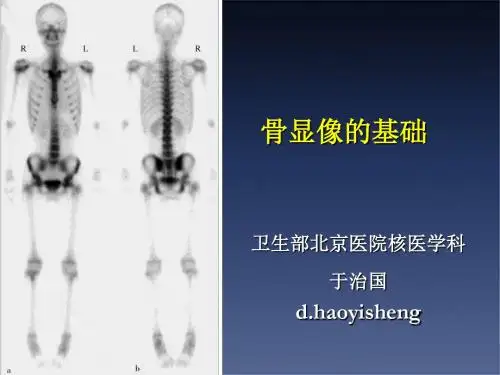
核医学科ect显像
核医学ECT一般可以检查骨骼系统、心血管系统,以及全身各个器官,如脑、脾脏、甲状腺、肾脏等,ECT是发射型计算机断层显像的英文缩写,是核医学独特的检查项目,比如SPECT-CT和PET-CT都属于核医学ECT检查范畴。
核医学ECT等放射性核素显像的原理,是建立在器官组织血流、功能和代谢变化的基础上,不仅能够显示脏器和病变的位置、形态、大小等解剖结构,更重要的是可以同时提供有关脏器、组织的血流、代谢等方面的信息,甚至是分子水平的代谢和生化信息,对于异常病变探测的灵敏度高,可以在疾病早期尚未发生形态结构改变时诊断疾病。
因此,核医学ECT可以检查的疾病很多,可以检查骨骼系统,进行全身骨扫描,检查有没有出现骨转移瘤,以及骨肿瘤的累及范围,还可以用于检查心血管疾病,心脏显像可以评估心肌缺血的情况,另外核医学ECT还可以用于脑血流的显像、脾脏显像、甲状腺显像以及肾脏显像等,可以适用的范围比较广。