免疫磁珠说明书
- 格式:doc
- 大小:87.00 KB
- 文档页数:7
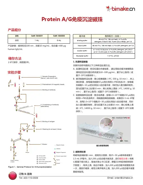
Protein A/G免疫沉淀磁珠Figure 1. General Protocol for ImmunoprecipitationcomplexSDS-PAGE loading buffer Neutralize bufferMagnetic Beads antibodyMagnetic Separator Remove supernatant Pipette Repeat45产品组分产品参数:磁珠粒径100 nm,浓度10 mg/mL,结合量>400 μg human IgG/mL2-8℃保存,保质期2年。
储存方法实验步骤1. 抗原样品制备本操作说明书提供以下三种样品处理方法。
2. 磁珠预处理将磁珠漩涡振荡1 min,使其充分混悬;取25~50 µL磁珠悬液置于1.5 mL EP管中。
加入200 µL结合缓冲液洗涤,进行磁性分离(将离心管置于磁力架上,管底对准①卡口压紧,静置2分钟或待磁珠吸附于管壁),吸弃上清。
抽出②磁条,加入200 µL结合缓冲液重复洗涤一次,插回②磁条,磁性分离并吸弃上清。
加入200 µL结合缓冲液重悬磁珠备用。
血清样品处理:若目标蛋白丰度较高, 建议用结合缓冲液稀释血清样品至目标蛋白终浓度为10~100 µg/mL,置于冰上备用(或置于-20℃长期保存)。
悬浮细胞样品处理:离心收集细胞(4℃, 500 g, 10 min),弃上清后称重,按每毫克细胞50 µL的比例用1×PBS洗涤2次;按每毫克细胞5~10 µL的比例加入结合缓冲液,同时加入蛋白酶抑制剂,混匀后置于冰上处理10 min;离心收集上清液(4℃, 14000 g, 10 min),置于冰上备用(或置于-20℃长期保存)。
贴壁细胞样品处理:移去培养基,按每1.0×10⁵个细胞150 µL的比例用1×PBS洗涤两次;用细胞刮棒刮脱细胞,收集至1.5 mL EP管内,按每1.0×10⁵个细胞20~30 µL的比例加入结合缓冲液,同时加入蛋白酶抑制剂,混匀后置于冰上处理10 min;离心收集上清液(4℃, 14000 g, 10 min),置于冰上备用(或置于-20℃长期保存)。

AMMS TM CD3/CD28抗体偶联磁珠说明书产品名称通用名称:CD3/CD28抗体偶联磁珠英文名称:Anti-human CD3/CD28 monoclonal antibody beads适用范围AMMS TM CD3/CD28磁珠适用于人T细胞的分离,激活和扩增。
适用于直肠癌、乳腺癌、肺癌、肾癌、淋巴瘤、白血病、多发性骨髓瘤、恶性黑色素瘤、卵巢癌等多种肿瘤的细胞免疫治疗临床研究。
AMMS CD3/CD28磁珠提供了一种不需要抗原呈递细胞和抗原就能激活和扩增调节T细胞的简单的方法。
通过在磁珠上组合抗CD3和抗CD28抗体,就能提供调节T细胞激活和扩增需要的初级和协同刺激信号。
使用说明一、AMMS TM CD3/CD28磁珠的清洗:1.1 在小管里重悬磁珠(也就是说,涡旋超过30s,或者颠倒混匀5min)1.2 将确定体积的磁珠转移到管子中。
1.3 加入等体积的PBS缓冲液含1%的HAS,或者至少1ml的体积,进行重悬。
1.4把管子放在一个磁铁上1min,随后弃去上清。
1.5将管子从磁铁上转移下来,用相同体积的PBS缓冲液含1%的HAS重悬磁珠(第二步最初体积的磁珠)。
二、磁珠分离和扩增CD3+的T细胞注意:由于单核细胞在37℃能够快速的吞噬磁珠,因此就降低了所能够接触到T细胞磁珠的绝对数量,进而降低了T细胞活化和扩增的能力。
2.1 通过流式或者是其他的方法确定样本中CD3+T细胞的比例。
2.2 对于Ficoll分离的PBMC,将细胞轻柔的重悬于含1%HAS的PBS缓冲液中,并调节细胞密度为2-5×10⁷个/mL,此处注意,总的细胞数量不要超过2×108个。
2.3 将磁珠和细胞比例为3:1,加入已经洗过的AMMS TM CD3/CD28磁珠。
2.4 将样品置于1转的摇床上,在4℃-25℃条件下孵育30min,在每次的试验中需要在4℃-25℃范围内优化最适的孵育温度。
2.5用无血清培养基或者含1%HAS的PBS缓冲液稀释磁珠和细胞的混合物,来保证磁铁的分选体积。
免疫磁珠说明书Contents1. Description1.1 Principle of the MACS? Separation1.2 Background information1.3 Applications1.4 Reagent and instrument requirements试剂和仪器的要求2. Protocol2.1 Sample preparation样品制备2.2 Magnetic labeling磁性标记2.3 Magnetic separation磁性分离3. Example of a separation using the CD133 MicroBead Kit4. References1. DescriptionComponents 2 mL CD133 MicroBeads, human:MicroBeads conjugated to monoclonal antihumanCD133 antibodies (isotype: mouse IgG1,clone AC133).2 mL FcR Blocking Reagent, human Specificity CD133 antigen, epitope (CD133/1)1.Capacity For 2亊10?total cells, up to 100 separations.Product format CD133 MicroBeads are supplied in buffercontaining stabilizer and 0.05% sodium azide.Storage Store protected from light at 2?8 °C. Do not freeze. The expiration date is indicated on the vial label.1.1 Principle of the MACS? SeparationFirst, the CD133+ cells are magnetically labeled with CD133 MicroBeads. Then, the cell suspension is loaded onto a MACS?Column, which is placed in the magnetic field of a MACS Separator.The magnetically labeled CD133+ cells are retained withinthecolumn. The unlabeled cells run through; this cell fraction is thus depleted of CD133+ cells. After removing the column fromthe magnetic field, the magnetically retained CD133+ cells can beeluted as the positively selected cell fraction. To increase the purity,the positively selected cell fraction containing the CD133+ cells isseparated over a second column.1.2 Background informationThe CD133 MicroBead Kit is a magnetic labeling system designedfor the positive selection of CD133+ cells. It allows the single-stepisolation of nonhematopoietic and early hematopoietic progenitors and stem cells. The CD133 molecule is a 5-transmembrane cellsurface antigen with a molecular weight of 117 kD.2 The CD133/1(clone AC133) antibody recognizes epitope 1 of the CD133 antigen.1In the hematopoietic system, CD133 expression is restricted to asubset of CD34bright stem and progenitor cells in human fetal liver,bone marrow, cord blood, and peripheral blood.3 Isolated fromhematopoietic sources, CD133+ cells can become adherentand arereported to become CD133–during culture?. The CD34+CD133+cell population, which includes CD34+CD38–cells, was shown tobe capable of repopulating NOD/SCID mice.?Recently, CD133has also been found to be expressed on circulating endothelialprogenitor cells?,?and fetal neural stem cells?,?as well as on othertissue-specific stem cells, such as renal1?and prostate11 stem cells.Lately, when isolated from tumor tissue, the CD133+ populationcan be enriched for tumor-initiating cells.11-1?CD133 MicroBeadshave been used to isolate adult stem cells from cord blood and asa starting population for reprograming towards iPS cells.1?CD133expression has been found on undifferentiated human ES cells.Therefore CD133 MicroBeads could be used for enrichment ordepletion of these cells.1?1.3 Applications●Positive selection or depletion of cells expressing human CD133antigen.●Isolation or depletion of CD133+ cells from peripheral bloodmononuclear cells (PBMCs) or single-cell suspensions from tissue.●CD133+ cells are used in basic stem cell research, stem cellevaluation, stem cell expansion, research in hematological malignancies, stem cell plasticity, and potential cellulartherapies as well as in tissue regeneration and cancer research.1.4 Reagent and instrument requirements●Buffer: Prepare a solution containing phosphate-buffered saline(PBS), pH 7.2, 0.5% bovine serum albumin (BSA), and 2 mM EDTA by diluting MACS BSA Stock Solution (# 130?091-376) 1:20 with autoMACS Rinsing Solution (# 130-091-222). Keep buffer cold (2?8 °C). Degas buffer before use, as air bubbles could block the column.▲Note: EDTA can be replaced by other supplements such as anticoagulant citrate dextrose formula-A (ACD-A) or citrate phosphate dextrose (CPD). BSA can be replaced by other proteins such as human serum albumin, human serum, or fetal bovine serum (FBS). Buffers or media containing Ca2+ or Mg2+ are not recommended for use.●(Optional) Fluorochrome-conjugated antibodies forflow cytometric analysis, e.g., CD133/2 (293C3)-PE(# 130-090-853), CD133/2 (293C3)-APC (# 130-090-854),CD133/2 (293C3)?Biotin, CD34-FITC (# 130-081-001),CD34?APC (# 130-090-954), or CD34-PE (# 130-081-002). Formore information about fluorochrome-conjugatedantibodiessee .●MACS Columns and MACS Separators: CD133+ cells can beEnriched浓缩by using MS, LS, or XS Columns or depleted withthe use of LD, CS, or D Columns. Cells which strongly express the CD133 antigen can also be depleted using MS, LS, or XS Columns. Positive selection正向or depletion can also be performedby using the autoMACS Pro or the autoMACS Separator.▲Note: Column adapters are required to insert certain columns into the VarioMACS?or SuperMACS?Separators. For details see the respective MACS Separator data sheet.●(Optional) Propidium Iodide Solution (# 130-093-233) or7-AAD for flow cytometric exclusion of dead cells.●(Optional) Dead Cell Removal Kit (# 130-090-101) for thedepletion of dead cells.●(Optional) Pre-Separation Filters (# 130-041-407) to removecell clumps团.2. Protocol2.1 Sample preparationWhen working with anticoagulated peripheral blood or buffy coat,peripheral blood mononuclear cells (PBMCs) should be isolated bydensity gradient centrifugation, for example, using Ficoll-Paque?.▲Note: To remove platelets after density gradient separation, resuspend cell pellet in buffer and centrifuge at 200×g for 10?15 minutes at 20 °C. Carefully aspirate supernatant. Repeat washing step.When working with tissues or lysed blood, prepare a single-cellsuspension using standard methods.For details see the protocols section at /protocols.For preparation of cord blood cells, bone marrow cells, or cellsfrom leukapheresis material, please refer to the sample preparationprotocols at /protocols.▲Dead cells may bind non-specifically to MACS MicroBeads.To remove dead cells, we recommend using density gradient centrifugation or the Dead Cell Removal Kit (# 130-090-101).2.2 Magnetic labeling▲Work fast, keep cells cold, and use pre-cooled solutions. This willprevent capping of antibodies on the cell surface and non-specificcell labeling.▲V olumes for magnetic labeling given below are for up to 10?total cells. When working with fewer than 10?cells, use the same volumes as indicated. When working with higher cell numbers,scale up all reagent volumes and total volumes accordingly (e.g.for 2亊10?total cells, use twice the volume of all indicated reagent volumes and total volumes).▲For optimal performance it is important to obtain a single?cell suspension before magnetic labeling. Pass cells through 30 μm nylonmesh (Pre-Separation Filters, # 130-041-407) to remove cell clumpswhich may clog the column. Moisten filter with buffer before use.▲The recommended incubation temperature is 2–8 °C. Workingon ice may require increased incubation times. Higher temperaturesand/or longer incubation times may lead to non-specific cell labeling.1. Determine确定cell number.2. Centrifuge cell suspension细胞悬液at 300×g for 10 minutes. Aspirate supernatant completely.吸取上清3. Resuspend cell pellet in 300 μL of buffer per 10?total cells.4. Add 100 μL of FcR Blocking Reagent per 10?total cells.5. Add 100 μL of CD133 MicroBeads per 10?total cells.6. Mix well and incubate for 30 minutes in the refrigerator(2?8 °C).7. (Optional) Add staining antibodies, e.g., 50 μL of CD133/2(293C3)-PE (# 130-090-853), and incubate for 5 minutes in thedark in the refrigerator (2?8 °C).8. Wash cells by adding 1?2 mL of buffer per 10?cells andcentrifuge at 300×g for 10 minutes. Aspirate supernatantcompletely.9. Resuspend up t o 10?cells in 500 μL of buffer.▲Note: For higher cell numbers, scale up buffer volume accordingly.▲Note: For depletion with LD Columns, resuspend up to 1.25亊10?cells in 500 μL of buffer.10. Proceed to magnetic separation (2.3).2.3 Magnetic separation▲Choose an appropriate MACS Column and MACS Separatoraccording to the number of total cells and the number of CD133+cells. For details see table in section 1.4.▲Always wait until the column reservoir is empty before proceedingto the next step.Magnetic separation with MS or LS Columns▲To achieve highest purities, perform two consecutive columnruns.1. Place column in the magnetic field of a suitable MACS Separator.For details see the respective MACS Column data sheet.2. Prepare column by rinsing with the appropriate amount ofbuffer:MS: 500 μL LS: 3 mL3. Apply cell suspension onto the column. Collect flow-throughcontaining unlabeled cells.4. Wash column with the appropriate amount of buffer. Collectunlabeled cells that pass through and combine with the effluentfrom step 3.MS: 3×500 μL LS: 3×3 mL▲Note: Perform washing steps by adding buffer aliquots only when the column reservoir is empty.5. Remove column from the separator and place it on a suitablecollection tube.▲Note: To perform a secon d column run, you may elute the cells directly from the first onto the second, equilibrated column instead of a collection tube.6. Pipette the appropriate amount of buffer onto the column.Immediately flush out the magnetically labeled cells by firmly pushing the plunger into the column.MS: 1 mL LS: 5 mL7. To increase purity of CD133+ cells, enrich the eluted fractionover a second MS or LS Column. Repeat the magneticseparation procedure as described in steps 1 to 6 by using a new column.Magnetic separation with XS ColumnsFor instructions on the column assembly and the separation refer tothe XS Column data sheet.Depletion with LD Columns1. Place LD Column in the magnetic field of a suitable MACSSeparator. For details see LD Column data sheet.2. Prepare column by rinsing with 2 mL of buffer.3. Apply cell suspension onto the column.4. Collect unlabeled cells that pass through and washcolumn with 2×1 mL of buffer. Collect total effluent;this is the unlabeled cell fraction. Perform washingsteps by adding buffer two times. Only add new buffer when the column reservoir is empty.Depletion with CS Columns1. Assemble CS Column and place it in the magnetic field of asuitable MACS Separator. For details see CS Column datasheet.2. Prepare column by filling and rinsing with 60 mL of buffer.Attach a 22G flow resistor to the 3-way stopcock of theassembled column. For details see CS Column data sheet.3. Apply cell suspension onto the column.4. Collect unlabeled cells that pass through and wash columnwith 30 mL buffer from the top. Collect total effluent; this is the unlabeled cell fraction.Depletion with D ColumnsFor instructions on column assembly and separation refer to theD Column data sheet.Magnetic separation with the autoMACS? Pro Separator or theautoMACS? Separator▲Refer to the respective user manual for instructions on how to use the autoMACS Pro Separator or the autoMACS Separator.▲Buffers used for operating the autoMACS Pro Separator or the autoMACS Separator should have a temperature o f ≥10 °C.▲Program choice depends on the isolation strategy, the strengthof magnetic labeling, and the frequency of magnetically labeled cells. For details refer to the section describing the cell separation programs in the respective user manual.Magnetic separation with the autoMACS? Pro Separator1. Prepare and prime the instrument.2. Apply tube containing the sample and provide tubes for collecting the labeled and unlabeled cell fractions. Place sample tube in row A of the tube rack and the fractioncollection tubes in rows B and C.3. For a standard separation choose one of the following programs:Positive selection from peripheral blood, bone marrow, or leukapheresis: “Posseld”Collect positive fraction in row C of the tube rack.Positive selection from cord blood: “Posseld2”Collect positive fraction in row C of the tube rack.Depletion: “Depletes”Collect negative fraction in row B of the tube rack.Magnetic separation with the autoMACS? Separator1. Prepare and prime the instrument.2. Apply tube containing the sample and provide tubes forcollecting the labeled and unlabeled cell fractions. Place sample tube at the uptake port and the fraction collection tubes at port neg1 and port pos 2.3. For a standard separation choose one of the following programs:Positive selection from peripheral blood, bone marrow, or leukapheresis: “Posseld”Collect positive fraction from outlet port pos 2.Positive selection from cord blood: “Posseld2”Collect positive fraction from outlet port pos 2.Depletion: “Depletes”Collect negative fraction from outlet port neg1.3. Example of a separation using the CD133MicroBead KitCD133+ hematopoietic stem and progenitor cells were isolated from non-mobilized human PBMCs using the CD133 MicroBead Kit, MS Columns, and a MiniMACS?Separator. Cells were fluorescently stained with CD34-FITC (# 130-081-001) and CD133/2 (293C3)-PE (# 130-090-853) and analyzed by flow cytometry. Cell debris and dead cells werde excluded from the analysis based on scatter signals and propidium iodide fluorescence.All protocols and data sheets are available at .WarningsReagents contain sodium azide. Under acidic conditions sodium azide yields hydrazoic acid, which is extremely toxic. Azide compounds should be diluted with running water before discarding. These precautions are recommended to avoid deposits in plumbing where explosive conditions may develop.WarrantyThe products sold hereunder are warranted only to be free from defects in workmanship and material at the time of delivery to the customer. Miltenyi Biotec GmbHmakes no warranty or representation, either expressed or implied, with respect tothe fitness of a product for a particular purpose. There are no warranties, expressedor implied, which extend beyond the technical specifications of the products.Miltenyi Biotec GmbH’s liability is limited to either replacement of the products or refund of the purchase price. Miltenyi Biotec GmbH is not liable for any property damage, personal injury or economic loss caused by the product.autoMACS and MACS are registered trademarks and MidiMACS, MiniMACS, OctoMACS, QuadroMACS, SuperMACS, and VarioMACS are trademarks of Miltenyi Biotec GmbH.Ficoll-Paque is a trademark of GE Healthcare companies.Copyright . 2009 Miltenyi Biotec GmbH. All rights reserved.。
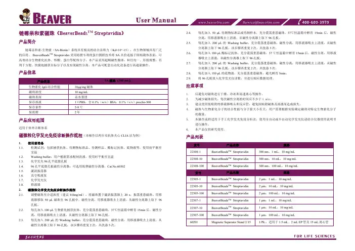
链霉亲和素磁珠(BeaverBeads TM Streptavidin )产品简介链霉亲和素-生物素(SA-Biotin )系统具有极高的结合亲和力(Kd=10^-15),在生物领域具有广泛的应用。
BeaverBeads TM Streptavidin 采用海狸专利的蛋白偶联技术将SA 共价连接于固相载体表面,可高效结合生物素化抗体、核酸、蛋白等配体分子。
本产品采用超顺磁性微球,粒径均一、形貌规整,有利于方便、快捷地捕获目标分子以及实现磁性分离。
本产品可配套自动化设备进行高通量操作。
产品信息产品信息 SA 磁珠(300 nm )生物素化IgG 结合性能 20μg/mg 磁珠 磁珠浓度 10 mg/mL 磁珠表面 亲水基团保存溶液 1×PBS ,含0.1%(w/v )BSA ,0.1%(v/v )proclin-300 保存条件 2-8 ℃ 保质期2年产品应用范围适用于体外诊断体系磁微粒化学发光免疫诊断操作流程(本操作以两步双抗体夹心CLIA 法为例)1. 使用前准备1.1. 检测试剂:包括捕获抗体、待测物标准品、待测样品、酶标记抗体、底物液等,使用前平衡至室温1.2. Washing buffer :用户根据需求配制洗液,使用时平衡至室温 1.3. 化学发光96孔平底微孔板1.4. 96孔平底微孔板磁性分离器:可选用海狸磁性分离器,Cat.No.60302 1.5. 漩涡振荡器 1.6. 真空吸液泵 1.7. 化学发光仪 1.8. 移液器2. 磁微粒化学发光免疫诊断操作流程2.1.调整磁珠至合适浓度(建议0.8mg/ml ),将磁珠置于漩涡振荡器上20 s ,振荡重悬磁珠。
用移液器移取50 μL 磁珠至96孔板中,磁性分离,用移液器吸去上清液,从磁性分离器上取下96孔板。
2.2. 每孔加入100 μL 生物素化捕获抗体,充分震荡重悬磁珠,37℃恒温箱中孵育15min 后,磁性分离,用移液器吸去上清液,从磁性分离器上取下96孔板。
flag磁珠说明书Anti-Flag磁珠,也称Anti-Flag免疫磁珠或Flag抗体磁珠,是由高品质的Flag 小鼠单克隆抗体与纳米级氨基磁珠共价偶联而成,可特异性地与动植物或微生物裂解液、血清、腹水等中含有Flag标签的蛋白结合,从而用于带有Flag标签的融合蛋白或其蛋白复合物的免疫沉淀(Immunoprecipitation, IP)、免疫共沉淀(Co-IP)或纯化。
Flag标签(Flag-tag)、Myc标签(Myc-tag)、HA标签(HA-tag)、His标签(His-tag)和GST标签(GST-tag)等是表达载体上最常见的一些标签,通过与这些标签的融合表达可以非常方便地检测目的蛋白及与目的蛋白相互结合的蛋白,也可以非常方便地用于目的蛋白的纯化。
Flag-tag是8个氨基酸残基(DYKDDDDK)组成的多肽,常用的形式有Flag和3X Flag,通过基因重组技术把Flag-tag的核酸序列与目的基因的5’端或3’端连接,就可以最终表达形成Flag-tag的目的蛋白。
Flag-tag具有以下优点:Flag-tag通常不会与目的蛋白相互作用,并且大多数情况下不会影响目的蛋白的功能;Flag-tag作为标签蛋白,后续通过Flag抗体(AF519)、Anti-Flag磁珠或Anti-Flag亲和凝胶(P2271)即可对目的基因的表达、定位及功能进行检测或对目的蛋白进行纯化、免疫沉淀或免疫共沉淀等;融合在N端的Flag标签,可被肠激酶(Enterokinase, EK)切除(DDDK),从而得到完美的没有标签的目的蛋白。
基于以上优点,Flag标签已被广泛应用于蛋白表达、纯化、鉴定、相互作用和功能等多方面的研究。
Anti-Flag Magnetic Beads (Anti-Flag磁珠),也被称为Anti-DYKDDDDK Magnetic Beads,可以特异性地结合Flag标签融合蛋白,并可以借助磁力架等磁分离设备非常便捷地应用于带有Flag标签的融合蛋白或其蛋白复合物的免疫沉淀或纯化等实验。
免疫共沉淀磁珠法-概述说明以及解释1.引言1.1 概述概述部分可以简要介绍免疫共沉淀磁珠法的定义和原理。
免疫共沉淀磁珠法是一种利用磁珠载体对特定抗原/抗体进行选择性结合的技术,通过磁场的作用实现对特定分子的快速分离纯化。
这种方法在生物学、医学和生物化学等领域有着广泛的应用,并且具有操作简便、效率高、成本低的优势。
在接下来的文章内容中,我们将对免疫共沉淀磁珠法的方法介绍、应用领域以及优势和局限性进行详细阐述。
1.2 文章结构文章结构部分的内容可以包括对整篇文章的结构和内容进行简要介绍,让读者对文章有一个整体的了解。
可以包括文章的各个章节内容和重点,以及各个部分之间的逻辑关系和连接。
此部分可以是对整篇文章的概述和导读,帮助读者更好地理解和阅读整篇文章。
部分的内容1.3 目的本文旨在介绍免疫共沉淀磁珠法在生物学和医学领域的应用和发展现状。
通过对该方法的详细介绍和分析,旨在帮助读者了解其原理、操作步骤和相关领域的实际应用。
同时,通过评估该方法的优势和局限性,旨在为研究人员提供参考,以便更好地应用和改进该方法。
最终目的是促进免疫共沉淀磁珠法在生物医学研究和临床诊断中的应用,并为未来的研究方向和方法改进提供启示。
2.正文2.1 方法介绍免疫共沉淀磁珠法是一种利用磁珠载体进行免疫共沉淀的方法,在生物医学领域有着广泛的应用。
该方法可以分为直接法和间接法两种。
直接法是指将特异性抗体直接连接到磁珠表面,然后将混合物中的抗原与抗体结合,再利用外加磁场将磁珠与非特异性物质分离。
该方法操作简单,灵敏度高,适用于大多数免疫学试验。
间接法是指首先将特异性抗体连接到磁珠表面,然后将待检测样品中的抗原与抗体结合,再加入第二抗体结合抗原,最后利用外加磁场将磁珠与非特异性物质分离。
该方法对于多肽和蛋白质分析有着较高的灵敏度和特异性。
免疫共沉淀磁珠法具有操作简便、试验时间短、结果准确等优点,同时也存在着磁珠分离效率与特异性抗体结合的选择性等局限性。
AMMS TM CD3/CD28抗体偶联磁珠说明书产品名称通用名称:CD3/CD28抗体偶联磁珠英文名称:Anti-human CD3/CD28 monoclonal antibody beads适用范围AMMS TM CD3/CD28磁珠适用于人T细胞的分离,激活和扩增。
适用于直肠癌、乳腺癌、肺癌、肾癌、淋巴瘤、白血病、多发性骨髓瘤、恶性黑色素瘤、卵巢癌等多种肿瘤的细胞免疫治疗临床研究。
AMMS CD3/CD28磁珠提供了一种不需要抗原呈递细胞和抗原就能激活和扩增调节T细胞的简单的方法。
通过在磁珠上组合抗CD3和抗CD28抗体,就能提供调节T细胞激活和扩增需要的初级和协同刺激信号。
使用说明一、AMMS TM CD3/CD28磁珠的清洗:1.1 在小管里重悬磁珠(也就是说,涡旋超过30s,或者颠倒混匀5min)1.2 将确定体积的磁珠转移到管子中。
1.3 加入等体积的PBS缓冲液含1%的HAS,或者至少1ml的体积,进行重悬。
1.4把管子放在一个磁铁上1min,随后弃去上清。
1.5将管子从磁铁上转移下来,用相同体积的PBS缓冲液含1%的HAS重悬磁珠(第二步最初体积的磁珠)。
二、磁珠分离和扩增CD3+的T细胞注意:由于单核细胞在37℃能够快速的吞噬磁珠,因此就降低了所能够接触到T细胞磁珠的绝对数量,进而降低了T细胞活化和扩增的能力。
2.1 通过流式或者是其他的方法确定样本中CD3+T细胞的比例。
2.2 对于Ficoll分离的PBMC,将细胞轻柔的重悬于含1%HAS的PBS缓冲液中,并调节细胞密度为2-5×10⁷个/mL,此处注意,总的细胞数量不要超过2×108个。
2.3 将磁珠和细胞比例为3:1,加入已经洗过的AMMS TM CD3/CD28磁珠。
2.4 将样品置于1转的摇床上,在4℃-25℃条件下孵育30min,在每次的试验中需要在4℃-25℃范围内优化最适的孵育温度。
2.5用无血清培养基或者含1%HAS的PBS缓冲液稀释磁珠和细胞的混合物,来保证磁铁的分选体积。

免疫磁珠链霉亲和素磁珠(免疫捕捉、纯化)100~500nm免疫磁珠 |链霉亲和素磁珠(免疫捕捉、纯化) 100~500nm关键词:免疫磁珠、链霉亲和素磁珠(免疫捕捉、纯化) 100~500nm免疫磁珠、链霉亲和素磁珠 (免疫捕捉、纯化) 1umSera-Mag™ 链霉亲和素磁珠和 SpeedBeads链霉亲和素磁珠可用于捕获生物素化靶分子,进行快速和可靠的蛋白质组学分析。
Sera-Mag™ and Sera-mag SpeedBeads 链霉亲和素磁珠,对于生物素化靶分子显示出较高亲和力和灵敏度,具有快速反应动力学,从而可以提高基因组学和蛋白质组学应用的通量和精密度。
性质:具有较低(2500 至3500 pmol/mg)、中等(3500 至4500 pmol/mg)或较高(4500 至 5500 pmol/mg)生物素结合载量,优化测定开发较低解离常数确保紧密的配基结合较低非特异性结合可实现更高的精密度和准确度结合了大比表面积、高灵敏度、物理性能稳定性和快速反应动力学的特点均匀的链霉亲和素涂层可获得可靠结果粒径均一,1um 的粒径可提供较大比表面积和出色的批次间重现性。
Sera-Mag™ SpeedBeads 和Sera-Mag™ 链霉亲和素磁珠,不仅有快速反应动力学,而且有较低的非特异性结合,可提高免疫测定和分子生物学应用(如用于基因组学和蛋白质组学的样品制备和测定开发)中的通量和精密度。
【成分】亲和素包覆的磁性氧化铁纳米球,含1%BSA的硼酸缓冲液【性状】黑褐色悬液,长时间静置可分层【特点与用途】链霉亲和素磁珠由超顺磁性微球与高纯度链霉亲和素共价结合而成。
链霉亲和素-生物素(SA-Biotin)系统具有极高的结合亲和力(Kd=10^-15),在生物领域具有广泛的应用。
本产品采用超顺磁性微球,粒径均一、形貌规整,有利于方便、快捷地捕获目标分子以及实现磁性分离。
【基本使用方法】在室温条件下,将生物素化抗体与链酶亲和素磁性微球混合,反应时间30~40min,制备成磁性微球探针;将制备好的磁性微球探针与带检测分离的样品混合,再用磁力架进行磁性分离清洗。
说明书Pierce™交联磁珠式免疫沉淀/免疫共沉淀试剂盒888052309.4货号描述88805 Pierce交联磁珠式免疫沉淀/免疫共沉淀试剂盒,包含足够完成40个反应的试剂,每个反应使用25 µL磁珠试剂盒组成:Pierce 蛋白 A/G 磁珠1 mL,贮存于含有 0.05% NaN3的水中,浓度为10 mg/mLPierce 免疫沉淀裂解/漂洗缓冲液,2 × 50 mL,pH 7.4,0.025M Tris,0.15M NaCl,0.001M EDTA,1% NP40,5% 甘油20×交联缓冲液,25 mL, 稀释后含10 mM 磷酸钠, 150 mM NaCl;pH 7.2DSS(辛二酸二琥珀酰亚胺酯),No-Weigh™形式, 8 × 2 mg 微型管中和液,1.0 mL,pH 8.5洗脱液,25 mL,pH 2.0电泳上样缓冲液,非还原性,(5×), 5 mL,pH 6.8,0.3M Tris·HCl,5% SDS,50%甘油,泳道标记示踪染料贮存条件:收到本产品后于4°C贮存。
冰盒运输。
目录产品简介 (2)流程简述 (2)重要产品信息 (2)所需额外试剂和仪器 (3)Pierce经典磁珠式免疫沉淀试剂盒流程 (3)A. 将抗体结合于蛋白A/G磁珠上 (3)B. 交联结合的抗体 (4)C.哺乳动物细胞裂解 (4)D. 手动抗原免疫沉淀 (5)E. 自动免疫沉淀 (5)问题解决 (7)网站附加信息 (7)Thermo Scientific KingFisher 仪器的常见问题 (8)Thermo Scientific相关产品 (8)产品简介Thermo Scientific™ Pierce™ 交联磁珠式免疫沉淀/免疫共沉淀试剂盒通过共价键将抗体交联于Thermo Scientific™ Pierce™ 蛋白 A/G磁珠上能够高效完成抗原免疫沉淀(IP)及免疫共沉淀(Co-IP)实验。
德国美天旎Miltenyi细胞分选磁珠CD90(Thy1.2)MicroBeads小鼠CD90(Thy1.2)微珠用于从淋巴和非淋巴组织单细胞悬液或外周血中分选或去除小鼠T细胞。
CD90表达于胸腺细胞、外周T细胞、一些上皮内T 细胞上,在骨髓早期造血干细胞上有低水平表达。
在小鼠中CD90识别的T细胞群与表达CD3的细胞群基本相同。
应用:CD90微珠分选的T细胞可用于分析小鼠肺纤维化模型中细胞因子的表达,或者将野生型和基因敲除小鼠的T细胞过继性转移至免疫缺陷小鼠体内,研究其细胞因子的表达。
另外,CD90微珠还可用于分选肿瘤浸润性T细胞,研究其免疫治疗潜能或者功能特性。
分选柱:阳性分选:MS、LS、XS或autoMACSTM分选柱。
去除分选:LD、CS、D或autoMACS分选柱。
有关磁珠的知识:MACS磁珠是一种与高度特异性单克隆抗体相偶联的超顺磁化微粒,用于目的细胞或者去除细胞的磁性标记。
磁珠直径约有50nm,比细胞小200多倍,体积为细胞的百万分之一,光学显微镜下不可见。
磁珠由多聚糖和氧化铁组成,无毒性,对细胞无损伤,可以生物降解。
MACS微珠与流式细胞仪兼容,不会影响细胞的光散射特性;磁性标记只占用20-30%的结合位点,不影响细胞的荧光抗体标记。
此外,MACS磁珠可以最大限度地避免细胞活化;无需解离磁珠,可以直接进行后续实验:如流式细胞仪分析或分选、细胞培养、分子生物学研究、回输给人或者动物。
MACS磁珠主要有三种:直标磁珠、间标磁珠、多选磁珠。
其中间标磁珠有抗免疫球蛋白磁珠、抗生物素磁珠或链霉亲和素磁珠、抗荧光素磁珠。
多选磁珠是专门为分选细胞亚群而研制的一种磁珠。
这种磁珠通过特殊的方式与抗体偶联,在第一次阳性分选完成后,与细胞结合的多选磁珠可以被解离试剂剪切下来,阳性分选的细胞可以进行再次阳性分选或者去除分选。
Anti-FLAG(DYKDDDDK)免疫磁珠Cat#:MIP0064(本品仅用于科学研究,不可用于诊断及治疗)一.背景信息Flag-Tag(DYKDDDDK),常用于真核蛋白质重组表达标记。
Anti-FLAG(DYKDDDDK)免疫磁珠,由高品质的FLAG抗体与磁珠共价偶联制成, 具有高载量(至少为0.6mg protein/ml磁珠),高特异性,操作迅速便捷,性质稳定,可反复使用的特点,可用于FLAG标签融合蛋白的亲和纯化及免疫(共)沉淀。
二.性能指标应用范围:可用于Met修饰的N端FLAG融合蛋白(Met-FLAG–Protein),N端FLAG融合蛋白(FLAG–Protein)和C端FLAG融合蛋白(Protein-FLAG)的亲和纯化及免疫(共)沉淀。
抗体属性:小鼠单克隆抗体,IgG2a亚型。
磁珠属性:琼脂糖包裹的超顺磁珠,平均粒径50um。
载量:0.5mL纳米磁珠,共价偶联2mg Anti-FLAG单克隆抗体,可纯化或沉淀至少0.6mg FLAG融合蛋白。
强度:竞争洗脱,可反复使用5次以上。
成分:0.5mL共价偶联Anti-FLAG单克隆抗体的磁珠, 溶于1ml PBS中。
保存方法:在加入了0.2‰叠氮钠的PBS保存液中,4℃可保存1年。
三.说明书阅读指南四.用前必读1.以下试剂及设备,需要客户自备,试剂推荐配方见说明书附件1, 推荐蛋白质制备方案见附件2。
磁力架1个,1.5mL 离心管若干细胞裂解液,酸性洗脱液,中和液,PBS,2x蛋白上样缓冲液, 3xFLAG多肽(货号Q7007)待测抗原样品及检测抗体2.注意事项(开箱前必读)1)本产品在在加入了0.2‰叠氮钠的PBS保存液中,4℃可保存1年,冷藏条件下运输。
2)勿离心、冷冻、干燥磁珠,勿使用超声处理磁珠,勿使酸处理磁珠时间超过10min。
3)混匀磁珠时,请采用移液枪轻柔吹打,柔和涡旋,上下颠倒及摇床混匀等方法。
勿使用超声等方法。
版本2022/09/08(02)
链霉亲和素纳米磁珠(100-300nm )说明书1/1链霉亲和素纳米磁珠(100-300nm )产品说明书
【产品名称】链霉亲和素纳米磁珠(100-300nm )
【英文名称】Streptavidin Nano Magnetic Beads (100-300nm)
【订货信息】
纳米磁珠/纯水【简介】
东纳生物科技有限公司提供链霉亲和素纳米磁珠(100-300nm ),链霉亲和素磁珠可与生物素化的多肽、蛋白、抗体等生物配体高效偶联,应用于免疫分析、基因分析和纯化PCR 产物等。
【产品参数】
图1:100nm 链霉亲和素磁珠扫描电镜(左图,100±10nm )、200nm 链霉亲和素磁珠扫描电镜(中图,
220±30nm )以及300nm 链霉亲和素磁珠扫描电镜(右图,310±20nm )。
【注意事项】
1.磁珠取用前应充分混匀,防止取用改变磁珠浓度,避免长时间超声对磁珠表面破坏;
2.磁珠取用后,使用前请进行磁分离并用纯水或所用缓冲溶液清洗2-3遍;
3.磁珠使用和保存过程中应避免冻融。
【生产单位】
公司名称
南京东纳生物科技有限公司地址
南京市江宁区龙眠大道568号南京生命科技小镇5号楼北楼6楼邮政编码
210000电话号码
025********电子邮箱
**************公司网站。
免疫磁珠制备1. 简介免疫磁珠是一种常用的实验工具,用于从混合物中高效地富集、分离和纯化目标分子。
它主要由磁性颗粒和特异性抗体构成,能够通过特异性的抗原-抗体相互作用捕获目标分子,并通过磁力快速分离。
本文将详细介绍免疫磁珠制备的步骤和相关注意事项,帮助读者全面了解免疫磁珠制备的过程。
2. 免疫磁珠制备步骤2.1 材料准备在进行免疫磁珠制备之前,需要准备以下材料:•磁性颗粒:选择合适尺寸和表面修饰的磁性颗粒,如Fe3O4或CoFe2O4等。
•活化剂:例如EDC (1-ethyl-3-(3-dimethylaminopropyl) carbodiimide) 和 NHS (N-hydroxysuccinimide),用于激活颗粒表面上的羧基。
•抗体:选择与目标分子特异性结合的抗体。
2.2 磁性颗粒修饰2.2.1 活化剂激活将适量的EDC和NHS加入磁性颗粒悬浮液中,使其浓度分别达到一定比例(通常为10-50 mM)。
将悬浮液在室温下轻轻摇动或搅拌,保持反应30分钟至1小时。
2.2.2 抗体偶联将抗体加入到活化剂激活的磁性颗粒悬浮液中,使其与磁性颗粒表面上的活化剂反应。
反应时间通常为1-4小时,温度可以根据实验需求在室温或4°C下进行。
反应结束后,通过磁力分离将未偶联的抗体和其他杂质去除。
2.3 免疫磁珠包装与保存将制备好的免疫磁珠用适当的缓冲液洗涤,并调整至合适的浓度。
接着,可以选择将免疫磁珠包装成小管装进行保存,或直接使用于实验。
3. 注意事项在免疫磁珠制备过程中,需要注意以下几点:3.1 杂质去除在制备免疫磁珠的过程中,必须确保将未偶联的抗体、活化剂和其他杂质充分去除,以避免对实验结果产生干扰。
3.2 存储条件制备好的免疫磁珠应保存在适当的温度和缓冲液条件下,避免受潮、受热或冷冻等不利因素。
同时,注意避免与金属物质接触,以防止颗粒表面氧化。
3.3 实验设计在使用免疫磁珠进行实验时,需要根据具体实验目的和样品特性进行合理的实验设计。
志贺氏菌属免疫磁球分离检测试剂说明书【产品名称】通用名称:志贺氏菌属免疫磁球分离检测试剂英文名称:Immunomagnetic separation (IMS) regent anti-Shigella【用途】用于食品、饮用水等环境样品中已增菌的志贺氏菌的快速分离检测。
【检验原理】免疫磁球表面偶联有志贺氏菌特异抗体,可与样本中的志贺氏菌表面抗原进行特异性结合,带有靶物质的磁球在磁场作用下使目标菌与样本中的其它物质快速分离,分离纯度高,保留靶物质活性,从而达到分离检测志贺氏菌的目的。
【产品特征】志贺氏菌免疫磁球,粒径180nm左右,为小粒径的纳米免疫微球,由于具有更好的溶液稳定性,表现出超高的微生物捕获效率。
【储藏条件】志贺氏菌免疫磁球产品请置于2~8℃保存。
【适用仪器】磁力分选装置(可使用磁铁或磁板、或选择购买磁力架)【使用方法】1、待检样品准备,进行10-12h的增菌。
2、取一支无菌2mL离心管,加入1mL增菌液。
3、取出志贺氏菌免疫磁球,在漩涡振荡器上振荡5s至磁球全部重悬。
4、吸取20μL志贺氏菌免疫磁球加入到含有1mL增菌液的离心管中,颠倒混合后将离心管放在摇床中,震荡混合30min。
5、将加有磁球的样品管放在磁力分选装置上,静置15min,小心吸走管中的液体。
6、取1ml 1×PBS溶液洗涤磁球,静置3min,小心吸走多余液体。
重复洗涤两次,最后加入1mL洗脱缓冲液重悬磁球。
7、将磁球全部吸出在选择性培养基上涂布,37℃培养箱培养18-24h。
8、观察选择性培养基上的菌落生长情况。
【注意事项】1.请严格按照说明书操作,避免样品交叉污染,所有实验步骤均需戴手套。
2.在15-25℃室温操作磁分离实验,所有的试剂在使用前请恢复到室温。
3.使用前应保证磁球充分悬浮在溶液中。
4.对于富含油脂或粘滞的特殊样品,需在增菌后24h用1×PBS 稀释一定倍数才能作为免疫磁分离的待检样品。
Contents1. Description1.1 Principle of the MACS® Separation1.2 Background information1.3 Applications1.4 Reagent and instrument requirements试剂和仪器的要求2. Protocol2.1 Sample preparation样品制备2.2 Magnetic labeling磁性标记2.3 Magnetic separation磁性分离3. Example of a separation using the CD133 MicroBead Kit4. References1. DescriptionComponents 2 mL CD133 MicroBeads, human:MicroBeads conjugated to monoclonal antihumanCD133 antibodies (isotype: mouse IgG1,clone AC133).2 mL FcR Blocking Reagent, human Specificity CD133 antigen, epitope (CD133/1)1.Capacity For 2亊10⁹total cells, up to 100 separations.Product format CD133 MicroBeads are supplied in buffercontaining stabilizer and 0.05% sodium azide.Storage Store protected from light at 2−8 °C. Do not freeze. The expiration date is indicated on the vial label.1.1 Principle of the MACS® SeparationFirst, the CD133+ cells are magnetically labeled with CD133MicroBeads. Then, the cell suspension is loaded onto a MACS®Column, which is placed in the magnetic field of a MACS Separator.The magnetically labeled CD133+ cells are retained within thecolumn. The unlabeled cells run through; this cell fraction isthus depleted of CD133+ cells. After removing the column fromthe magnetic field, the magnetically retained CD133+ cells can beeluted as the positively selected cell fraction. To increase the purity,the positively selected cell fraction containing the CD133+ cells isseparated over a second column.1.2 Background informationThe CD133 MicroBead Kit is a magnetic labeling system designedfor the positive selection of CD133+ cells. It allows the single-stepisolation of nonhematopoietic and early hematopoietic progenitors and stem cells. The CD133 molecule is a 5-transmembrane cellsurface antigen with a molecular weight of 117 kD.2 The CD133/1(clone AC133) antibody recognizes epitope 1 of the CD133 antigen.1In the hematopoietic system, CD133 expression is restricted to asubset of CD34bright stem and progenitor cells in human fetal liver,bone marrow, cord blood, and peripheral blood.3 Isolated fromhematopoietic sources, CD133+ cells can become adherent and arereported to become CD133–during culture⁴. The CD34+CD133+cell population, which includes CD34+CD38–cells, was shown tobe capable of repopulating NOD/SCID mice.⁵Recently, CD133has also been found to be expressed on circulating endothelialprogenitor cells⁶,⁷and fetal neural stem cells⁸,⁹as well as on othertissue-specific stem cells, such as renal1⁰and prostate11 stem cells.Lately, when isolated from tumor tissue, the CD133+ populationcan be enriched for tumor-initiating cells.11-1⁵CD133 MicroBeadshave been used to isolate adult stem cells from cord blood and asa starting population for reprograming towards iPS cells.1⁶CD133expression has been found on undifferentiated human ES cells.Therefore CD133 MicroBeads could be used for enrichment ordepletion of these cells.1⁷1.3 Applications●Positive selection or depletion of cells expressing human CD133antigen.●Isolation or depletion of CD133+ cells from peripheral bloodmononuclear cells (PBMCs) or single-cell suspensions fromtissue.●CD133+ cells are used in basic stem cell research, stem cellevaluation, stem cell expansion, research in hematologicalmalignancies, stem cell plasticity, and potential cellulartherapies as well as in tissue regeneration and cancer research.1.4 Reagent and instrument requirements●Buffer: Prepare a solution containing phosphate-buffered saline(PBS), pH 7.2, 0.5% bovine serum albumin (BSA), and 2 mMEDTA by diluting MACS BSA Stock Solution (# 130‑091-376)1:20 with autoMACS Rinsing Solution (# 130-091-222). Keepbuffer cold (2−8 °C). Degas buffer before use, as air bubblescould block the column.▲Note: EDTA can be replaced by other supplements such as anticoagulant citrate dextrose formula-A (ACD-A) or citrate phosphate dextrose (CPD). BSA can be replaced by other proteins such as human serum albumin, human serum, or fetal bovine serum (FBS). Buffers or media containing Ca2+ or Mg2+ are not recommended for use.●(Optional) Fluorochrome-conjugated antibodies forflow cytometric analysis, e.g., CD133/2 (293C3)-PE(# 130-090-853), CD133/2 (293C3)-APC (# 130-090-854),CD133/2 (293C3)‑Biotin, CD34-FITC (# 130-081-001),CD34‑APC (# 130-090-954), or CD34-PE (# 130-081-002). Formore information about fluorochrome-conjugated antibodiessee .●MACS Columns and MACS Separators: CD133+ cells can beEnriched浓缩by using MS, LS, or XS Columns or depleted withthe use of LD, CS, or D Columns. Cells which strongly expressthe CD133 antigen can also be depleted using MS, LS, or XSColumns. Positive selection正向or depletion can also be performedby using the autoMACS Pro or the autoMACS Separator.▲Note: Column adapters are required to insert certain columns into the VarioMACS™or SuperMACS™Separators. For details see the respective MACS Separator data sheet.●(Optional) Propidium Iodide Solution (# 130-093-233) or7-AAD for flow cytometric exclusion of dead cells.●(Optional) Dead Cell Removal Kit (# 130-090-101) for thedepletion of dead cells.●(Optional) Pre-Separation Filters (# 130-041-407) to removecell clumps团.2. Protocol2.1 Sample preparationWhen working with anticoagulated peripheral blood or buffy coat,peripheral blood mononuclear cells (PBMCs) should be isolated bydensity gradient centrifugation, for example, using Ficoll-Paque™.▲Note: To remove platelets after density gradient separation, resuspend cell pellet in buffer and centrifuge at 200×g for 10−15 minutes at 20 °C. Carefully aspirate supernatant. Repeat washing step.When working with tissues or lysed blood, prepare a single-cellsuspension using standard methods.For details see the protocols section at /protocols.For preparation of cord blood cells, bone marrow cells, or cellsfrom leukapheresis material, please refer to the sample preparationprotocols at /protocols.▲Dead cells may bind non-specifically to MACS MicroBeads.To remove dead cells, we recommend using density gradientcentrifugation or the Dead Cell Removal Kit (# 130-090-101).2.2 Magnetic labeling▲Work fast, keep cells cold, and use pre-cooled solutions. This willprevent capping of antibodies on the cell surface and non-specificcell labeling.▲V olumes for magnetic labeling given below are for up to10⁸total cells. When working with fewer than 10⁸cells, use the same volumes as indicated. When working with higher cell numbers,scale up all reagent volumes and total volumes accordingly (e.g.for 2亊10⁸total cells, use twice the volume of all indicated reagent volumes and total volumes).▲For optimal performance it is important to obtain a single‑cell suspension before magnetic labeling. Pass cells through 30 μm nylonmesh (Pre-Separation Filters, # 130-041-407) to remove cell clumpswhich may clog the column. Moisten filter with buffer before use.▲The recommended incubation temperature is 2–8 °C. Workingon ice may require increased incubation times. Higher temperaturesand/or longer incubation times may lead to non-specific celllabeling.1. Determine确定cell number.2. Centrifuge cell suspension细胞悬液at 300×g for 10 minutes. Aspirate supernatant completely.吸取上清3. Resuspend cell pellet in 300 μL of buffer per 10⁸total cells.4. Add 100 μL of FcR Blocking Reagent per 10⁸total cells.5. Add 100 μL of CD133 MicroBeads per 10⁸total cells.6. Mix well and incubate for 30 minutes in the refrigerator(2−8 °C).7. (Optional) Add staining antibodies, e.g., 50 μL of CD133/2(293C3)-PE (# 130-090-853), and incubate for 5 minutes in thedark in the refrigerator (2−8 °C).8. Wash cells by adding 1−2 mL of buffer per 10⁸cells andcentrifuge at 300×g for 10 minutes. Aspirate supernatantcompletely.9. Resuspend up to 10⁸cells in 500 μL of buffer.▲Note: For higher cell numbers, scale up buffer volume accordingly.▲Note: For depletion with LD Columns, resuspend up to 1.25亊10⁸cells in 500 μL of buffer.10. Proceed to magnetic separation (2.3).2.3 Magnetic separation▲Choose an appropriate MACS Column and MACS Separatoraccording to the number of total cells and the number of CD133+cells. For details see table in section 1.4.▲Always wait until the column reservoir is empty before proceedingto the next step.Magnetic separation with MS or LS Columns▲To achieve highest purities, perform two consecutive columnruns.1. Place column in the magnetic field of a suitable MACS Separator.For details see the respective MACS Column data sheet.2. Prepare column by rinsing with the appropriate amount ofbuffer:MS: 500 μL LS: 3 mL3. Apply cell suspension onto the column. Collect flow-throughcontaining unlabeled cells.4. Wash column with the appropriate amount of buffer. Collectunlabeled cells that pass through and combine with the effluentfrom step 3.MS: 3×500 μL LS: 3×3 mL▲Note: Perform washing steps by adding buffer aliquots only when the column reservoir is empty.5. Remove column from the separator and place it on a suitablecollection tube.▲Note: To perform a second column run, you may elute the cells directly from the first onto the second, equilibrated column instead of a collection tube.6. Pipette the appropriate amount of buffer onto the column.Immediately flush out the magnetically labeled cells by firmlypushing the plunger into the column.MS: 1 mL LS: 5 mL7. To increase purity of CD133+ cells, enrich the eluted fractionover a second MS or LS Column. Repeat the magneticseparation procedure as described in steps 1 to 6 by using anew column.Magnetic separation with XS ColumnsFor instructions on the column assembly and the separation refer tothe XS Column data sheet.Depletion with LD Columns1. Place LD Column in the magnetic field of a suitable MACSSeparator. For details see LD Column data sheet.2. Prepare column by rinsing with 2 mL of buffer.3. Apply cell suspension onto the column.4. Collect unlabeled cells that pass through and washcolumn with 2×1 mL of buffer. Collect total effluent;this is the unlabeled cell fraction. Perform washingsteps by adding buffer two times. Only add new buffer whenthe column reservoir is empty.Depletion with CS Columns1. Assemble CS Column and place it in the magnetic field of asuitable MACS Separator. For details see CS Column datasheet.2. Prepare column by filling and rinsing with 60 mL of buffer.Attach a 22G flow resistor to the 3-way stopcock of theassembled column. For details see CS Column data sheet.3. Apply cell suspension onto the column.4. Collect unlabeled cells that pass through and wash columnwith 30 mL buffer from the top. Collect total effluent; this isthe unlabeled cell fraction.Depletion with D ColumnsFor instructions on column assembly and separation refer to theD Column data sheet.Magnetic separation with the autoMACS® Pro Separator or theautoMACS® Separator▲Refer to the respective user manual for instructions on how to use the autoMACS Pro Separator or the autoMACS Separator.▲Buffers used for operating the autoMACS Pro Separator or the autoMACS Separator should have a temperature of ≥10 °C.▲Program choice depends on the isolation strategy, the strengthof magnetic labeling, and the frequency of magnetically labeled cells. For details refer to the section describing the cell separation programs in the respective user manual.Magnetic separation with the autoMACS® Pro Separator1. Prepare and prime the instrument.2. Apply tube containing the sample and provide tubes for collecting the labeled and unlabeled cell fractions. Placesample tube in row A of the tube rack and the fractioncollection tubes in rows B and C.3. For a standard separation choose one of the following programs:Positive selection from peripheral blood, bone marrow, or leukapheresis: “Posseld”Collect positive fraction in row C of the tube rack.Positive selection from cord blood: “Posseld2”Collect positive fraction in row C of the tube rack.Depletion: “Depletes”Collect negative fraction in row B of the tube rack.Magnetic separation with the autoMACS® Separator1. Prepare and prime the instrument.2. Apply tube containing the sample and provide tubes for collecting the labeled and unlabeled cell fractions. Placesample tube at the uptake port and the fraction collectiontubes at port neg1 and port pos 2.3. For a standard separation choose one of the following programs:Positive selection from peripheral blood, bone marrow, or leukapheresis: “Posseld”Collect positive fraction from outlet port pos 2.Positive selection from cord blood: “Posseld2”Collect positive fraction from outlet port pos 2.Depletion: “Depletes”Collect negative fraction from outlet port neg1.3. Example of a separation using the CD133MicroBead KitCD133+ hematopoietic stem and progenitor cells were isolated from non-mobilized human PBMCs using the CD133 MicroBead Kit, MS Columns, and a MiniMACS™Separator. Cells were fluorescently stained with CD34-FITC (# 130-081-001) and CD133/2 (293C3)-PE (# 130-090-853) and analyzed by flow cytometry. Cell debris and dead cells werde excluded from the analysis based on scatter signals and propidium iodide fluorescence.All protocols and data sheets are available at .WarningsReagents contain sodium azide. Under acidic conditions sodium azide yields hydrazoic acid, which is extremely toxic. Azide compounds should be diluted with running water before discarding. These precautions are recommended to avoid deposits in plumbing where explosive conditions may develop.WarrantyThe products sold hereunder are warranted only to be free from defects in workmanship and material at the time of delivery to the customer. Miltenyi Biotec GmbHmakes no warranty or representation, either expressed or implied, with respect tothe fitness of a product for a particular purpose. There are no warranties, expressedor implied, which extend beyond the technical specifications of the products.Miltenyi Biotec GmbH’s liability is limited to either replacement of the products or refund of the purchase price. Miltenyi Biotec GmbH is not liable for any property damage, personal injury or economic loss caused by the product.autoMACS and MACS are registered trademarks and MidiMACS, MiniMACS, OctoMACS, QuadroMACS, SuperMACS, and VarioMACS are trademarks of Miltenyi Biotec GmbH.Ficoll-Paque is a trademark of GE Healthcare companies.Copyright . 2009 Miltenyi Biotec GmbH. All rights reserved.。