病理学英文课件:呼吸系统疾病
- 格式:pptx
- 大小:70.20 MB
- 文档页数:174
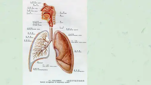
Diseases of Respiratory System9.1.1 Acute tracheobronchitisAcute catarrhal tracheobronchitis.The inflammatory exudate on the mucosal surface is chiefly a stringy,basophilic mucus only scantily mixed with leukocytes. Acute suppurative tracheobronchitis.There is a significant element of leukocytic infiltration.Acute ulcerative tracheobronchitis.The inflammatory reaction is more intense, with necrosis of the mucosa inareas, it constitutes an ulcerative form.9.1.2 Acute bronchiolitisThe bronchioli mucosa is hyperemia and swelling with a lymphomonocytic and leukocyticinfiltration of the submucosa accompanied by overproduction of mucous secretions. Bronchiolitis obliterans is characterized bypolypoid masses of organizing inflammatoryexudates and granulation tissue extendingfrom alveoli into bronchioles1. Bacteria pneumonia (1) Lobar pneumoniaan acute bacterial infection of alarge portion of a lobe or of an entire lobe•fibrinous inflammation •Symptoms:abrupt onset,high fever,shaking chills,pleuritic chest pain,a productive mucopurulent cough(“rusty”sputum)congestion stage(2)red hepatization:3rd-4th days gross●the lobe distinctly red, firm, and airless with a liver-like consistency●an overlying fibrinous or fibrinosuppurative pleuritisLMalveolar space: a flock of RBC, packed with fibrin nets which stream from one alveolus through the pores of kohn into adjacent alveoli, neutrophilsLobar pneumonia (gray hepatization). The upper lobe is uniformly consolidated.Lobar pneumonia(gray hepatization).Lobar pneumonia (resolution stage) exudates within the alveoli are enzymatically digested and either resorbed or expectorated, leaving the basic architecture intact.Lobar pneumonia(carnification)(2) Lobular pneumonia( Bronchopneumonia )●clinic: infants, the aged, illness (much more prevalent at the extremes of age)●patchy distribution,a purulent inflammation that centered bronchiolesEtiology and pathogenesisPathogens: staphylococci, pneumococci, streptococci, influenzae haemophilusInduce factors: cold, heart failureInfection ways: respiratory tract, bloodLobular pneumonia (Scattered foci of consolidation are centered on bronchi and bronchioles)Foci of inflammatory consolidation are distributed in patches through one or several lobes, in severe cases the foci may confluent, producing the appearance of a lobar consolidationlobular pneumonia (A suppurative, neutrophil-rich exudates fills the bronchi, bronchioles, and adjacent alveolar spaces)lobular pneumoniaInterstitial pneumonia. The alveolar septa are widened and edematous and infiltrated with mononuclear cells.viral inclusion body is round or oval shape, erythrocyte-like in size, eosinophilic cytoplasmic or nuclearSARS (severe acute respiratory syndrome)Pathogen: SARS associated cornonavirusLM:diffuse alveolar damage in varying phages oforganization3. Mycoplasmal pneumonia4. Pneumocystis pneumoniapneumocystis9.2 Chronic obstructivepulmonary diseaseA group of conditions that share a major symptom dyspnea and are accompanied by chronic or recurrent obstruction to airflow within the lung9.2.1 Chronic bronchitisA persistent productive cough forat least 3 consecutive months in at least 2 consecutive yearsEtiology1. Infectionvirus/ bacteria2. Physical chemical factors(1)smoking (2)air pollution(3)cold (4)others: neuroendocrine, nutritionChronic bronchitis (degeneration, necrosis of the bronchial epithelium with loss of ciliated cells)Mucous glandular metaplasia9.2.2 Pulmonary emphysemaA condition of the lung characterized byabnormal permanent enlargement of the airspaces distal to the terminalbronchioles accompanied by destruction of their walls.Alveolar emphysema. Pale, voluminous.Alveolar emphysema (marked enlargement of airspaces, with thinning and destruction of alveolar septa. )2.interstitial emphysema3.others:paracicatrical emphysema bullae lung emphysema senile emphysema compensatory emphysema。