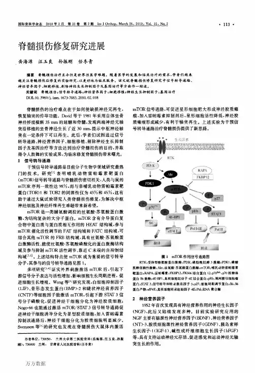
脊髓损伤后细胞凋亡基因调控研究进展
- 格式:pdf
- 大小:293.16 KB
- 文档页数:3
山东医药2021年第61卷第17期间充质干细胞外泌体修复脊髓损伤作用机制的研究进展马麟,张晓勃,赵光海,巩朝阳,张海鸿兰州大学第二医院,兰州730030摘要:脊髓损伤(SCI)通常会导致不可逆的神经退行性改变并影响终生,但目前缺乏有效治疗策略。
间充质干细胞外泌体可通过促进血管生成、促进轴突生长、调节炎症反应、调节免疫反应及抑制细胞凋亡等方式修复SCI,或可能成为SCI患者治疗的新选择。
关键词:间充质干细胞;外泌体;脊髓损伤;脊髓修复;作用机制doi:10.3969/j.issn.1002-266X.2021.17.024中图分类号:R744.9;R651.2文献标志码:A文章编号:1002-266X(2021)17-0089-03脊髓损伤(SCI)是一种破坏性神经退行性疾病,临床上目前缺乏对该病有效的治疗方法,而纳米技术和再生医学策略为新型疗法的开发带来了希望。
干细胞可通过替代丢失或受损的细胞为神经元提供营养支持,改善脊髓的微环境,从而促进受损轴突再生,加快SCI修复[1]。
间充质干细胞(MSCs)可来自骨髓、脂肪、脐带血和胎盘等多种组织,具有归巢、增殖、分化、分泌和免疫调节的功能,是动物研究和人类临床试验中最常用的干细胞[2]。
外泌体是释放到细胞膜外的纳米级囊泡,含有大量复杂分子如蛋白质、脂类和各种核酸,而这些分子的特性与它们的来源细胞有关[3]。
MSCs外泌体(MSCs-Exo)的生物学功能与MSCs相似,但MSCs-Exo更稳定,不会引发机体免疫排斥反应;其具有易分离的特点,故可用于将遗传物质或药物转运至靶细胞;并且尺寸相对较小,故能渗透血脑屏障到达中枢神经系统损伤部位[4-5]。
因此,MSCs-Exo是无细胞治疗的合适选择。
多项研究显示,MSCs-Exo在SCI修复中有巨大潜力。
本文就MSCs-Exo修复SCI的作用机制综述如下。
1MSCs-Exo通过促进血管生成修复SCI血管生成是SCI修复的关键,局部血管丢失与血脑屏障损伤引起的破坏可导致缺血和炎症反应,从而引发脊髓神经组织的综合性损伤[6]。
生物技术进展 2023 年 第 13 卷 第 2 期 240 ~ 246Current Biotechnology ISSN 2095‑2341进展评述ReviewsNrf2在脊髓损伤后铁死亡的研究进展曹静钰1 , 刘承梅2* , 祁晨旭1 , 杜开颜1 , 陈蒙1 , 侯思伟21.河南中医药大学,郑州 450046;2.河南中医药大学第一附属医院,郑州 450099摘要:铁死亡是脊髓损伤(spinal cord injury ,SCI )后神经元细胞损伤的重要病理机制之一,近几年受到国内外广泛关注,但并未取得突破性进展。
目前,有研究发现氧化应激在SCI 病理生理过程中发挥着重要作用。
核因子E2相关因子2(nuclear factor erythroid -2 related factor 2, Nrf2)因具有抗氧化应激作用并参与SCI 后神经修复而成为研究热点,但其调控铁死亡发生发展的机制尚不明确。
简要介绍了Nrf2的基础结构及其下游靶基因,总结了铁死亡的相关机制并探索了Nrf2在SCI 治疗中的潜在作用,以期为进一步研究其与铁死亡在SCI 发生发展中的关系和探索SCI 治疗的新策略提供思路。
关键词:脊髓损伤;铁死亡;核因子E2相关因子2;氧化应激DOI :10.19586/j.2095‑2341.2022.0178 中图分类号:R651.2 文献标志码:AResearch Progress of Nrf2 in Ferroptosis After Spinal Cord InjuryCAO Jingyu 1 , LIU Chengmei 2 * , QI Chenxu 1 , DU Kaiyan 1 , CHEN Meng 1 , HOU Siwei 21.Henan University of Chinese Medicine , Zhengzhou 450046, China ;2.The First Affiliated Hospital of Henan University of Chinese Medicine , Zhengzhou 450099, ChinaAbstract :Ferroptosis is one of the important pathological mechanisms of neuronal cell injury after spinal cord injury (SCI ). It has received widespread attention at home and abroad in recent years , but no effective breakthrough has been achieved. In recent years , it has been found that oxidative stress plays an important role in the pathophysiological process. Nuclear factor E2-related factor 2 (Nrf2) has become a hot topic of research because of its anti -oxidative stress effect and involvement in neural repair after SCI , but its mechanism of regulating the development of ferroptosis is still unclear. In this paper , we briefly described the basic structure of Nrf2 and its downstream target genes , introduced the mechanism of ferroptosis and explored the potential role of Nrf2in SCI treatment , and provided ideas for further study of its relationship with iron death in the development of SCI and explore new strategies for SCI treatment.Key words :spinal cord injury ; ferroptosis ; nuclear factor E2-related factor 2; oxidative stress脊髓损伤(spinal cord injury ,SCI )属于中枢神经系统(central nervous system ,CNS )的一种创伤性疾病,损伤发生后可导致神经组织的感觉或运动功能不可逆受损。
外泌体治疗脊髓损伤的研究进展史冬玲;何冰倩;戴灵豪【摘要】Exosomes are membrane vesicles secreted by cells into the extracellular environment. Exosomes are a new cell component in the treatment of spinal cord injury. Exosomes are secreted by mesenchymal stem cells have anti-apoptotic, anti-inflammatory and pro-angiogenic effects in the treatment of spinal cord injury, which play a key role in nerve cell-cell communication and can delivery exogenous genetic material. Exosomes can penetrate blood-brain barrier and are more stable than their parent cells, reducing the safety risks in the administration of viable cells.%外泌体是细胞分泌到胞外环境的膜囊泡,是治疗脊髓损伤的一种新策略.间充质干细胞分泌的外泌体在脊髓损伤的治疗中具有抗凋亡、抗炎和促血管生成的作用,同时能促进神经细胞间的交流,并可作为外源性遗传物质的载体.与其亲代细胞相比,外泌体可穿越血脑屏障,更加稳定,降低了施用活细胞所固有的安全风险.【期刊名称】《基础医学与临床》【年(卷),期】2018(038)006【总页数】4页(P849-852)【关键词】脊髓损伤;外泌体;间充质干细胞【作者】史冬玲;何冰倩;戴灵豪【作者单位】浙江中医药大学中医药科学院,浙江杭州311400;浙江中医药大学中医药科学院,浙江杭州311400;浙江中医药大学中医药科学院,浙江杭州311400【正文语种】中文【中图分类】R341 脊髓损伤研究的现状脊髓损伤是脊柱损伤严重的并发症之一。
前列腺素E1对大鼠脊髓损伤后PARP-1表达摘要】目的:观察前列腺素E1对大鼠脊髓损伤后多聚ADP核糖聚合酶(PPRP-1) 表达的影响。
方法成年SD大鼠30只,采用Allen重物打击法制作大鼠脊髓损伤模型,随机分为前列腺素E1治疗组及对照组各15只。
治疗组每日尾静脉注射前列腺素E15ug/d,对照组尾静脉注射等量生理盐水。
取损伤段脊髓组织,免疫组化检测6小时及12小时后PPRP-1的表达变化。
结果 6小时及12小时后,前列腺素E1治疗组PARP-1表达均低于对照组(P<0.05)。
结论前列腺素E1可减少大鼠继发性脊髓损伤时的氧化应激反应,可能作为脊髓损伤的一种有潜力的新药。
【关键词】前列腺素E1;脊髓损伤;多聚ADP核糖聚合酶【中图分类号】R352【文献标识码】A【文章编号】1005-0515(2011)03-0010-01继发性脊髓损伤与脊髓序列性组织自毁性破坏发生的继发性性损伤相关,其中,氧化应激反应在损伤的继发性病理变化中有着重要作用[1]。
多聚ADP核糖聚合酶(poly(ADP-ribose)polymerase PARP)是一种DNA修复酶,DNA受到氧化损伤时被活化,参与脊髓继发性损伤[2]。
手术、药物及基因治疗的运用对脊髓损伤带来一定的效果,但脊髓损伤所致的致残率仍相当高。
前列腺素E1 (prostaglandin E1, PGE1) 是一种具有高度生物活性物质,它可舒张血管平滑肌,降低外周阻力,扩张微血管,改善全身血流动力学,并可抑制血小板聚集。
本实验通过观察PARP-1的表达,探讨PGE1对脊髓继发性损害的保护作用。
1材料与方法1.1材料:成年健康雄性SD大鼠30只,3~4月龄,体重(250±30) g,由湖北省实验动物中心提供。
实验器材为Allen打击器,FA2004A电子天平,OLYMPUS显微镜,荧光显微镜。
具体试剂及来源如下:PGE1注射液(北京泰德公司),多聚ADP核糖聚合酶(PARP-1)兔抗大鼠多克隆抗体(eBioscience公司),FITC标记的山羊抗兔抗体IgG(北京中杉金桥生物技术公司)。
脊髓损伤治疗的方法与进展[摘要] 现代医学对脊髓损伤的研究主要包括两大方面:西医药物治疗研究和中医药物治疗研究。
在动物实验基础上,临床观察损伤早期大剂量甲基强的松治疗有效;其他药物,如纳络酮、促甲状腺释放激素、自由基消除剂维生素E、超氧化物歧化因子和别嘌呤醇可减轻脊髓损伤程度。
组织及细胞的移植也一直是研究者探索的热点,如骨髓基质干细胞移植,以及基因治疗的实验研究。
但目前这些研究大多仍处于实验阶段。
近年来,随着现代医学科学的发展,运用中西医结合方法,发挥祖国医药学的巨大潜力,中药治疗脊髓原发性损伤和防治继发性损伤已取得了一些进展。
[关键词] 脊髓损伤西医治疗中医治疗一、西医治疗1、激素治疗1.1 甲基强的松龙甲基强的松龙(MP)是在20 世纪90 年代起开始应用于临床急性脊髓损伤的治疗,认为激素能减轻脊髓损伤后的继发水肿。
但近年来经过各国学者的深入研究,认为MP具有多方面的神经保护作用,包括改善微循环、抑制脂质过氧化、减少细胞钙内流、维持神经元兴奋性、降低一氧化氮活酶活性、抑制神经细胞凋亡等[1]。
马延超等[2]用甲基强的松龙对大鼠Allen’s急性脊髓损伤模型进行了实验,结果发现:甲基强的松龙能促进大鼠脊髓损伤后微管相关蛋白-2(microtubule associated protein-2, MAP-2)的表达和运动功能的回复,甲基强的松龙对MAP-2表达的上调和其能促进脊髓运动功能的恢复可能存在关联。
并推测其机制可能与其能减轻脊髓继发性损伤、保护残存的神经元有关,最终是通过与细胞内的糖皮质激素受体结合,转运至细胞核内与DNA上的特异性结构结合形成复合物,从而调控和MAP-2合成有关基因的表达。
甲基强的松龙除了对于残存神经元的保护作用以外,对于轴突再生局部的神经胶质细胞也能发生作用,诱导神经胶质分泌神经营养因子[3],从而改变了局部轴突再生的微环境。
另外,甲基强的松龙还具有抑制钙蛋白酶的作用,从而减少了MAP -2的降解[4]。
山西医科大学硕士学位论文大鼠皮质脊髓束和红核脊髓束定位损伤后运动皮质和红核神经细胞凋亡及NT-3表达的变化姓名:王茂申请学位级别:硕士专业:骨外科学指导教师:孙天胜20100315 山Ifl『医科人学硕上学位论文大鼠皮质脊髓束和红核脊髓束定位损伤后运动皮质和红核神经细胞凋亡及NT-3表达的变化。
研究生:王茂专业:骨外科导师:孙天胜摘要目的:1.观察分别损伤大鼠双侧皮质脊髓背侧束和红核脊髓束后相应的运动皮质内的贝茨细胞和红核细胞凋亡的形态及数量的变化。
2.观察分别损伤大鼠双侧皮质脊髓背侧束和红核脊髓束后相应的运动皮质内的贝茨细胞和红核细胞神经营养因子NT一3的表达变化。
3.分析两者的变化是否能反映进化论的观点,即进化低的结构的修复能力大于进化高的结构。
方法:分别建立SD大鼠双侧皮质脊髓背侧束损伤模型和双侧红核脊髓束损伤模型,采用原位细胞凋亡TUNEL法检测两组术后1d、3d、7d、14d大脑运动皮质及红核中神经细胞凋亡的形态及凋亡指数的变化;采用免疫组织化学方法检测神经营养因子NT一3的阳性细胞数及表达变化。
结果:1.皮质脊髓背侧束损伤和红核脊髓束损伤后原位细胞凋亡TUNEL法检测神经细胞凋亡结果:不同时问点大脑皮质及红核均有凋亡细胞分布,细胞核呈紫色及黄褐色双重染色,形态不一;损伤后1d,皮质脊髓背侧束损伤组的运动皮质及红核脊髓束损伤组红核的神经细胞凋亡数量增加不明显;损伤后3d,两组相应的大脑皮质及红核有大量凋亡细胞出现,并持续至14d,RST损伤组凋亡指数较dCST损伤组低(P<0.05);各时间点凋亡细胞数量与假手术组相比均增加(P<0.05);凋亡神经细胞在伤后14d较7d少,RST损伤组凋亡指数在7d及14d均较dCST损伤组低(P<0.05)。
2.免疫组织化学方法检测神经营养因子NT一3结果:NT一3在两组损伤后不同时间点在相应的运动皮质和红核均有表达;与假手术组相比,皮质脊髓背侧束损伤组于术后7d,NT一3在运动皮质表达达高峰;红核脊髓束组于术后3d,NT一3在红核表达达高峰,后缓慢下降。
急性脊髓损伤后线粒体的功能变化程刚【摘要】Mitochondria is one of the most vulnerable organelles. Mitochondrial oxidative phosphorylation provides most of the energy required by cell activity. Post-traumatic changes of mitochondrial function directly affect the function and survival of nerve cells. Therefore, understanding the molecular mechanism behind the functional changes of mitochondria in secondary spinal cord injury is important for the treatment and prognosis of spinal cord injury. This article presents an overview on the mechanisms of mitochondrial injury, in such aspects as energy metabolism, reactive oxygen species generation, calcium overload.%线粒体是对各种损伤最为敏感的细胞器之一;细胞活动所需绝大多数的能量都是由线粒体氧化磷酸化提供.外伤后线粒体功能的改变直接影响到神经细胞的功能和存活.因此,了解继发性脊髓损伤中线粒体的功能变化的分子机制,对于脊髓损伤的治疗和预后具有重要意义.文中从线粒体能量代谢、活性氧生成、钙超载等角度对线粒体损伤机制进行综述.【期刊名称】《医学研究生学报》【年(卷),期】2011(024)008【总页数】4页(P866-869)【关键词】急性脊髓损伤;线粒体;活性氧;钙超载【作者】程刚【作者单位】210029南京,南京医科大学附属第一医院骨脊柱科【正文语种】中文【中图分类】R7440 引言急性脊髓损伤(acute spinal cord injury,ASCI)是一种后果极其严重的创伤,虽然在过去的几十年里其死亡率从 50%已经下降了到目前的 6%[1]。
LSD1抑制介导脊髓损伤早期神经元自噬影响调亡的实验研究摘要:长链非编码RNA调节蛋白1(LSD1)是一种与神经元自噬和细胞凋亡密切相关的蛋白。
本研究旨在探究LSD1是否介导脊髓损伤(SM)早期神经元自噬影响调亡,以期为神经系统损伤治疗提供新思路。
采用大鼠SM模型,结合行为学、免疫组化、荧光定量PCR、Western blot, electron microscopy等多种实验手段,研究LSD1的表达、神经元自噬、凋亡以及细胞核内甲基化等相关变化。
结果显示,SM早期,LSD1表达显著升高,伴随着神经元自噬和细胞凋亡的增加,脊髓神经元核内甲基化水平降低。
应用化学药剂抑制LSD1后发现,LSD1抑制可以显著抑制SM早期神经元凋亡,且据文献报道,LSD1抑制可诱导细胞自噬,对神经元自噬的影响也值得研究。
但本文的数据不支持LSD1介导神经元自噬,进一步研究可考虑LSD1在细胞凋亡通路其他分子途径中介的作用。
综上所述,本研究表明LSD1介导SM早期神经元凋亡的发生,抑制LSD1可为提高神经元存活率提供新的治疗方案。
关键词:LSD1, 自噬, 细胞凋亡, 脊髓损伤(SM)Abstract:Lysine-specific demethylase 1 (LSD1) is a proteinclosely related to neuronal autophagy and apoptosis. This study aims to investigate whether LSD1 mediates the effect of early nerve cell autophagy on apoptosis after spinal cord injury (SCI), in order to provide new ideas for the treatment of neurological damage. Using a rat model of SCI, a combination of behavioral tests, immunohistochemistry, quantitative PCR, Western blot analysis, electron microscopy, and other experimental methods, the expression of LSD1, neuronal autophagy, apoptosis, and nuclear methylation changes were studied. The results showed that LSD1 expression significantly increased in the early stage of SCI, accompanied by an increase in neuronal autophagy and apoptosis, and a decrease in nuclear methylationlevels in spinal cord neurons. It was found that pharmacological inhibition of LSD1 significantly inhibited the early apoptosis of nerve cells after SCI. According to previous literature reports, LSD1inhibition can induce autophagy in cells, but this study did not find that LSD1 mediates neuronal autophagy. Therefore, further research can considerthe role of LSD1 in other molecular pathways of thecell apoptosis pathway. In summary, this study shows that LSD1 is involved in the early apoptosis of spinal cord neurons after SCI, and the inhibition of LSD1 may provide a new therapeutic strategy to improve neuronal survival.Keywords: LSD1, autophagy, apoptosis, spinal cordinjury (SCISpinal cord injury (SCI) is a devastating event that often leads to irreversible damage and functional impairments. One of the major causes of secondary damage after SCI is neuronal apoptosis. Therefore, identifying the molecular mechanisms of neuronal apoptosis and developing therapeutic strategies to prevent or reduce apoptosis are critical for promoting neuronal survival and improving functional outcomes after SCI.In recent years, there has been growing interest inthe role of histone methylation in regulating gene expression and cellular processes, including apoptosis. LSD1, also known as KDM1A, is a lysine-specific demethylase that removes mono- and di-methyl groups from histone H3 lysine 4 (H3K4) and regulates gene transcription. Several studies have reported the involvement of LSD1 in various cellular processes, including differentiation, proliferation, and apoptosis. However, the role of LSD1 in neuronal apoptosis after SCI is not well understood.In this study, the researchers investigated theexpression and role of LSD1 in the apoptosis of spinal cord neurons after SCI. Using a rat model of SCI, they found that LSD1 expression was significantly upregulated in the spinal cord tissue at 24 hoursafter injury. Furthermore, immunofluorescence staining revealed that LSD1 was mainly expressed in neurons and colocalized with caspase-3, a key executor of apoptosis.To further investigate the role of LSD1 in neuronal apoptosis after SCI, the researchers used a specific inhibitor of LSD1, tranylcypromine (TCP), to block LSD1 activity in vitro and in vivo. They found that TCP treatment significantly reduced the number of TUNEL-positive apoptotic cells and increased the expression of the anti-apoptotic protein Bcl-2 in the spinal cord tissue after SCI. Moreover, TCP treatment improved the functional outcomes of the rats, as assessed by the Basso, Beattie, and Bresnahan (BBB) locomotor scale.Interestingly, the researchers also investigated the role of LSD1 in autophagy, a cellular process that disposes of damaged organelles and proteins and promotes cell survival. They found that LSD1inhibition did not induce autophagy in spinal cord neurons after SCI, suggesting that LSD1 mediatesneuronal apoptosis through other molecular pathways.In conclusion, this study provides evidence that LSD1 is involved in the early apoptosis of spinal cord neurons after SCI and that LSD1 inhibition may provide a new therapeutic strategy to improve neuronal survival. Future research can further investigate the molecular mechanisms by which LSD1 regulates apoptosis and identify other potential targets for therapeutic interventions after SCIFurther studies can also explore the optimal time and dosage for LSD1 inhibition therapy after SCI, as well as its potential effects on motor function recovery and tissue repair.Moreover, this study provides a new perspective on the role of histone demethylation in mediating neuronal apoptosis after SCI. While previous studies mainly focused on the involvement of histone acetylation and DNA methylation, this study suggests that histone demethylation may also play a critical role in regulating gene expression and cell fate after SCI.Overall, this study sheds light on the potential therapeutic benefits of LSD1 inhibition in SCI and highlights the need for further research to fullyelucidate its molecular mechanisms and clinical applications. With the development of more specificand effective LSD1 inhibitors, it is possible to translate these findings into clinical practice and improve the long-term outcomes of SCI patientsIn addition to the potential therapeutic benefits of LSD1 inhibition, it is important to consider the limitations and challenges in developing this approach for SCI treatment. One major challenge is thespecificity of LSD1 inhibitors, as they may alsoaffect other histone demethylases or other cellular pathways leading to off-target effects. Thus, it is crucial to develop more selective and specific LSD1 inhibitors to minimize adverse effects.Another challenge is the complexity of SCI pathophysiology, which involves multiple cellular and molecular mechanisms. LSD1 inhibition may be effective in targeting one specific aspect of SCI, such as neuronal survival or glial scar formation, but may not be sufficient to address all aspects of SCI. Therefore, combination therapies targeting multiple pathways may be necessary for effective SCI treatment.In addition, the optimal timing and duration of LSD1 inhibition after SCI requires further investigation.The study by Wu et al. demonstrated the efficacy of LSD1 inhibition in promoting neuronal survival and reducing glial scar formation when administered soon after SCI. However, the long-term effects of LSD1 inhibition and the potential consequences of chronic inhibition require further study.Furthermore, the use of LSD1 inhibition in SCI treatment raises ethical and safety concerns, as it involves the manipulation of gene expression in living organisms. Therefore, it is important to consider the potential risks and benefits of LSD1 inhibition and to ensure that it is used safely and responsibly.In conclusion, the study by Wu et al. highlights the potential therapeutic benefits of LSD1 inhibition in SCI and provides insights into its molecular mechanisms. LSD1 inhibition offers a promising approach for promoting neuronal survival and reducing glial scar formation after SCI, but further researchis needed to optimize its efficacy, specificity, and safety. With continued investigation and advancement of LSD1 inhibitors, it is possible to translate these findings into effective SCI treatments that improve the quality of life for patientsIn conclusion, the inhibition of LSD1 has been identified as a potential therapeutic strategy for the treatment of spinal cord injury. This approach has been shown to promote neuronal survival and reduce glial scar formation through its molecular mechanisms. However, further research is needed to fully understand the efficacy, specificity, and safety of LSD1 inhibitors in SCI. With ongoing study and development, LSD1 inhibition may offer a promising avenue for improving the quality of life for patients with SCI。
·综述·小胶质细胞在脊髓损伤中的作用机制研究进展夏宇,丁璐,邓宇斌作者单位中山大学附属第七医院科研中心深圳518107基金项目国家自然科学基金项目(No.82071362)收稿日期2022-04-25通讯作者邓宇斌dengyub@摘要脊髓损伤(spinal cord injury ,SCI )是由于外力或非外力作用造成脊柱骨、韧带及神经结构的破坏,并伴随着损伤部位以下躯干与四肢的感觉运动功能障碍,其致残率高。
小胶质细胞作为中枢神经系统固有的免疫细胞,在SCI 后接受损伤信号,发挥分泌因子及吞噬作用,同时和神经元、星形胶质细胞、少突胶质细胞及其它细胞与非细胞成分发生反应。
目前研究显示,小胶质细胞具有多态性和多功能性,参与SCI 的病理生理过程,包括炎症、疤痕形成和疼痛。
本综述结合前期课题组星形胶质细胞研究基础,通过总结近年来小胶质细胞在SCI 过程中功能的研究文献,为SCI 疾病进展研究提供新的思路与方向。
关键词脊髓损伤;小胶质细胞;星形胶质细胞中图分类号R741;R741.02;R744文献标识码A DOI 10.16780/ki.sjssgncj.20220376本文引用格式:夏宇,丁璐,邓宇斌.小胶质细胞在脊髓损伤中的作用机制研究进展[J].神经损伤与功能重建,2023,18(10):593-596.脊髓损伤(spinal cord injury ,SCI )是由于外力或非外力作用造成脊柱骨、韧带及神经结构的破坏,并伴随着损伤部位以下躯干与四肢的感觉运动功能障碍,每年全球约70万例新发病例,致残率高[1,2]。
神经功能障碍是导致SCI 高残障率的基础。
除了神经元的死亡、突触连接的丢失等原发性损伤,小胶质细胞作为胶质细胞的一员参与激活炎症级联反应,造成继发性损伤[3]。
1小胶质细胞的定义小胶质细胞作为中枢神经系统(central nervous system ,CNS )固有的免疫细胞,是神经组织中唯一来源于中胚层的细胞[4]。
microRNA在脊柱脊髓损伤中作用的研究现状与进展李贺祥;丛岩;王佳瑶;邱涛【摘要】脊髓损伤(SCI)是当今全球性的医学难题之一,致残率及病死率极高,当前临床治疗手段有限且疗效欠佳.SCI后复杂的病理生理过程使得神经功能障碍进一步加重,脊髓再生和功能恢复困难.近年来的研究发现,基因表达的改变在SCI中起到了重要作用,microRNA (miRNA)是一类在转录后水平负性调控基因表达的非编码单链RNA小分子,是基因表达调控的重要机制之一,在细胞分化、生长、增殖和神经活动等多种神经生物学过程及病理过程中起重要作用.在SCI后miRNA主要通过调节中性粒细胞和炎性反应通路在炎性应答中起到了重要作用;在细胞凋亡过程中,因miRNA表达的改变可能同时刺激和抑制凋亡而显得其功能复杂;miRNA也可作用于星形胶质细胞调节胶质瘢痕的进程,在促使神经元存活、轴突生长和促进SCI 部位再生进程中发挥重要作用.总之,对神经系统疾病中miRNA作用机制的研究不仅为基因治疗SCI的研究提供了一种新的思路,miRNA本身也可能成为临床治疗SCI的潜在靶点.【期刊名称】《重庆医学》【年(卷),期】2019(048)003【总页数】4页(P479-482)【关键词】微RNAs;脊髓损伤;脊柱损伤;基因表达;新靶点【作者】李贺祥;丛岩;王佳瑶;邱涛【作者单位】贵州医科大学临床医学院,贵阳550004;贵州医科大学临床医学院,贵阳550004;贵州医科大学临床医学院,贵阳550004;贵州医科大学临床医学院,贵阳550004【正文语种】中文【中图分类】R651.2作为中枢神经系统中较为严重的创伤之一,脊髓损伤(SCI)是脊柱骨折的严重并发症[1]。
其早期主要有因损伤组织缺血、水肿、氧自由基形成及炎性反应等多种因素联合作用引起的暂时性或永久性的病理生理改变,最终可导致肢体运动和感觉能力丧失[2],目前尚无令人满意的疗法[3]。
最近有研究表明,SCI的继发性损伤和损伤后神经再生过程受到microRNA(miRNA)的显著调控,对与SCI相关的miRNA的研究可以提高对疾病进程的认识,从而为临床医生提供治疗SCI及预测临床预后的机会[4]。