repertoire of microglial and macrophage responses after spinal cord injury
- 格式:pdf
- 大小:827.91 KB
- 文档页数:12
Toxicologic Pathology,35:495–516,2007Copyright C by the Society of Toxicologic PathologyISSN:0192-6233print/1533-1601onlineDOI:10.1080/01926230701320337Invited ReviewApoptosis:A Review of Programmed Cell DeathS USAN E LMORENIEHS,Laboratory of Experimental Pathology,Research Triangle Park,North Carolina27709,USAA BSTRACTThe process of programmed cell death,or apoptosis,is generally characterized by distinct morphological characteristics and energy-dependent biochemical mechanisms.Apoptosis is considered a vital component of various processes including normal cell turnover,proper development and functioning of the immune system,hormone-dependent atrophy,embryonic development and chemical-induced cell death.Inappropriate apoptosis (either too little or too much)is a factor in many human conditions including neurodegenerative diseases,ischemic damage,autoimmune disorders and many types of cancer.The ability to modulate the life or death of a cell is recognized for its immense therapeutic potential.Therefore,research continues to focus on the elucidation and analysis of the cell cycle machinery and signaling pathways that control cell cycle arrest and apoptosis.To that end,thefield of apoptosis research has been moving forward at an alarmingly rapid rate.Although many of the key apoptotic proteins have been identified,the molecular mechanisms of action or inaction of these proteins remain to be elucidated.The goal of this review is to provide a general overview of current knowledge on the process of apoptosis including morphology,biochemistry,the role of apoptosis in health and disease,detection methods,as well as a discussion of potential alternative forms of apoptosis.Keywords.Apoptosis;programmed cell death;intrinsic/extrinsic pathway;granzyme A/B;perforin;autophagy.I NTRODUCTIONThe term apoptosis(a-po-toe-sis)wasfirst used in a now-classic paper by Kerr,Wyllie,and Currie in1972to describe a morphologically distinct form of cell death,although cer-tain components of the apoptosis concept had been explicitly described many years previously(Kerr et al.,1972;Paweletz, 2001;Kerr,2002).Our understanding of the mechanisms in-volved in the process of apoptosis in mammalian cells tran-spired from the investigation of programmed cell death that occurs during the development of the nematode Caenorhab-ditis elegans(Horvitz,1999).In this organism1090somatic cells are generated in the formation of the adult worm,of which131of these cells undergo apoptosis or“programmed cell death.”These131cells die at particular points during the development process,which is essentially invariant between worms,demonstrating the remarkable accuracy and control in this system.Apoptosis has since been recognized and ac-cepted as a distinctive and important mode of“programmed”cell death,which involves the genetically determined elim-ination of cells.However,it is important to note that other forms of programmed cell death have been described and other forms of programmed cell death may yet be discovered (Formigli et al.,2000;Sperandio et al.,2000;Debnath et al., 2005).Apoptosis occurs normally during development and aging and as a homeostatic mechanism to maintain cell populations in tissues.Apoptosis also occurs as a defense mechanism such as in immune reactions or when cells are damaged by diseaseAddress correspondence to:Susan Elmore,NIEHS,P.O.Box12233, Research Triangle Park,NC27709,USA;e-mail:elmore@or noxious agents(Norbury and Hickson,2001).Although there are a wide variety of stimuli and conditions,both phys-iological and pathological,that can trigger apoptosis,not all cells will necessarily die in response to the same stimulus.Irradiation or drugs used for cancer chemotherapy results in DNA damage in some cells,which can lead to apoptotic death through a p53-dependent pathway.Some hormones,such as corticosteroids,may lead to apoptotic death in some cells(e.g.,thymocytes)although other cells are unaffected or evenstimulated.Some cells express Fas or TNF receptors that can lead to apoptosis via ligand binding and protein cross-linking.Other cells have a default death pathway that must be blocked bya survival factor such as a hormone or growth factor.Thereis also the issue of distinguishing apoptosis from necrosis, two processes that can occur independently,sequentially,as well as simultaneously(Hirsch,1997;Zeiss,2003).In some cases it’s the type of stimuli and/or the degree of stimuli that determines if cells die by apoptosis or necrosis.At low doses,a variety of injurious stimuli such as heat,radiation,hypoxiaand cytotoxic anticancer drugs can induce apoptosis but these same stimuli can result in necrosis at higher doses.Finally, apoptosis is a coordinated and often energy-dependent pro-cess that involves the activation of a group of cysteine pro-teases called“caspases”and a complex cascade of events that link the initiating stimuli to thefinal demise of the cell.Morphology of ApoptosisLight and electron microscopy have identified the various morphological changes that occur during apoptosis(Hacker, 2000).During the early process of apoptosis,cell shrinkage 495496ELMORE T OXICOLOGIC P ATHOLOGYand pyknosis are visible by light microscopy(Kerr et al., 1972).With cell shrinkage,the cells are smaller in size,the cy-toplasm is dense and the organelles are more tightly packed. Pyknosis is the result of chromatin condensation and this is the most characteristic feature of apoptosis.On histologic examination with hematoxylin and eosin stain,apoptosis in-volves single cells or small clusters of cells.The apoptotic cell appears as a round or oval mass with dark eosinophilic cytoplasm and dense purple nuclear chromatin fragments (Figure1).Electron microscopy can better define the sub-cellular changes.Early during the chromatin condensation phase,the electron-dense nuclear material characteristically aggregates peripherally under the nuclear membrane al-though there can also be uniformly dense nuclei(Figures 2A,2B).Extensive plasma membrane blebbing occurs followed by karyorrhexis and separation of cell fragments into apoptotic bodies during a process called“budding.”Apoptotic bodies consist of cytoplasm with tightly packed organelles with or without a nuclear fragment(Figure2C).The organelle in-tegrity is still maintained and all of this is enclosed within an intact plasma membrane.These bodies are subsequently phagocytosed by macrophages,parenchymal cells,or neo-plastic cells and degraded within phagolysosomes(Figure 2D).Macrophages that engulf and digest apoptotic cells are called“tingible body macrophages”and are frequently found within the reactive germinal centers of lymphoid follicles or occasionally within the thymic cortex.The tingible bodies are the bits of nuclear debris from the apoptotic cells.There is essentially no inflammatory reaction associated with the process of apoptosis nor with the removal of apoptotic cells because:(1)apoptotic cells do not release their cellular con-stituents into the surrounding interstitial tissue;(2)they are quickly phagocytosed by surrounding cells thus likely pre-venting secondary necrosis;and,(3)the engulfing cells do not produce anti-inflammatory cytokines(Savill and Fadok, 2000;Kurosaka et al.,2003).Distinguishing Apoptosis from NecrosisThe alternative to apoptotic cell death is necrosis,which is considered to be a toxic process where the cell is a passive victim and follows an energy-independent mode of death.But since necrosis refers to the degradative processes that occur after cell death,it is considered by some to be an inappro-priate term to describe a mechanism of cell death.Oncosis is therefore used to describe a process that leads to necrosis with karyolysis and cell swelling whereas apoptosis leads to cell death with cell shrinkage,pyknosis,and karyorrhexis. Therefore the terms“oncotic cell death”and“oncotic necro-sis”have been proposed as alternatives to describe cell death that is accompanied by cell swelling,but these terms are not widely used at this time(Majno and Joris,1995;Levin et al., 1999).Although the mechanisms and morphologies of apopto-sis and necrosis differ,there is overlap between these two processes.Evidence indicates that necrosis and apoptosis represent morphologic expressions of a shared biochemi-cal network described as the“apoptosis-necrosis continuum”(Zeiss,2003).For example,two factors that will convert an ongoing apoptotic process into a necrotic process include a decrease in the availability of caspases and intracellular ATPT ABLE1.—Comparison of morphological features of apoptosis and necrosis.Apoptosis NecrosisSingle cells or small clusters of cells Often contiguous cellsCell shrinkage and convolution Cell swellingPyknosis and karyorrhexis Karyolysis,pyknosis,andkaryorrhexisIntact cell membrane Disrupted cell membrane Cytoplasm retained in apoptotic bodies Cytoplasm releasedNo inflammation Inflammation usually present (Leist et al.,1997;Denecker et al.,2001).Whether a cell dies by necrosis or apoptosis depends in part on the nature of the cell death signal,the tissue type,the developmental stage of the tissue and the physiologic milieu(Fiers et al.,1999;Zeiss, 2003).Using conventional histology,it is not always easy to dis-tinguish apoptosis from necrosis,and they can occur simul-taneously depending on factors such as the intensity and du-ration of the stimulus,the extent of ATP depletion and the availability of caspases(Zeiss,2003).Necrosis is an uncon-trolled and passive process that usually affects largefields of cells whereas apoptosis is controlled and energy-dependent and can affect individual or clusters of cells.Necrotic cell injury is mediated by two main mechanisms;interference with the energy supply of the cell and direct damage to cell membranes.Some of the major morphological changes that occur with necrosis include cell swelling;formation of cytoplasmic vac-uoles;distended endoplasmic reticulum;formation of cyto-plasmic blebs;condensed,swollen or ruptured mitochondria; disaggregation and detachment of ribosomes;disrupted or-ganelle membranes;swollen and ruptured lysosomes;and eventually disruption of the cell membrane(Kerr et al.,1972; Majno and Joris,1995;Trump et al.,1997).This loss of cell membrane integrity results in the release of the cytoplasmic contents into the surrounding tissue,sending chemotatic sig-nals with eventual recruitment of inflammatory cells.Because apoptotic cells do not release their cellular constituents into the surrounding interstitial tissue and are quickly phagocy-tosed by macrophages or adjacent normal cells,there is es-sentially no inflammatory reaction(Savill and Fadok,2000; Kurosaka et al.,2003).It is also important to note that pykno-sis and karyorrhexis are not exclusive to apoptosis and can be a part of the spectrum of cytomorphological changes that occurs with necrosis(Cotran et al.,1999).Table1compares some of the major morphological features of apoptosis and necrosis.Is Apoptosis an Irreversible Process?Many of the genes that control the killing and engulfment processes of programmed cell death have been identified,and the molecular mechanisms underlying these processes have proven to be evolutionarily conserved(Metzstein et al.,1998). Until recently,apoptosis has traditionally been considered an irreversible process with caspase activation committing a cell to death and the engulfment genes serving the purpose of dead cell removal.However,the uptake and clearance of apoptotic cells by macrophages may involve more than just the removal of cell debris.Hoeppner et al.have shown that blocking engulfment genes in C.elegans embryos enhances cell survival when cells are subjected to weak pro-apoptotic signals(Hoeppner et al.,2001).V ol.35,No.4,2007REVIEW OF APOPTOSIS497F IGURE1.—Figure1A is a photomicrograph of a section of exocrine pancreas from a B6C3F1mouse.The arrows indicate apoptotic cells that are shrunken with condensed cytoplasm.The nuclei are pyknotic and fragmented.Note the lack of inflammation.Figure1B is an image of myocardium from a14week-old rat treated with ephedrine(25mg/kg)and caffeine(30mg/kg).Within the interstitial space there are apoptotic cells with condensed cytoplasm,condensed and hyperchromatic chromatin and fragmented nuclei(long arrows).Admixed with the apoptotic bodies are macrophages,some with engulfed apoptotic bodies(arrowheads)(Howden et al.,2004,by permission of the American Journal of Physiology).Figure1C is a photomicrograph of normal thymus tissue from a control Sprague–Dawley rat for comparison.Figure ID illustrates sheets of apoptotic cells in the thymus from a rat that was treated with dexamethasone to induce lymphocyte apoptosis.Under physiological conditions,apoptosis typically affects single cells or small clusters of cells.However,the degree of apoptosis in this treated rat is more severe due to the amount of dexamethasone administered(1mg/kg bodyweight)and the time posttreatment(12hours).The majority of lymphocytes are apoptotic although there are a few interspersed cells that are morphologically normal and most likely represent lymphoblasts or macrophages.The apoptotic lymphocytes are small and deeply basophilic with pyknotic and often-fragmented nuclei.Macrophages are present with engulfed cytoplasmic apoptotic bodies(arrows).H&E.498ELMORE T OXICOLOGIC PATHOLOGYF IGURE 1.—(Continued)Reddien et al.demonstrated that,in C.elegans ,mutations that cause partial loss of function of killer genes allow the sur-vival of some cells that are programmed to die via apoptosis,and mutations in engulfment genes enhance the frequency of this cell survival.Moreover,mutations in engulfment genes alone allowed the survival and differentiation of some cells that were otherwise destined to die via apoptosis (Reddien et al.,2001).These findings suggest that genes that medi-ate corpse removal can also function to actively kill cells.In other words,the engulfing cells may act to ensure that cellsV ol.35,No.4,2007REVIEW OF APOPTOSIS499F IGURE2.—Figure2A is a transmission electron micrograph(TEM)of the normal thymus tissue depicted in Figure1C.The lymphocytes are closely packed,have large nuclei and scant cytoplasm.Figure2B is a TEM of apoptotic thymic lymphocytes in an early phase of apoptosis with condensed and peripheralized chromatin. The cytoplasm is beginning to condense and the cell outlines are irregular.The arrow indicates a fragmented section of nucleus and the arrowhead most likely indicates an apoptotic body that seems to contain predominantly cytoplasm without organelles or nuclear material.Figure2C illustrates an apoptotic lymphocyte in the process of“budding”or extrusion of membrane-bound cytoplasm containing organelles(arrow).Once the budding has occurred,this extruded fragment will be an“apoptotic body.”These apoptotic bodies are membrane-bound and thus do not release cytoplasmic contents into the interstitium.Macrophages or other adjacent healthy cells subsequently engulf the apoptotic bodies.For these reasons,apoptosis does not incite an inflammatory reaction.Figure2D is a TEM of a section of thymus with lymphocytes in various stages of apoptosis.The large cell in the center of the photomicrograph is a macrophage with engulfed intracytoplasmic apoptotic bodies. This macrophage is also called a“tingible body macrophage.”The arrowhead indicates a lymphocyte in an advanced stage of apoptosis with nuclear fragmentation.500ELMORE T OXICOLOGIC PATHOLOGYF IGURE 2.—(Continued)triggered to undergo apoptosis will die rather than recover after the initial stages of death.In vertebrates,there is some evidence of a potential role for macrophages in promoting the death of cells in some tis-sues.Elimination of macrophages in the anterior chamber of the rat eye resulted in the survival of vascular endothe-lial cells that normally undergo apoptosis (Diez-Roux and Lang,1997).Other studies have demonstrated that inhibition of macrophages can disrupt the remodeling of tissues in the mouse eye or in the tadpole tail during regression;two pro-cesses that involve apoptosis (Lang and Bishop,1993;Little and Flores,1993).Geske and coworkers (2001)demonstrated that early p53-induced apoptotic cells can be rescued from the apoptotic program if the apoptotic stimulus is removed.V ol.35,No.4,2007REVIEW OF APOPTOSIS 501Their research suggests that DNA repair is activated early in the p 53-induced apoptotic process and that this DNA repair may be involved in reversing the cell death pathway in some circumstances.Mechanisms of ApoptosisThe mechanisms of apoptosis are highly complex and so-phisticated,involving an energy-dependent cascade of molec-ular events (Figure 3).To date,research indicates that there are two main apoptotic pathways:the extrinsic or death receptor pathway and the intrinsic or mitochondrial path-way.However,there is now evidence that the two path-ways are linked and that molecules in one pathway can influence the other (Igney and Krammer,2002).There is an additional pathway that involves T-cell mediated cyto-toxicity and perforin-granzyme-dependent killing of the cell.The perforin/granzyme pathway can induce apoptosis via ei-ther granzyme B or granzyme A.The extrinsic,intrinsic,and granzyme B pathways converge on the same terminal,or ex-ecution pathway.This pathway is initiated by the cleavage of caspase-3and results in DNA fragmentation,degradation of cytoskeletal and nuclear proteins,cross-linking of pro-teins,formation of apoptotic bodies,expression of ligands for phagocytic cell receptors and finally uptake by phago-cytic cells.The granzyme A pathway activates a parallel,caspase-independent cell death pathway via single stranded DNA damage (Martinvalet et al.,2005).Biochemical Features:Apoptotic cells exhibit several biochemical modifications such as protein cleavage,protein cross-linking,DNA breakdown,and phagocytic recognition that together result in the distinctive structural pathology de-scribed previously (Hengartner,2000).Caspases are widely expressed in an inactive proenzyme form in most cellsand F IGURE 3.—Schematic representation of apoptotic events.The two main pathways of apoptosis are extrinsic and intrinsic as well as a perforin/granzyme pathway.Each requires specific triggering signals to begin an energy-dependent cascade of molecular events.Each pathway activates its own initiator caspase (8,9,10)which in turn will activate the executioner caspase-3.However,granzyme A works in a caspase-independent fashion.The execution pathway results in characteristic cytomorphological features including cell shrinkage,chromatin condensation,formation of cytoplasmic blebs and apoptotic bodies and finally phagocytosis of the apoptotic bodies by adjacent parenchymal cells,neoplastic cells or macrophages.once activated can often activate other pro-caspases,allow-ing initiation of a protease cascade.Some pro-caspases can also aggregate and autoactivate.This proteolytic cascade,in which one caspase can activate other caspases,amplifies the apoptotic signaling pathway and thus leads to rapid cell death.Caspases have proteolytic activity and are able to cleave proteins at aspartic acid residues,although different caspases have different specificities involving recognition of neigh-boring amino acids.Once caspases are initially activated,there seems to be an irreversible commitment towards cell death.To date,ten major caspases have been identified and broadly categorized into initiators (caspase-2,-8,-9,-10),ef-fectors or executioners (caspase-3,-6,-7)and inflammatory caspases (caspase-1,-4,-5)(Cohen,1997;Rai et al.,2005).The other caspases that have been identified include caspase-11,which is reported to regulate apoptosis and cytokine maturation during septic shock,caspase-12,which mediates endoplasmic-specific apoptosis and cytotoxicity by amyloid-β,caspase-13,which is suggested to be a bovine gene,and caspase-14,which is highly expressed in embryonic tissues but not in adult tissues (Hu et al.,1998;Nakagawa et al.,2000,Koenig et al.,2001;Kang et al.,2002).Extensive protein cross-linking is another characteristic of apoptotic cells and is achieved through the expression and activation of tissue transglutaminase (Nemes et al.,1996).DNA breakdown by Ca 2+-and Mg 2+-dependent endonucle-ases also occurs,resulting in DNA fragments of 180to 200base pairs (Bortner et al.,1995).A characteristic “DNA lad-der”can be visualized by agarose gel electrophoresis with an ethidium bromide stain and ultraviolet illumination.Another biochemical feature is the expression of cell sur-face markers that result in the early phagocytic recogni-tion of apoptotic cells by adjacent cells,permitting quick502ELMORE T OXICOLOGIC P ATHOLOGY T ABLE2.—Extrinsic pathway proteins,abbreviations,and alternate nomenclature.Abbreviation Protein Name Select Alternate NomenclatureTNF-αTumor necrosis factor alpha TNF ligand,TNFA,cachectinTNFR1Tumor necrosis factor receptor1TNF receptor,TNFRSF1A,p55TNFR,CD120aFasL Fatty acid synthetase ligand Fas ligand,TNFSF6,Apo1,apoptosis antigen ligand1,CD95L,CD178,APT1LG1 FasR Fatty acid synthetase receptor Fas receptor,TNFRSF6,APT1,CD95Apo3L Apo3ligand TNFSF12,Apo3ligand,TWEAK,DR3LGDR3Death receptor3TNFRSF12,Apo3,WSL-1,TRAMP,LARD,DDR3Apo2L Apo2ligand TNFSF10,TRAIL,TNF-related apoptosis inducing ligandDR4Death receptor4TNFRSF10A,TRAILR1,APO2DR5Death receptor5TNFRS10B,TRAIL-R2,TRICK2,KILLER,ZTNFR9FADD Fas-associated death domain MORT1TRADD TNF receptor-associated death domain TNFRSF1A associated via death domainRIP Receptor-interacting protein RIPK1DED Death effector domain Apoptosis antagonizing transcription factor,CHE1caspase-8Cysteinyl aspartic acid-protease8FLICE,FADD-like Ice,Mach-1,Mch5c-FLIP FLICE-inhibitory protein Casper,I-FLICE,FLAME-1,CASH,CLARP,MRITReferences:1.Human Protein Reference Database / .2.ExPASy Proteomics Server / .phagocytosis with minimal compromise to the surround-ing tissue.This is achieved by the movement of the nor-mal inward-facing phosphatidylserine of the cell’s lipid bilayer to expression on the outer layers of the plasma mem-brane(Bratton et al.,1997).Although externalization of phosphatidylserine is a well-known recognition ligand for phagocytes on the surface of the apoptotic cell,recent stud-ies have shown that other proteins are also be exposed on the cell surface during apoptotic cell clearance.These include Annexin I and calreticulin.Annexin V is a recombinant phosphatidylserine-binding protein that interacts strongly and specifically with phos-phatidylserine residues and can be used for the detection of apoptosis(Van Engeland et al.,1998;Arur et al.,2003).Cal-reticulin is a protein that binds to an LDL-receptor related protein on the engulfing cell and is suggested to cooperate with phosphatidylserine as a recognition signal(Gardai et al., 2005).The adhesive glycoprotein,thrombospondin-1,can be expressed on the outer surface of activated microvascular en-dothelial cells and,in conjunction with CD36,caspase-3-like proteases and other proteins,induce receptor-mediated apop-tosis(Jimenez et al.,2000).Extrinsic Pathway:The extrinsic signaling pathways that initiate apoptosis involve transmembrane receptor-mediated interactions.These involve death receptors that are members of the tumor necrosis factor(TNF)receptor gene superfamily (Locksley et al.,2001).Members of the TNF receptor fam-ily share similar cyteine-rich extracellular domains and have a cytoplasmic domain of about80amino acids called the “death domain”(Ashkenazi and Dixit,1998).This death do-main plays a critical role in transmitting the death signal from the cell surface to the intracellular signaling pathways.To date,the best-characterized ligands and corresponding death receptors include FasL/FasR,TNF-α/TNFR1,Apo3L/DR3, Apo2L/DR4and Apo2L/DR5(Chicheportiche et al.,1997; Ashkenazi et al.,1998;Peter and Kramer,1998;Suliman et al.,2001;Rubio-Moscardo et al.,2005).The sequence of events that define the extrinsic phase of apoptosis are best characterized with the FasL/FasR and TNF-α/TNFR1models.In these models,there is clustering of receptors and binding with the homologous trimeric lig-and.Upon ligand binding,cytplasmic adapter proteins are re-cruited which exhibit corresponding death domains that bind with the receptors.The binding of Fas ligand to Fas receptor results in the binding of the adapter protein FADD and the binding of TNF ligand to TNF receptor results in the bind-ing of the adapter protein TRADD with recruitment of FADD and RIP(Hsu et al.,1995;Grimm et al.,1996;Wajant,2002). FADD then associates with procaspase-8via dimerization of the death effector domain.At this point,a death-inducing signaling complex(DISC)is formed,resulting in the auto-catalytic activation of procaspase-8(Kischkel et al.,1995). Once caspase-8is activated,the execution phase of apop-tosis is triggered.Death receptor-mediated apoptosis can be inhibited by a protein called c-FLIP which will bind to FADD and caspase-8,rendering them ineffective(Kataoka et al., 1998;Scaffidi,1999).Another point of potential apoptosis regulation involves a protein called Toso,which has been shown to block Fas-induced apoptosis in T cells via inhibi-tion of caspase-8processing(Hitoshi et al.,1998).Table2 lists the major extrinsic pathway proteins with common ab-breviations and some of the alternate nomenclature used for each protein.Perforin/granzyme Pathway:T-cell mediated cytotoxic-ity is a variant of type IV hypersensitivity where sensitized CD8+cells kill antigen-bearing cells.These cytotoxic T lym-phocytes(CTLs)are able to kill target cells via the extrinsic pathway and the FasL/FasR interaction is the predominant method of CTL-induced apoptosis(Brunner et al.,2003). However,they are also able to exert their cytotoxic effects on tumor cells and virus-infected cells via a novel pathway that involves secretion of the transmembrane pore-forming molecule perforin with a subsequent exophytic release of cy-toplasmic granules through the pore and into the target cell (Trapani and Smyth,2002).The serine proteases granzyme A and granzyme B are the most important component within the granules(Pardo et al.,2004).Granzyme B will cleave proteins at aspartate residues and will therefore activate pro-caspase-10and can cleave factors like ICAD(Inhibitor of Caspase Activated DNAse)(Sakahira et al.,1998).Reports have also shown that granzyme B can utilize the mitochondrial pathway for amplification of the death signal by specific cleavage of Bid and induction of cytochrome c release(Barry and Bleackley,2002;Russell andV ol.35,No.4,2007REVIEW OF APOPTOSIS503Ley,2002).However,granzyme B can also directly activate caspase-3.In this way,the upstream signaling pathways are bypassed and there is direct induction of the execution phase of apoptosis.It is suggested that both the mitochondrial pathway and direct activation of caspase-3are critical for granzyme B-induced killing(Goping et al.,2003).Recentfindings indi-cate that this method of granzyme B cytotoxicity is critical as a control mechanism for T cell expansion of type2helper T(Th2)cells(Devadas et al.,2006).Moreover,findings in-dicate that neither death receptors nor caspases are involved with the T cell receptor-induced apoptosis of activated Th2 cells because blocking their ligands has no effect on apop-tosis.On the other hand,Fas-Fas ligand interaction,adapter proteins with death domains and caspases are all involved in the apoptosis and regulation of cytotoxic Type1helper cells whereas granzyme B has no effect.Granzyme A is also important in cytotoxic T cell induced apoptosis and activates caspase independent pathways.Once in the cell,granzyme A activates DNA nicking via DNAse NM23-H1,a tumor suppressor gene product(Fan et al., 2003).This DNAse has an important role in immune surveil-lance to prevent cancer through the induction of tumor cell apoptosis.The nucleosome assembly protein SET normally inhibits the NM23-H1gene.Granzyme A protease cleaves the SET complex thus releasing inhibition of NM23-H1,re-sulting in apoptotic DNA degradation.In addition to inhibit-ing NM23-H1,the SET complex has important functions in chromatin structure and DNA repair.The proteins that make up this complex(SET,Ape1,pp32,and HMG2)seem to work together to protect chromatin and DNA structure (Lieberman and Fan,2003).Therefore,inactivation of this complex by granzyme A most likely also contributes to apop-tosis by blocking the maintenance of DNA and chromatin structure integrity.Intrinsic Pathway:The intrinsic signaling pathways that initiate apoptosis involve a diverse array of non-receptor-mediated stimuli that produce intracellular signals that act directly on targets within the cell and are mitochondrial-initiated events.The stimuli that initiate the intrinsic pathway produce intracellular signals that may act in either a positive or negative fashion.Negative signals involve the absence of certain growth factors,hormones and cytokines that can lead to failure of suppression of death programs,thereby trig-gering apoptosis.In other words,there is the withdrawal of factors,loss of apoptotic suppression,and subsequent activa-tion of apoptosis.Other stimuli that act in a positive fashion include,but are not limited to,radiation,toxins,hypoxia, hyperthermia,viral infections,and free radicals.All of these stimuli cause changes in the inner mito-chondrial membrane that results in an opening of the mi-tochondrial permeability transition(MPT)pore,loss of the mitochondrial transmembrane potential and release of two main groups of normally sequestered pro-apoptotic proteins from the intermembrane space into the cytosol (Saelens et al.,2004).Thefirst group consists of cytochrome c,Smac/DIABLO,and the serine protease HtrA2/Omi(Cai et al.,1998;Du et al.,2000;Loo et al.,2002;Garrido et al.,2005).These proteins activate the caspase-dependent mitochondrial pathway.Cytochrome c binds and activates Apaf-1as well as procaspase-9,forming an“apoptosome”(Chinnaiyan,1999;Hill et al.,2004).The clustering of procaspase-9in this manner leads to caspase-9activation.Smac/DIABLO and HtrA2/Omi are re-ported to promote apoptosis by inhibiting IAP(inhibitors of apoptosis proteins)activity(van Loo et al.,2002a;Schimmer, 2004).Additional mitochondrial proteins have also been identified that interact with and suppress the action of IAP however gene knockout experiments suggest that binding to IAP alone may not be enough evidence to label a mitochon-drial protein as“pro-apoptotic”(Ekert and Vaux,2005). The second group of pro-apoptotic proteins,AIF,endonu-clease G and CAD,are released from the mitochondria dur-ing apoptosis,but this is a late event that occurs after the cell has committed to die.AIF translocates to the nucleus and causes DNA fragmentation into∼50–300kb pieces and condensation of peripheral nuclear chromatin(Joza et al., 2001).This early form of nuclear condensation is referred to as“stage I”condensation(Susin et al.,2000).Endonu-clease G also translocates to the nucleus where it cleaves nuclear chromatin to produce oligonucleosomal DNA frag-ments(Li et al.,2001).AIF and endonuclease G both function in a caspase-independent manner.CAD is subsequently re-leased from the mitochondria and translocates to the nucleus where,after cleavage by caspase-3,it leads to oligonucleo-somal DNA fragmentation and a more pronounced and ad-vanced chromatin condensation(Enari et al.,1998).This later and more pronounced chromatin condensation is referred to as“stage II”condensation(Susin et al.,2000).The control and regulation of these apoptotic mitochon-drial events occurs through members of the Bcl-2family of proteins(Cory and Adams,2002).The tumor suppressor pro-tein p53has a critical role in regulation of the Bcl-2family of proteins,however the exact mechanisms have not yet been completely elucidated(Schuler and Green,2001).The Bcl-2 family of proteins governs mitochondrial membrane perme-ability and can be either pro-apoptotic or anti-apoptotic.To date,a total of25genes have been identified in the Bcl-2fam-ily.Some of the anti-apoptotic proteins include Bcl-2,Bcl-x, Bcl-XL,Bcl-XS,Bcl-w,BAG,and some of the pro-apoptotic proteins include Bcl-10,Bax,Bak,Bid,Bad,Bim,Bik,and Blk.These proteins have special significance since they can determine if the cell commits to apoptosis or aborts the pro-cess.It is thought that the main mechanism of action of the Bcl-2family of proteins is the regulation of cytochrome c re-lease from the mitochondria via alteration of mitochondrial membrane permeability.A few possible mechanisms have been studied but none have been proven definitively.Mitochondrial damage in the Fas pathway of apoptosis is mediated by the caspase-8cleav-age of Bid(Li et al.,1998;Esposti,2002).This is one exam-ple of the“cross-talk”between the death-receptor(extrinsic) pathway and the mitochondrial(intrinsic)pathway(Igney and Krammer,2002).Serine phosphorylation of Bad is asso-ciated with14-3-3,a member of a family of multifunctional phosphoserine binding molecules.When Bad is phosphory-lated,it is trapped by14-3-3and sequestered in the cytosol but once Bad is unphosphorylated,it will translocate to the mitochondria to release cytochrome C(Zha,et al.,1996). Bad can also heterodimerize with Bcl-Xl or Bcl-2,neutral-izing their protective effect and promoting cell death(Yang。

小胶质细胞极化在脑缺血中的研究进展杨程显; 包新杰; 王任直【期刊名称】《《中国医药导报》》【年(卷),期】2019(016)030【总页数】4页(P34-37)【关键词】小胶质细胞; 极化; 脑缺血; 药物【作者】杨程显; 包新杰; 王任直【作者单位】中国医学科学院北京协和医院神经外科北京100730【正文语种】中文【中图分类】R743.3脑缺血是血栓形成或外源性栓子导致颅内动脉梗塞,造成脑灌注不足,从而引起神经功能障碍的一类疾病。
急性期脑缺血治疗包括静脉溶栓和机械取栓,但治疗时间窗短,大部分患者在发病后无法及时接受治疗。
大量研究探寻亚急性期和慢性期脑缺血的有效治疗方法,希望改善甚至恢复患者的神经功能。
小胶质细胞是脑内最重要的天然免疫细胞,在脑缺血发病后率先被激活。
受微环境调控影响,小胶质细胞极化形成不同的表型,兼有损伤和修复的潜能。
这一独特的生物学现象对阐明脑缺血发病机制和发现新药物靶点具有重要意义。
1 小胶质细胞的发育和分布脑内小胶质细胞发育和分布的研究较为清楚。
在胚胎发育第8 天,卵黄囊中原始髓系祖细胞开始迁移至脑实质组织,逐渐发育形成小胶质细胞[1]。
Stremmel等[2]发现卵黄囊来源的巨噬细胞前体,在胚胎发育形成血管的有限时间窗内随血流迁移至胎儿体内,这一细胞迁移过程在胚胎发育第10.5 天达到最高峰。
在不同脑区之间,小胶质细胞的比例差异较大,波动在0.3%~16.9%。
在大鼠脑内,小胶质细胞主要分布在黑质、皮层等脑灰质中,而人的小胶质细胞主要分布在脑白质[3]。
2 小胶质细胞的生理功能小胶质细胞通过快速伸缩“分枝”结构,监测脑内微环境,吞噬病原微生物和坏死细胞。
此外,小胶质细胞在突触发育、功能和可塑性方面可发挥重要调节作用。
在正常大鼠脑组织中,Wake 等[4]利用双光子活体成像技术发现小胶质细胞在迁移过程中可与神经元突触结构接触。
神经元越活跃,小胶质细胞与之接触的频率越高。
在脑发育过程中,小胶质细胞会吞噬和清除突触结构,这一生理现象被称为“突触修剪”,对突触结构的成熟和维持具有重要意义[5]。

2021年第4期广东化工第48卷总第438期 · 115 · 中性粒细胞与淋巴细胞比率在脑出血中的研究进展刘志恒,刘劲睿*(佳木斯大学附属第一医院,黑龙江佳木斯154000)[摘要]自发性脑出血约占所有急性脑血管事件的10 %至30 %,免疫反应早期由血肿成分触发,可增加脑内损伤。
评估炎症的外周生物标志物有助于增加对ICH所致损伤机制的了解,并提供有关疾病的生理病理过程的信息。
本综述的目的是突出现有的证据,说明免疫反应的相关病理生理机制,以及中性粒细胞/淋巴细胞比率与急性ICH患者临床结果之间的关系,期望为确定神经保护的新的潜在靶点,并制定新的治疗策略提供新思路。
[关键词]脑出血;中性粒细胞;免疫反应[中图分类号]TQ [文献标识码]A [文章编号]1007-1865(2021)04-0115-02Progress in the Study of the Ratio of Neutrophils to Lymphocytes in CerebralHemorrhageLiu Zhengheng, Liu Jinrui*(The First Affiliated Hospital of Jiamusi University, Jiamusi 154000, China)Abstract: Spontaneous intracerebral haemorrhage accounts for about 10 % to 30 % of all acute cerebrovascular events, and an early immune response triggered by hematoma components can increase brain damage. Evaluation of peripheral biomarkers of inflammation helps to increase understanding of the mechanisms of ICH-induced injury and provides information on the physiological and pathological processes of the disease.The purpose of this review is to provide evidence on the pathophysiology of immune responses and the relationship between neutrophil/lymphocyte ratios and clinical outcomes in patients with acute ICH, and to provide new insights into new potential targets for neuroprotection and new treatment strategies.Keywords: Cerebral hemorrhage;Neutrophils;Immune response脑卒中是指归因于脑血管的一类由中枢神经系统急性局灶性损伤造成神经系统缺陷的疾病。

吴娟,刘冲,张艳;免疫调节R743.3A1004-0188(2021)03-0257-03doi:10.3969/j.issn.1004-0188.2021.03.022小胶质细胞是中枢神经系统的主要免疫细胞,在神经发育中扮演着重要的角色,而且在机体稳态、损伤或病理状态下也发挥着重要的作用。
小胶质细胞表面的髓样细胞触发受体2(triggering receptor expressed on myeloid cells2,TREM2)是免疫球蛋白超家族的成员之一,是一种跨膜细胞免疫调节受体,它参与了小胶质细胞的增殖、存活、迁移、吞噬及炎症调节等功能。
对TREM2的研究多集中在神经退行性疾病[1],包括阿尔兹海默病(AD),Nasu-Hakola病(NHD),肌萎缩侧索硬化症(ALS)等,最近发现TREM2在缺血性脑卒中中也发挥着重要的作用。
TREM2的表达增加可以减轻脑缺血再灌注损伤,有助于神经功能的恢复[2-3],这可能和TREM2可以调节缺血后的炎症反应,介导小胶质细胞向髓鞘碎片和凋亡细胞的迁移和吞噬有关。
因此,深入研究TREM2的免疫调节机制,可能对改善缺血性脑卒中的预后有着重要的意义。
小胶质细胞是大脑内固有的免疫细胞,是中枢神经系统防御损伤的第一道防线,在大脑免疫反应中起着至关重要的作用。
小胶质细胞与其他髓样细胞一样,它们能够转化为吞噬细胞,因此被称为“脑巨噬细胞”。
迄今为止,许多研究已经阐明了小胶质细胞在缺血性脑卒中的病理机制中的作用。
它既具有趋化性,又可分泌促炎性和抗炎性细胞因子、生长因子、趋化因子、蛋白酶和神经营养蛋白[4-5]。
目前,对小胶质细胞的调控已作为缺血性脑卒中一种新的治疗靶点。
随着对Nasu-Hakola病(多囊性脂膜性骨增生伴硬化性白质脑病)的研究,人们开始认识到TREM2在小胶质细胞功能中的重要性。
TREM2是一种分子量为26kDa的单程跨膜受体,由细胞外V型免疫球蛋白(Ig)结构域、赖氨酸残基的跨膜区以及无任何转导活化信号作用的短细胞质尾巴3部分构成,在脑组织中,仅表达于小胶质细胞。

纳米生物学英文单词nanometer 纳米nanoparticle 纳米粒子nanomateria 纳米材料lnanobiology 纳米生物学nanotechnology 纳米技术nanocapsule 纳米胶囊nanocapsulation 纳米胶囊化nanocolloidal 纳米溶胶nanosphere 纳米球AFM atomic force microscope 原子力显微镜STM scanning tunneling microscope 扫描扫显微镜nanolabel 纳米标记nano-drug 纳米药物nano-medicine 纳米医药nano-carrier 纳米载体controlled-releaseing system 控制释放系统micro emulsion微乳biodegradable可降解的liposome 脂质体lipid vehicle 脂质小泡magnetic nano particle 磁性纳米微粒solid lipid nanoparticle 固体脂质纳米粒emulsification-evaporation technique 乳化蒸发法high pressure homogenization technique 高压均质法nano-precipitation 纳米沉淀envelop包封disperse 分散drug delivery system 药物递送系统drug incorparation 药物掺入nanostructure 纳米结构nanocrystal 纳米晶体nanosized 纳米尺寸diffusion 扩散diameter 直径polydispersity 多分散性surfactant 表面活性剂self-microemulsion drug delivery system自乳化药物递送系统micelle 胶束molecular cluster 分子簇amphilic 亲脂性的catanionic surfactant 阳离子表面活性剂anionic surfactant 阴离子表面活性剂amphoteric surfactant 两性表面活性剂amphipathic 两亲性disperse system 分散系统aggregate 凝聚reticuloendothelial system 网状内皮系统macrophage 巨噬细胞polylactic acid 聚乳酸poly(lectide-co-glycolide 乳酸、羟基乙酸共聚物poly(D, L-lactide-co-glycolide D,L-乳酸、羟基乙酸共聚物latex 乳液microencapsulattion 微囊包裹chitosan 壳聚糖poly ethylene glycol 聚乙二醇polyethyleneeinine 聚乙二氨oligonucleotide 寡核苷酸colloid 溶胶conjugate 偶连sustained release 持续释放long circulation 长循环gene delivery 基因递送drug-loaded 载药的spray-drying 喷雾干燥phagocytic 吞噬性uptake 吸收gene transfer 基因转导entry 进入lipid fusion 脂质融合cationic liposome 阳离子脂质体non-viral gene transfer system 非病毒基因递送系统polycation liposome 多聚阳离子脂质体glycosylated 糖基化modified 修饰targeting 靶向immunoliposome 免疫脂质体gelator 明胶organogel 有机凝胶cross-link 交联reverse aerogel 反相气凝胶sol-gel 溶胶-凝胶法gelatin 明胶magnetic microsphere 磁性微球magnetic nanoparticle 磁性纳米粒magnetic capsule 磁性微囊magnetic nanosphere 磁性毫微球magnetic liposome 磁性脂质体magnetic emulsion 磁性乳液magnetic starch microsphere 磁性淀粉微球magnetic albumin nanosphere磁性白蛋白毫微球biocompatibility 生物相溶性immunomagnetic microsphere免疫磁性微球immunomagnetic bead 免疫磁性微球superparamagnetic iron oxide 超顺磁性铁氧化物ferrocolloid 铁溶胶bioseperation 生物分离vector 载体graft 偶联bioavailability 生物利用度complexelectrochemical biosensor 电化学生物传感器optical biosenser 光学生物传感器thermal biosensor 热生物传感器piezoelectric biosensor 压电生物传感器intelligent microreactor 智能微反应器reversed micelle 反相胶束nano bioprobe 生物探针biochip 生物芯片microfluidic chip 微流芯片gene chip 基因芯片。

医学细胞生物学专业英语词汇* acrocentric chromosome 近端着丝粒染色体 actin 肌动蛋白 actin filament 肌动蛋白丝 actinomycin D 放线菌素D activator 活化物 active transport 主动运输 adenine 腺嘌呤 adenosine monophosphate, AMP 腺苷一磷酸, 腺苷酸 adenyl cyclase, AC 腺苷酸环化酶 adhesion plaque 黏着斑agranular endoplasmic reticulum 无颗粒内质网 Alzheimer disease 阿尔茨海默病 amino acid 氨基酸 aminoacyl site, A site 氨基酰位,A位 amitosis; direct division 无丝分裂;直接分裂 amphipathic molecule 双型性分子anaphase 后期anchoring junction 锚定连接 annular granule 孔环颗粒 anticoding strand 反编码链 antigen 抗原antiparallel 逆平行性 apoptic body 凋亡小体 apoptosis 凋亡assembly 组装aster 星体asymmetry 不对称性autolysis 自溶作用 autophagolysosome 自噬性溶酶体 autophagy 自噬作用autoradiography 放射自显影技术 autosome 常染色体 B lymphocyte B淋巴细胞bacteria 细菌 base substitution 碱基替换 belt desmosome 带状桥粒bioblast 生命小体 biological macromolecule 生物大分子 biomembrane 生物膜biotechnology 生物技术 bivalent 二价体 breakage 断裂 cadherin 钙粘连素calmodulin, CaM 钙调蛋白 cAMP 环一磷酸腺苷 cAMP-dependent protein kinase 环一磷酸腺苷依赖型蛋白激酶capping 戴帽 carrier protein 载体蛋白 cat cry syndrome 猫叫综合症cell division cycle gene CDC基因 cell 细胞 cell and molecular biology 细胞分子生物学 cell biology 细胞生物学 cell coat; glycocalyx 细胞衣;糖萼 cell culture 细胞培养 cell cycle 细胞周期cell cycle-regulating protein 细胞周期调节蛋白 cell cycle time 细胞周期时间 cell determination 细胞决定 cell differentiation 细胞分化 cell division cycle, CDC 细胞分裂周期 cell division cycle gene, CDC gene 细胞分裂周期基因 cell engineering 细胞工程 cell fractionation 细胞分级分离cell fusion 细胞融合 cell junction 细胞连接 cell line 细胞系 cell membrane; plasma membrane 细胞膜;质膜 cell plate 细胞板 cell proliferation 细胞增殖 cell recognition 细胞识别 cell surface antigen 细胞表面抗原 cell theory 细胞学说 cell strain 细胞株 cell aging 细胞衰老cell synchronization 细胞同步化 cellular oxidation 细胞氧化 cellular respiration 细胞呼吸 central granule 中央颗粒 centromere 着丝粒 chalone 抑素 channel protein 通道蛋白 chemiosmotic hypothesis 化学渗透假说chiasmata 交叉 cholesterol 胆固醇chromatid 染色单体 chromatin 染色质 chromomere 染色粒 chromosome 染色体 chromosome arm 染色体臂 chromosome banding 染色体带 chromosome disease 染色体病 chromosome engineering 染色体工程 chromosome scaffold 染色体支架 chromosome syndrome 染色体综合症 cis Golgi network 顺面高尔基网状结构 cisterna(pl. cisternae)扁平囊 clathrin 笼蛋白 clone 克隆coated pit 有被小窝 coated vesicle 包被小泡 coding strand 编码链 codon 密码子 codon degeneracy 密码子兼并性 coenzyme 辅酶 collagenfibronectin, FN 纤连蛋白 communication junction 通讯连接 complementation 互补性condensation stage 凝集期 confocal laser scanning microscope 共焦激光扫描显微镜 connexin 连接子 constitutive heterochromatin 结构异染色质continuous microtubules 极微管 converting enzyme 转变酶crista(pl. cristae)嵴 cyanine 胞嘧啶 cyclin 细胞周期素cydoeximide 放线菌酮 cytidine monophosphate, CMP 胞苷一磷酸,胞苷酸cytokinesis 细胞质分裂 cytology 细胞学 cytoplasm 细胞质 cytoplasm engineering 细胞质工程 cytoplasm substitution 细胞质代换 cytoplasmic plaque 胞质斑 cytoskeleton 细胞骨架 dark field microscope 暗视野显微镜dedifferentiation 去分化 degeneracy 兼并 deletion 缺失 density gradient centrifugation 密度梯度离心 deoxyadenosine monophosphate, dAMP 脱氧腺苷酸 deoxycytidine monophosphate, dCMP 脱氧胞苷酸 deoxyguanosine monophosphate, dGMP 脱氧鸟苷酸 deoxyribonucleic acid, DNA 脱氧核糖核酸deoxythymidine monophosphate, dTMP 脱氧胸苷酸 desmosome 桥粒 diakinesis 终变期 differential centrifugation 差速离心 differential expression 差异性表达 differentiation induction 分化诱导 differentiation inhibition 分化抑制 diplococcus pneumonia 肺炎双球菌diplotene 双线期 disassembly 去组装 DNA probe DNA探针 DNA synthesis phase DNA合成期 dosage compensation 剂量补偿 doublet 二联管 duplication 重复 effector 效应器 electric coupling 电偶联 electron microscope 电子显微镜 elementary particle 基粒 eletronfusion 电融合 elongation factor, EF 延长因子 embryonic induction 胚胎诱导作用 endocytosis 内吞作用endolysosome 内体性溶酶体 endomembrane system 内膜系统 endoplasmic reticulum, ER 内质网 enhancer 增强子 enzyme 酶 equatorial plane 赤道面eucaryotes 真核生物 euchromatin 常染色质 eukaryotic cell 真核细胞exocytosis 胞吐作用 exon 外显子 extracellular matrix, ECM 细胞外基质extrinsic; peripheral protein 外在蛋白;外周蛋白 F body 荧光小体facilitated diffusion 易化扩散 facultative heterochromatin 兼性异染色质 fibrillar component 原纤维成分 fibronectin, FN 纤粘连蛋白 fibrous actin, F-actin 纤维状肌动蛋白 flanking sequence 侧翼顺序 fluid mosaic model 液态镶嵌模型 fluorescence microscope 荧光显微镜 fluorescence recovery after 荧光漂白恢复 photobleaching, FRAPfork-initiation protein 叉起始蛋白 frameshift mutation 移码突变 free cell 游离细胞 free diffusion 自由扩散 free energy 自由能galactocerebroside 半乳糖脑苷脂 ganglioside 神经节苷脂 gap junction 间隙连接 gene 基因 gene cluster 基因簇 gene engineering 基因工程 gene expression 基因表达 gene family 基因家族 gene mutation 基因突变 genetic code 遗传密码 genetic message 遗传信息 genome 基因组 genome engineering 染色体工程 genomic DNA library 基因组DNA文库glycogen storage disease type? ?型糖原蓄积病 glycolipid 糖脂glycoprotein 糖蛋白 glycosaminoglycan, GAG 氨基聚糖 glycosylation 糖基化Golgi apparatus 高尔基器 Golgi body 高尔基体 Golgi complex 高尔基复合体granular component 颗粒成分 granular drop 脱粒 granular endoplasmic reticulum 颗粒内质网 growth factor 生长因子 GT-AG rule GT-AG法则guanine 鸟嘌呤 guanosine monophosphate, GMP 鸟苷一磷酸,鸟苷酸hemidesmosome 半桥粒 hereditary factor 遗传因子 heterochromatin 异染色质heterogeneous nuclear RNA, hnRNA 不均一核RNA heterokaryon 异核体heterophagolysosome 异噬性溶酶体 heterophagy 异噬作用 heteropyknosis 异固缩 highly repetitive sequence 高度重复序列 histone 组蛋白 holoenzyme全酶 homokaryon 同核体 housekeeping gene 管家基因 housekeeping protein管家蛋白human leukocyte antigen, HLA 人白细胞抗原 hyaluronic acid, HA 透明质酸 hybrid cell 杂交细胞 hyperdiploid 超二倍体 hypodiploid 亚二倍体immunofluorescence microscopy 免疫荧光显微镜技术 immunoglobulin 免疫球蛋白 in vitro 离体的 in vivo 体内的 inactive X hypothesis 失活X假说inborn errors of metabolism 先天性代谢缺陷病 inducer 诱导物 induction 诱导 inhibitor of mitotic factor, IMF 有丝分裂因子抑制物 initiation factor, IF 起始因子 inner membrane 内膜 inner nuclear membrane 内层核膜insertion sequence, IS 插入顺序 Integral protein 整合蛋白 integrin 整连蛋白 inter membrane space; outer chamber 膜间腔;外室 intercellular communication 细胞间通讯 intercristal space; inner chamber 嵴间腔;内室intermediate filament 中间纤维 internal membrane 内膜 internal reticular apparatus 内网器 interphase 间期 interstitial deletion 中间缺失interzonal microtubules 区间微管intracristal space 嵴内腔 intra-nucleolar chromatin 核仁内染色质intrinsic; integral protein 内在蛋白;整合蛋白 intron 内含子 inversion倒位 inverted repetitive sequence 倒位重复顺序 ionic channel 离子通道ionic coupling 离子偶联 jumping gene 跳跃基因 karyotype 核型 kinetochore 着丝点 kinetochore microtubules 动粒微管Klinefelter’s syndrome 先天性睾丸发育不全症 lagging strand 后随链 laminin, LN 层粘连蛋白 lateral diffusion 侧向扩散 leading strand 前导链 leptotene 细线期 ligand; chemical signal 配体;化学信号 light microscope 光学显微镜 linear polymer 线性多聚体 linker 连接线 liposome 脂质体 liquid crystal 液晶 lowdensity lipoprotein, LDL 低密度脂蛋白 luxury gene 奢侈基因 luxuryprotein 奢侈蛋白 lymphokine 淋巴激活素 lymphotoxin 淋巴毒素lysosome 溶酶体 major histocompatibility complex, MHC 组织相容性复合体 malignancy 恶性 matrical granule 基质颗粒 matrix 基质 matrix fibronectin, mFN 基质纤连蛋白 maturation-prompting factor, MPF 成熟促进因子 medial Golgi stack 高尔基中间囊膜 meiosis 减数分裂 membrane antigen 膜抗原 membrane carbohydrate 膜碳水化合物 membrane flow 膜流 membrane lipid 膜脂 membrane protein 膜蛋白 membrane receptor 膜受体 membranous structure 膜相结构 messenger RNA 信使核糖核酸 mesosome 中间体 metabolic coupling 代谢偶联 metacentric chromosome 中央着丝粒染色体 metaphase 中期micelle 微团 microfilament 微丝 microscopy 显微镜技术 microsome 微粒体microtrabecular lattice 微梁网格 microtubule 微管 microtubule associated protein, MAP 微管结合蛋白 microtubule organizing centers, MTOC 微管组织中心microvillus 微绒毛 middle repetitive sequence 中度重复序列 miniband 微带 missense mutation 错义突变 mitochondria 线粒体 mitosis 有丝分裂mitosis phase 有丝分裂期 mitotic apparatus 有丝分裂器 mitotic factor, MF 有丝分裂因子 mobility 流动性 model for controlling gene expression 基因表达调控模型 molecular biology 分子生物学 molecular disease 分子病monopotent cell 单能细胞 monosomy 单体性 multiple coiling model 多级螺旋模型 multipotent cell 多能细胞 myasthenia gravis 重症肌无力症 mycoplasma 支原体 myofibrils 肌原纤维 necrosis 坏死 neuropeptide 神经肽 non-continuation 不连续性 non-histone 非组蛋白 non-membranous structure 非膜相结构 nonsense mutation 无义突变 nuclear envelope 核被膜 nuclear lamina 核纤层 nuclear matrix 核基质nuclear pore 核孔 nuclear pore complex 核孔复合体 nuclear sap 核液nuclear sex 核性别 nuclear skeleton 核骨架 nucleic acid 核酸 nucleic acid hybridization 核酸分子杂交 nucleo-cytoplasmic ratio 核质比 nucleoid 类核体 nucleoids 拟核 nucleolar associated chromatin 核仁相随染色质nucleolar organizing region 核仁组织区 nucleolus 核仁 nucleosome 核小体nucleotide 核苷酸 nucleosome core 核小体核心 nucleus 细胞核 nucleus transplantation 核移植法 nucleus-cytoplasm hybrid 核质杂种 Okazaki fragment 岗崎片段 oligomer fibronectin,oFN 寡聚纤连蛋白 oncogene 癌基因operator gene 操纵基因 operon 操纵子 operon theory 操纵子学说 organelle 细胞器 origin 起点 outer membrane 外膜 outer nuclear membrane 外层核膜overlapping gene 重叠基因 oxidative phosphorylation 氧化磷酸化pachytene 粗线期 pairing stage 配对期 partial monosome 部分单体 partial trisomy 部分三体 passive transport 被动运输 patching 成斑现象 peptide bond 肽键 peptidyl site, P site 肽基位;P位 perinuclear space 核间隙perinucleolar chromatin 核仁周围染色质 peripheral granule 周边颗粒peripheral protein 外周蛋白 permeability 通透性 peroxisome; microbody 过氧化物酶体;微体 phagocytosis 吞噬作用 phagolysosome 吞噬性溶酶体phagosome 自噬体 phase contrast microscope 相差显微镜 phenylalanine hydroxylase, PAH 苯丙氨酸羟化酶 phenylketonuria, PKU 苯丙酮尿症phosphatidylinositol, PL 磷脂酰肌醇 phosphodiester bond 磷酸二酯键phosphodiesterase, PDE 磷酸二酯酶 phosphoglyceride 磷酸甘油酯phospholipase C,PLC 磷脂酶C phospholipid 磷脂 pinocytosis 胞饮作用pinocytotic vesicle 吞饮泡 plasma cell 浆细胞 plasma fibronectin, pFN 血浆纤连蛋白 plasmid 质粒 point mutation 点突变 polar microtubule 极间微管 polarizing microscope 偏光显微镜 polyadenylation 多聚腺苷酸反应polyploid 多倍体 polyribosome 多聚核糖体 premature condensed chromosome, PCC 早熟染色体 premeiosis interphase 减数分裂前间期 primary constriction 主缢痕 primary culture 原代培养 primary culture cell 原代细胞 programmed cell death 细胞程序性死亡 prokaryotes 原核生物 prokaryotic cell 原核细胞promotor 启动子 promotor gene 启动基因 prophase 前期 protein 蛋白质protein kinase C, PKC 蛋白激酶C proteoglycan, PG 蛋白聚糖 protofilament 原纤维 protooncogene 原癌基因 protoplasm 原生质 purine 嘌呤碱 pyrimidine 嘧啶碱receptor mediated endocytosis 受体介导的内吞作用 reciprocal translocation 相互易位 recombinant DNA technology 重组DNA技术recombination nodules 重组小节 recombination stage 重组期 recondensation stage 再凝集期 redifferentiation 再分化 regulator gene 调节基因 release factor, RF 释放因子 replication 复制 replication eyes 复制眼 replication fork 复制叉 replicon 复制子 repressor 阻碍物 resolving power 分辨力residual body 残体 respiratory chain 呼吸链 restriction endonuclease 限制性内切核酸酶 restriction point 限制点 reverse transcription 逆转录 rho factor, ρ ρ因子 ribonucleic acid, RNA 核糖核酸 ribophorin 核糖体结合蛋白 ribosomal RNA 核糖体核糖核酸 ribosome 核糖核蛋白体 RNA polymerase RNA聚合酶 rough endoplasmic reticulum, rER 粗面内质网 sac 扁平囊 same sense mutation 同义突变sarcoplasmic reticulum 肌质网 satellite 随体 scanning electron microscope 扫描电子显微镜 scanning tunneling microscope 扫描隧道电子显微镜 secondary constriction 次缢痕 secondary culture 传代培养semiautonomous organelle 半自主性的细胞器 semiconservative replication 半保留复制 semidiscontinuous replication 半不连续复制 sensor 感受器sequential expression 顺序表达 sex chromosome 性染色体 signal codon 信号密码子 signal hypothesis 信号肽假说 signal molecule 信号分子 signal peptide 信号肽 signal recognition particle, SPR 信号识别颗粒 simple diffusion 简单扩散 single sequence 单一序列 single-stranded DNA binding protein 单链DNA结合蛋白 singlet 单管 small nuclear RNA, snRNA 小分子细胞核RNA smooth endoplasmic reticulum, sER 滑面内质网 solenoid 螺线管sparsomycin 稀疏酶素 sphingomyelin 神经鞘磷脂 spindle 纺锤体 splicing 剪接 split gene 断裂基因start codon 起始密码子 stem cell 干细胞 stress fiber 张力基因structural gene 结构基因 submetacentric chromosome 亚中着丝粒染色体supersolenoid 超螺线管 suppressor tRNA 校正tRNA synapsis 联会synaptonemal complex 联会复合体 synkaryon 合核体 synonymous codon 同义密码子 synonymous mutation 同义突变 T lymphocyte T淋巴细胞 tailing 加尾telomere 端粒 telophase 末期 terminal deletion 末端缺失 terminalization 端化 terminator 终止子 tetrad 四分体 tetraploid 四倍体 thymine 胸腺嘧啶three dimensional structure,3D 三维结构 tight junction 紧密连接 tissue cell 组织细胞 tissue engineering 组织工程 totipotency 全能性 trans Golgi network 反面高尔基网状结构 transcribed spacer 转录间隔区transcription 转录 transdifferentiation 转分化 transfer RNA 转运核糖核酸 transformation 转化 transition 转换 translation 翻译 translocation 易位 transport protein 运输蛋白 transposition 转座 transversion 颠换transmission electron microscope 透视电子显微镜 tricarboxylic acid cycle 三羧酸循环 trigger protein 触发蛋白 triplet 三联管 triploid 三倍体triskelion 三臂蛋白 trisomy 三体 tubulin 微管蛋白 tumor necrosis factor 肿瘤坏死因子Turner’s syndrome 先天性卵巢发育不全症 tyrosinase, TN 酪氨酸酶 ultravoltage electron microscope 超高压电子显微镜 unit membrane 单位膜 untranscribed spacer 非转录间隔区 unwinding protein 解链蛋白 uracil 尿嘧啶 uridine monophosphate, UMP 尿苷一磷酸;尿苷酸 vacuole 大囊泡vector 载体vesicle 小囊泡 vinculin 粘着斑连接蛋白 wobble hypothesis 摇摆学说 X chromatin X染色质 Y chromatin Y染色质 zygotene 偶线期。
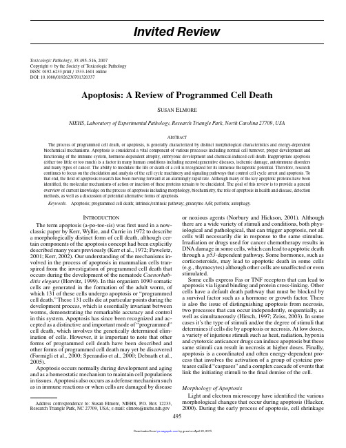
Chapter19Detection and Quantitative Analysis of Small RNAs by PCR Seungil Ro and Wei YanAbstractIncreasing lines of evidence indicate that small non-coding RNAs including miRNAs,piRNAs,rasiRNAs, 21U endo-siRNAs,and snoRNAs are involved in many critical biological processes.Functional studies of these small RNAs require a simple,sensitive,and reliable method for detecting and quantifying levels of small RNAs.Here,we describe such a method that has been widely used for the validation of cloned small RNAs and also for quantitative analyses of small RNAs in both tissues and cells.Key words:Small RNAs,miRNAs,piRNAs,expression,PCR.1.IntroductionThe past several years have witnessed the surprising discovery ofnumerous non-coding small RNAs species encoded by genomesof virtually all species(1–6),which include microRNAs(miR-NAs)(7–10),piwi-interacting RNAs(piRNAs)(11–14),repeat-associated siRNAs(rasiRNAs)(15–18),21U endo-siRNAs(19),and small nucleolar RNAs(snoRNAs)(20).These small RNAsare involved in all aspects of cellular functions through direct orindirect interactions with genomic DNAs,RNAs,and proteins.Functional studies on these small RNAs are just beginning,andsome preliminaryfindings have suggested that they are involvedin regulating genome stability,epigenetic marking,transcription,translation,and protein functions(5,21–23).An easy and sensi-tive method to detect and quantify levels of these small RNAs inorgans or cells during developmental courses,or under different M.Sioud(ed.),RNA Therapeutics,Methods in Molecular Biology629,DOI10.1007/978-1-60761-657-3_19,©Springer Science+Business Media,LLC2010295296Ro and Yanphysiological and pathophysiological conditions,is essential forfunctional studies.Quantitative analyses of small RNAs appear tobe challenging because of their small sizes[∼20nucleotides(nt)for miRNAs,∼30nt for piRNAs,and60–200nt for snoRNAs].Northern blot analysis has been the standard method for detec-tion and quantitative analyses of RNAs.But it requires a relativelylarge amount of starting material(10–20μg of total RNA or>5μg of small RNA fraction).It is also a labor-intensive pro-cedure involving the use of polyacrylamide gel electrophoresis,electrotransfer,radioisotope-labeled probes,and autoradiogra-phy.We have developed a simple and reliable PCR-based methodfor detection and quantification of all types of small non-codingRNAs.In this method,small RNA fractions are isolated and polyAtails are added to the3 ends by polyadenylation(Fig.19.1).Small RNA cDNAs(srcDNAs)are then generated by reverseFig.19.1.Overview of small RNA complementary DNA(srcDNA)library construction forPCR or qPCR analysis.Small RNAs are polyadenylated using a polyA polymerase.ThepolyA-tailed RNAs are reverse-transcribed using a primer miRTQ containing oligo dTsflanked by an adaptor sequence.RNAs are removed by RNase H from the srcDNA.ThesrcDNA is ready for PCR or qPCR to be carried out using a small RNA-specific primer(srSP)and a universal reverse primer,RTQ-UNIr.Quantitative Analysis of Small RNAs297transcription using a primer consisting of adaptor sequences atthe5 end and polyT at the3 end(miRTQ).Using the srcD-NAs,non-quantitative or quantitative PCR can then be per-formed using a small RNA-specific primer and the RTQ-UNIrprimer.This method has been utilized by investigators in numer-ous studies(18,24–38).Two recent technologies,454sequenc-ing and microarray(39,40)for high-throughput analyses of miR-NAs and other small RNAs,also need an independent method forvalidation.454sequencing,the next-generation sequencing tech-nology,allows virtually exhaustive sequencing of all small RNAspecies within a small RNA library.However,each of the clonednovel small RNAs needs to be validated by examining its expres-sion in organs or in cells.Microarray assays of miRNAs have beenavailable but only known or bioinformatically predicted miR-NAs are covered.Similar to mRNA microarray analyses,the up-or down-regulation of miRNA levels under different conditionsneeds to be further validated using conventional Northern blotanalyses or PCR-based methods like the one that we are describ-ing here.2.Materials2.1.Isolation of Small RNAs, Polyadenylation,and Purification 1.mirVana miRNA Isolation Kit(Ambion).2.Phosphate-buffered saline(PBS)buffer.3.Poly(A)polymerase.4.mirVana Probe and Marker Kit(Ambion).2.2.Reverse Transcription,PCR, and Quantitative PCR 1.Superscript III First-Strand Synthesis System for RT-PCR(Invitrogen).2.miRTQ primers(Table19.1).3.AmpliTaq Gold PCR Master Mix for PCR.4.SYBR Green PCR Master Mix for qPCR.5.A miRNA-specific primer(e.g.,let-7a)and RTQ-UNIr(Table19.1).6.Agarose and100bp DNA ladder.3.Methods3.1.Isolation of Small RNAs 1.Harvest tissue(≤250mg)or cells in a1.7-mL tube with500μL of cold PBS.T a b l e 19.1O l i g o n u c l e o t i d e s u s e dN a m eS e q u e n c e (5 –3 )N o t eU s a g em i R T QC G A A T T C T A G A G C T C G A G G C A G G C G A C A T G G C T G G C T A G T T A A G C T T G G T A C C G A G C T A G T C C T T T T T T T T T T T T T T T T T T T T T T T T T V N ∗R N a s e f r e e ,H P L CR e v e r s e t r a n s c r i p t i o nR T Q -U N I r C G A A T T C T A G A G C T C G A G G C A G GR e g u l a r d e s a l t i n gP C R /q P C Rl e t -7a T G A G G T A G T A G G T T G T A T A G R e g u l a r d e s a l t i n gP C R /q P C R∗V =A ,C ,o r G ;N =A ,C ,G ,o r TQuantitative Analysis of Small RNAs299 2.Centrifuge at∼5,000rpm for2min at room temperature(RT).3.Remove PBS as much as possible.For cells,remove PBScarefully without breaking the pellet,leave∼100μL of PBS,and resuspend cells by tapping gently.4.Add300–600μL of lysis/binding buffer(10volumes pertissue mass)on ice.When you start with frozen tissue or cells,immediately add lysis/binding buffer(10volumes per tissue mass)on ice.5.Cut tissue into small pieces using scissors and grind it usinga homogenizer.For cells,skip this step.6.Vortex for40s to mix.7.Add one-tenth volume of miRNA homogenate additive onice and mix well by vortexing.8.Leave the mixture on ice for10min.For tissue,mix it every2min.9.Add an equal volume(330–660μL)of acid-phenol:chloroform.Be sure to withdraw from the bottom phase(the upper phase is an aqueous buffer).10.Mix thoroughly by inverting the tubes several times.11.Centrifuge at10,000rpm for5min at RT.12.Recover the aqueous phase carefully without disrupting thelower phase and transfer it to a fresh tube.13.Measure the volume using a scale(1g=∼1mL)andnote it.14.Add one-third volume of100%ethanol at RT to the recov-ered aqueous phase.15.Mix thoroughly by inverting the tubes several times.16.Transfer up to700μL of the mixture into afilter cartridgewithin a collection bel thefilter as total RNA.When you have>700μL of the mixture,apply it in suc-cessive application to the samefilter.17.Centrifuge at10,000rpm for15s at RT.18.Collect thefiltrate(theflow-through).Save the cartridgefor total RNA isolation(go to Step24).19.Add two-third volume of100%ethanol at RT to theflow-through.20.Mix thoroughly by inverting the tubes several times.21.Transfer up to700μL of the mixture into a newfilterbel thefilter as small RNA.When you have >700μL of thefiltrate mixture,apply it in successive appli-cation to the samefilter.300Ro and Yan22.Centrifuge at10,000rpm for15s at RT.23.Discard theflow-through and repeat until all of thefiltratemixture is passed through thefilter.Reuse the collectiontube for the following washing steps.24.Apply700μL of miRNA wash solution1(working solu-tion mixed with ethanol)to thefilter.25.Centrifuge at10,000rpm for15s at RT.26.Discard theflow-through.27.Apply500μL of miRNA wash solution2/3(working solu-tion mixed with ethanol)to thefilter.28.Centrifuge at10,000rpm for15s at RT.29.Discard theflow-through and repeat Step27.30.Centrifuge at12,000rpm for1min at RT.31.Transfer thefilter cartridge to a new collection tube.32.Apply100μL of pre-heated(95◦C)elution solution orRNase-free water to the center of thefilter and close thecap.Aliquot a desired amount of elution solution intoa1.7-mL tube and heat it on a heat block at95◦C for∼15min.Open the cap carefully because it might splashdue to pressure buildup.33.Leave thefilter tube alone for1min at RT.34.Centrifuge at12,000rpm for1min at RT.35.Measure total RNA and small RNA concentrations usingNanoDrop or another spectrophotometer.36.Store it at–80◦C until used.3.2.Polyadenylation1.Set up a reaction mixture with a total volume of50μL in a0.5-mL tube containing0.1–2μg of small RNAs,10μL of5×E-PAP buffer,5μL of25mM MnCl2,5μL of10mMATP,1μL(2U)of Escherichia coli poly(A)polymerase I,and RNase-free water(up to50μL).When you have a lowconcentration of small RNAs,increase the total volume;5×E-PAP buffer,25mM MnCl2,and10mM ATP should beincreased accordingly.2.Mix well and spin the tube briefly.3.Incubate for1h at37◦C.3.3.Purification 1.Add an equal volume(50μL)of acid-phenol:chloroformto the polyadenylation reaction mixture.When you have>50μL of the mixture,increase acid-phenol:chloroformaccordingly.2.Mix thoroughly by tapping the tube.Quantitative Analysis of Small RNAs3013.Centrifuge at10,000rpm for5min at RT.4.Recover the aqueous phase carefully without disrupting thelower phase and transfer it to a fresh tube.5.Add12volumes(600μL)of binding/washing buffer tothe aqueous phase.When you have>50μL of the aqueous phase,increase binding/washing buffer accordingly.6.Transfer up to460μL of the mixture into a purificationcartridge within a collection tube.7.Centrifuge at10,000rpm for15s at RT.8.Discard thefiltrate(theflow-through)and repeat until allof the mixture is passed through the cartridge.Reuse the collection tube.9.Apply300μL of binding/washing buffer to the cartridge.10.Centrifuge at12,000rpm for1min at RT.11.Transfer the cartridge to a new collection tube.12.Apply25μL of pre-heated(95◦C)elution solution to thecenter of thefilter and close the cap.Aliquot a desired amount of elution solution into a1.7-mL tube and heat it on a heat block at95◦C for∼15min.Open the cap care-fully because it might be splash due to pressure buildup.13.Let thefilter tube stand for1min at RT.14.Centrifuge at12,000rpm for1min at RT.15.Repeat Steps12–14with a second aliquot of25μL ofpre-heated(95◦C)elution solution.16.Measure polyadenylated(tailed)RNA concentration usingNanoDrop or another spectrophotometer.17.Store it at–80◦C until used.After polyadenylation,RNAconcentration should increase up to5–10times of the start-ing concentration.3.4.Reverse Transcription 1.Mix2μg of tailed RNAs,1μL(1μg)of miRTQ,andRNase-free water(up to21μL)in a PCR tube.2.Incubate for10min at65◦C and for5min at4◦C.3.Add1μL of10mM dNTP mix,1μL of RNaseOUT,4μLof10×RT buffer,4μL of0.1M DTT,8μL of25mM MgCl2,and1μL of SuperScript III reverse transcriptase to the mixture.When you have a low concentration of lig-ated RNAs,increase the total volume;10×RT buffer,0.1M DTT,and25mM MgCl2should be increased accordingly.4.Mix well and spin the tube briefly.5.Incubate for60min at50◦C and for5min at85◦C toinactivate the reaction.302Ro and Yan6.Add1μL of RNase H to the mixture.7.Incubate for20min at37◦C.8.Add60μL of nuclease-free water.3.5.PCR and qPCR 1.Set up a reaction mixture with a total volume of25μL ina PCR tube containing1μL of small RNA cDNAs(srcD-NAs),1μL(5pmol of a miRNA-specific primer(srSP),1μL(5pmol)of RTQ-UNIr,12.5μL of AmpliTaq GoldPCR Master Mix,and9.5μL of nuclease-free water.ForqPCR,use SYBR Green PCR Master Mix instead of Ampli-Taq Gold PCR Master Mix.2.Mix well and spin the tube briefly.3.Start PCR or qPCR with the conditions:95◦C for10minand then40cycles at95◦C for15s,at48◦C for30s and at60◦C for1min.4.Adjust annealing Tm according to the Tm of your primer5.Run2μL of the PCR or qPCR products along with a100bpDNA ladder on a2%agarose gel.∼PCR products should be∼120–200bp depending on the small RNA species(e.g.,∼120–130bp for miRNAs and piRNAs).4.Notes1.This PCR method can be used for quantitative PCR(qPCR)or semi-quantitative PCR(semi-qPCR)on small RNAs suchas miRNAs,piRNAs,snoRNAs,small interfering RNAs(siRNAs),transfer RNAs(tRNAs),and ribosomal RNAs(rRNAs)(18,24–38).2.Design miRNA-specific primers to contain only the“coresequence”since our cloning method uses two degeneratenucleotides(VN)at the3 end to make small RNA cDNAs(srcDNAs)(see let-7a,Table19.1).3.For qPCR analysis,two miRNAs and a piRNA were quan-titated using the SYBR Green PCR Master Mix(41).Cyclethreshold(Ct)is the cycle number at which thefluorescencesignal reaches the threshold level above the background.ACt value for each miRNA tested was automatically calculatedby setting the threshold level to be0.1–0.3with auto base-line.All Ct values depend on the abundance of target miR-NAs.For example,average Ct values for let-7isoforms rangefrom17to20when25ng of each srcDNA sample from themultiple tissues was used(see(41).Quantitative Analysis of Small RNAs3034.This method amplifies over a broad dynamic range up to10orders of magnitude and has excellent sensitivity capable ofdetecting as little as0.001ng of the srcDNA in qPCR assays.5.For qPCR,each small RNA-specific primer should be testedalong with a known control primer(e.g.,let-7a)for PCRefficiency.Good efficiencies range from90%to110%calcu-lated from slopes between–3.1and–3.6.6.On an agarose gel,mature miRNAs and precursor miRNAs(pre-miRNAs)can be differentiated by their size.PCR prod-ucts containing miRNAs will be∼120bp long in size whileproducts containing pre-miRNAs will be∼170bp long.However,our PCR method preferentially amplifies maturemiRNAs(see Results and Discussion in(41)).We testedour PCR method to quantify over100miRNAs,but neverdetected pre-miRNAs(18,29–31,38). AcknowledgmentsThe authors would like to thank Jonathan Cho for reading andediting the text.This work was supported by grants from theNational Institute of Health(HD048855and HD050281)toW.Y.References1.Ambros,V.(2004)The functions of animalmicroRNAs.Nature,431,350–355.2.Bartel,D.P.(2004)MicroRNAs:genomics,biogenesis,mechanism,and function.Cell, 116,281–297.3.Chang,T.C.and Mendell,J.T.(2007)Theroles of microRNAs in vertebrate physiol-ogy and human disease.Annu Rev Genomics Hum Genet.4.Kim,V.N.(2005)MicroRNA biogenesis:coordinated cropping and dicing.Nat Rev Mol Cell Biol,6,376–385.5.Kim,V.N.(2006)Small RNAs just gotbigger:Piwi-interacting RNAs(piRNAs) in mammalian testes.Genes Dev,20, 1993–1997.6.Kotaja,N.,Bhattacharyya,S.N.,Jaskiewicz,L.,Kimmins,S.,Parvinen,M.,Filipowicz, W.,and Sassone-Corsi,P.(2006)The chro-matoid body of male germ cells:similarity with processing bodies and presence of Dicer and microRNA pathway components.Proc Natl Acad Sci U S A,103,2647–2652.7.Aravin,A.A.,Lagos-Quintana,M.,Yalcin,A.,Zavolan,M.,Marks,D.,Snyder,B.,Gaaster-land,T.,Meyer,J.,and Tuschl,T.(2003) The small RNA profile during Drosophilamelanogaster development.Dev Cell,5, 337–350.8.Lee,R.C.and Ambros,V.(2001)An exten-sive class of small RNAs in Caenorhabditis ele-gans.Science,294,862–864.u,N.C.,Lim,L.P.,Weinstein, E.G.,and Bartel,D.P.(2001)An abundant class of tiny RNAs with probable regulatory roles in Caenorhabditis elegans.Science,294, 858–862.gos-Quintana,M.,Rauhut,R.,Lendeckel,W.,and Tuschl,T.(2001)Identification of novel genes coding for small expressed RNAs.Science,294,853–858.u,N.C.,Seto,A.G.,Kim,J.,Kuramochi-Miyagawa,S.,Nakano,T.,Bartel,D.P.,and Kingston,R.E.(2006)Characterization of the piRNA complex from rat testes.Science, 313,363–367.12.Grivna,S.T.,Beyret,E.,Wang,Z.,and Lin,H.(2006)A novel class of small RNAs inmouse spermatogenic cells.Genes Dev,20, 1709–1714.13.Girard, A.,Sachidanandam,R.,Hannon,G.J.,and Carmell,M.A.(2006)A germline-specific class of small RNAs binds mammalian Piwi proteins.Nature,442,199–202.304Ro and Yan14.Aravin,A.,Gaidatzis,D.,Pfeffer,S.,Lagos-Quintana,M.,Landgraf,P.,Iovino,N., Morris,P.,Brownstein,M.J.,Kuramochi-Miyagawa,S.,Nakano,T.,Chien,M.,Russo, J.J.,Ju,J.,Sheridan,R.,Sander,C.,Zavolan, M.,and Tuschl,T.(2006)A novel class of small RNAs bind to MILI protein in mouse testes.Nature,442,203–207.15.Watanabe,T.,Takeda, A.,Tsukiyama,T.,Mise,K.,Okuno,T.,Sasaki,H.,Minami, N.,and Imai,H.(2006)Identification and characterization of two novel classes of small RNAs in the mouse germline: retrotransposon-derived siRNAs in oocytes and germline small RNAs in testes.Genes Dev,20,1732–1743.16.Vagin,V.V.,Sigova,A.,Li,C.,Seitz,H.,Gvozdev,V.,and Zamore,P.D.(2006)A distinct small RNA pathway silences selfish genetic elements in the germline.Science, 313,320–324.17.Saito,K.,Nishida,K.M.,Mori,T.,Kawa-mura,Y.,Miyoshi,K.,Nagami,T.,Siomi,H.,and Siomi,M.C.(2006)Specific asso-ciation of Piwi with rasiRNAs derived from retrotransposon and heterochromatic regions in the Drosophila genome.Genes Dev,20, 2214–2222.18.Ro,S.,Song,R.,Park, C.,Zheng,H.,Sanders,K.M.,and Yan,W.(2007)Cloning and expression profiling of small RNAs expressed in the mouse ovary.RNA,13, 2366–2380.19.Ruby,J.G.,Jan,C.,Player,C.,Axtell,M.J.,Lee,W.,Nusbaum,C.,Ge,H.,and Bartel,D.P.(2006)Large-scale sequencing reveals21U-RNAs and additional microRNAs and endogenous siRNAs in C.elegans.Cell,127, 1193–1207.20.Terns,M.P.and Terns,R.M.(2002)Small nucleolar RNAs:versatile trans-acting molecules of ancient evolutionary origin.Gene Expr,10,17–39.21.Ouellet,D.L.,Perron,M.P.,Gobeil,L.A.,Plante,P.,and Provost,P.(2006)MicroR-NAs in gene regulation:when the smallest governs it all.J Biomed Biotechnol,2006, 69616.22.Maatouk,D.and Harfe,B.(2006)MicroR-NAs in development.ScientificWorldJournal, 6,1828–1840.23.Kim,V.N.and Nam,J.W.(2006)Genomics of microRNA.Trends Genet,22, 165–173.24.Bohnsack,M.T.,Kos,M.,and Tollervey,D.(2008)Quantitative analysis of snoRNAassociation with pre-ribosomes and release of snR30by Rok1helicase.EMBO Rep,9, 1230–1236.25.Hertel,J.,de Jong, D.,Marz,M.,Rose,D.,Tafer,H.,Tanzer, A.,Schierwater,B.,and Stadler,P.F.(2009)Non-codingRNA annotation of the genome of Tri-choplax adhaerens.Nucleic Acids Res,37, 1602–1615.26.Kim,M.,Patel,B.,Schroeder,K.E.,Raza,A.,and Dejong,J.(2008)Organization andtranscriptional output of a novel mRNA-like piRNA gene(mpiR)located on mouse chro-mosome10.RNA,14,1005–1011.27.Mishima,T.,Takizawa,T.,Luo,S.S.,Ishibashi,O.,Kawahigashi,Y.,Mizuguchi, Y.,Ishikawa,T.,Mori,M.,Kanda,T., and Goto,T.(2008)MicroRNA(miRNA) cloning analysis reveals sex differences in miRNA expression profiles between adult mouse testis and ovary.Reproduction,136, 811–822.28.Papaioannou,M.D.,Pitetti,J.L.,Ro,S.,Park, C.,Aubry, F.,Schaad,O.,Vejnar,C.E.,Kuhne, F.,Descombes,P.,Zdob-nov, E.M.,McManus,M.T.,Guillou, F., Harfe,B.D.,Yan,W.,Jegou,B.,and Nef, S.(2009)Sertoli cell Dicer is essential for spermatogenesis in mice.Dev Biol,326, 250–259.29.Ro,S.,Park,C.,Sanders,K.M.,McCarrey,J.R.,and Yan,W.(2007)Cloning and expres-sion profiling of testis-expressed microRNAs.Dev Biol,311,592–602.30.Ro,S.,Park,C.,Song,R.,Nguyen,D.,Jin,J.,Sanders,K.M.,McCarrey,J.R.,and Yan, W.(2007)Cloning and expression profiling of testis-expressed piRNA-like RNAs.RNA, 13,1693–1702.31.Ro,S.,Park,C.,Young,D.,Sanders,K.M.,and Yan,W.(2007)Tissue-dependent paired expression of miRNAs.Nucleic Acids Res, 35,5944–5953.32.Siebolts,U.,Varnholt,H.,Drebber,U.,Dienes,H.P.,Wickenhauser,C.,and Oden-thal,M.(2009)Tissues from routine pathol-ogy archives are suitable for microRNA anal-yses by quantitative PCR.J Clin Pathol,62, 84–88.33.Smits,G.,Mungall,A.J.,Griffiths-Jones,S.,Smith,P.,Beury,D.,Matthews,L.,Rogers, J.,Pask, A.J.,Shaw,G.,VandeBerg,J.L., McCarrey,J.R.,Renfree,M.B.,Reik,W.,and Dunham,I.(2008)Conservation of the H19 noncoding RNA and H19-IGF2imprint-ing mechanism in therians.Nat Genet,40, 971–976.34.Song,R.,Ro,S.,Michaels,J.D.,Park,C.,McCarrey,J.R.,and Yan,W.(2009)Many X-linked microRNAs escape meiotic sex chromosome inactivation.Nat Genet,41, 488–493.Quantitative Analysis of Small RNAs30535.Wang,W.X.,Wilfred,B.R.,Baldwin,D.A.,Isett,R.B.,Ren,N.,Stromberg, A.,and Nelson,P.T.(2008)Focus on RNA iso-lation:obtaining RNA for microRNA (miRNA)expression profiling analyses of neural tissue.Biochim Biophys Acta,1779, 749–757.36.Wu,F.,Zikusoka,M.,Trindade,A.,Das-sopoulos,T.,Harris,M.L.,Bayless,T.M., Brant,S.R.,Chakravarti,S.,and Kwon, J.H.(2008)MicroRNAs are differen-tially expressed in ulcerative colitis and alter expression of macrophage inflam-matory peptide-2alpha.Gastroenterology, 135(1624–1635),e24.37.Wu,H.,Neilson,J.R.,Kumar,P.,Manocha,M.,Shankar,P.,Sharp,P.A.,and Manjunath, N.(2007)miRNA profiling of naive,effec-tor and memory CD8T cells.PLoS ONE,2, e1020.38.Yan,W.,Morozumi,K.,Zhang,J.,Ro,S.,Park, C.,and Yanagimachi,R.(2008) Birth of mice after intracytoplasmic injec-tion of single purified sperm nuclei and detection of messenger RNAs and microR-NAs in the sperm nuclei.Biol Reprod,78, 896–902.39.Guryev,V.and Cuppen,E.(2009)Next-generation sequencing approaches in genetic rodent model systems to study func-tional effects of human genetic variation.FEBS Lett.40.Li,W.and Ruan,K.(2009)MicroRNAdetection by microarray.Anal Bioanal Chem.41.Ro,S.,Park,C.,Jin,JL.,Sanders,KM.,andYan,W.(2006)A PCR-based method for detection and quantification of small RNAs.Biochem and Biophys Res Commun,351, 756–763.。
巨噬细胞刺激因子(MSF)与巨噬细胞抑制因子(MIF)对视网膜节细胞轴突再生的影响岑令平;梁嘉健;张铭志【摘要】目的研究巨噬细胞刺激因子(macrophage stimulating factor,MSF)及巨噬细胞抑制因子(macrophage inhibitory factor,MIF)对视网膜节细胞(retinal ganglion cell,RGC)轴突再生的影响.方法实验性研究.费希尔大鼠9只,施行视神经损伤术7d后取出眼球并剥离视网膜,把剥离下来的视网膜平铺,放射状剪成8片,每一片视网膜粘铺于已包被好的培养板孔中,分为对照组、MSF组、MIF组,分别加入培养基及MSF/MIF等处理因素试剂;培养7d后对视网膜培养块进行固定及免疫荧光染色,分别计数RGC再生神经轴突的数量及长度和外迁巨噬细胞的数量.结果与对照组相比,MSF及MIF可以一定程度上分别增多及减少迁移巨噬细胞的数量(P>0.05),与此同时,MIF可以减少RGC再生神经轴突的数量及长度(P<0.05),MSF虽未能增加神经突的长度,但还是有效地增加了再生神经突的数量(P<0.05).结论MSF可以促进RGC神经轴突的再生,与此相反MIF可以抑制神经轴突的再生.【期刊名称】《中国中医眼科杂志》【年(卷),期】2016(026)003【总页数】4页(P150-153)【关键词】视网膜节细胞;巨噬细胞;视神经损伤;存活;神经再生【作者】岑令平;梁嘉健;张铭志【作者单位】汕头大学·香港中文大学联合汕头国际眼科中心,广东汕头515041;汕头大学·香港中文大学联合汕头国际眼科中心,广东汕头515041;汕头大学·香港中文大学联合汕头国际眼科中心,广东汕头515041【正文语种】中文【中图分类】R774视神经损伤严重影响视觉功能,甚至可以导致视力丧失而致盲。
视神经损伤会引起大量视网膜神经节细胞(retinal ganglion cell,RGC)继发性凋亡,从而导致视力损害。
RNA Biology 7:5, 621-627; September/October 2010; © 2010 Landes BioscienceReSeARch pApeRReSeARch pApeR*Correspondence to: Jörg Vogel and Mary A. O’Connell; Email: vogel@mpiib-berlin.mpg.de and M.O’Connell@ Submitted: 07/13/10; Accepted: 08/05/10Previously published online: /journals/rnabiology/article/13269DOI: 10.4161.rna.7.5.13269IntroductionIn mammals, ADAR1 and ADAR2, members of the adenosine deaminase family that act on double-stranded RNA (dsRNA), have been demonstrated to deaminate specific adenosines in miRNAs.1-6 This deamination usually results in a reduction in the mature miRNA levels as RNA editing and binding of ADARs can inhibit processing of pri-miRNA.7 RNA editing has also been shown to redirect miRNA activity when the editing event is located in the mature miRNA sequence.3Intuitively, when ADAR activity increases there is an expecta-tion of more frequent editing of miRNAs. It is well documented that ADAR1 is a ubiquitously expressed 110 kDa protein that is predominantly nuclear. However upon interferon induction a larger isoform of 150 kDa is expressed that shuttles between nucleus and cytoplasm (reviewed in ref. 8) and mice that are null for ADAR1 exhibit high levels of interferon suggesting that there may even be a feedback loop between ADAR1 and interferon levels.In this study we wanted to determine if an increase in ADAR1 levels due to interferon would affect mature miRNAs. We chose to study Salmonella infection as Samuels and colleagues have shown that mice infected with Salmonella have a large inductionThe main mediator of the lipopolysaccharide (LpS) response in macrophages is activation of Toll-like receptor 4 (TLR4). This generates interferon-β (INFβ) production that stimulates increased expression of the RNA editing enzyme ADAR1. To determine if there is an increase in RNA editing in mature miRNA in response to TLR4 activation upon Salmonella infection of macrophages we analyzed small RNA deep sequencing data. Interestingly, we found that direct infection of macrophage cell lines with Salmonella does not result in an increase of edited mature miRNA. Thus, despite elevated levels of ADAR1 during TLR4 activation of macrophages mediated by Salmonella infection, ADAR1 does not result in redirection of miRNA. The most common consequence of ADAR activity on miRNA is a reduction in the mature miRNA level due to interference with miRNA processing of pri-miRNA. however, we found very few miRNAs with reductions in level, and no significant difference between miRNAs previously reported to be edited and those reported to be not edited. In particular, we did not see significant decrease in mir-22 and mir-142, nor editing of pri-mir-22 or pri-mir-142 in infected RAW macrophages. Thus, ADAR1 has very little, if any, effect on the miRNA machinery following TL4 activation by Salmonella infection.Analysis of A to I editing of miRNA in macrophages exposed to salmonellaBret S.e. heale,1 Ana eulalio,2 Leon Schulte,2 Jörg Vogel 2,* and Mary A. O’connell 1,*1MRc human Genetics Unit; Institute of Genetics and Molecular Medicine; Western General hospital; edinburgh, UK; 2Max planck Institute for Infection Biology;Berlin, GermanyKey words: RNA editing, miRNA, ADAR, deep sequencingAbbreviations: LPS, lipopolysaccharide; TLR4, toll-like receptor 4; INF b , interferon-b ; ADAR, adenosine deaminase that act onRNA; dsRNA, double-stranded RNA; salmonella, Salmonella enterica serovar typhimuriumof ADAR1 transcript levels.9 The increase is due to the induction of an interferon inducible isoform of ADAR1.10 Furthermore, Rabinovici and colleagues demonstrated that editing activity of ADAR1 rises dramatically upon alveolar macrophage activation by LPS.11 Thus, we wanted to ascertain if there was an increase in editing of mature miRNA upon Salmonella infection of macro-phages. We also wanted to determine whether the consequences would be redirection or inhibition of pri-miRNA processing.This work also addresses the role ADAR1 may have in innate immunity. In many physiological pathways miRNAs are prominent players, and evidence is accumulating that miRNAs are regulated as part of the innate immune response. Notably, LPS simulation of macrophages alters the levels of mir-146a,12 mir-155,13,14 and mir-125b.14 An increased level of mir-146 has been directly linked to a reduction of IRAK1 and TRAF6, two important members of the TLR signaling pathway.12 Conversely, a decrease in mir-125b was hypothesized to be linked to changes in TNF α levels, as TNF α is a target gene for mir-125b,14 indi-cating that decreases in miRNA levels can also be important. These studies demonstrate the important role miRNAs have in the regulation of the innate immune response and motivate the examination of how interferon induction of ADAR1 may con-tribute miRNA regulation.do not rise in mature miRNA in macro-phages, despite induction of ADAR1 by Salmonella infection. Further, we exam-ined two miRNA transcripts reported to be edited by ADAR1 and did not observe an increase in editing. Overall, our results support the conclusion that inosine is not a common modification present in mature miRNA. This suggests that the miRNA machinery is resistant to increased ADAR activity caused by TLR4 activation.ResultsSequence analysis o f miRNAs fro m infected and no n infected macro phage cell lines. I nitially we wanted to verify that ADAR1 was present in RAW mac-rophage cells and that it was induced in RAW macrophages infected with Salmonella. We choose a 24 hr time point as it has been reported that ADAR1 induction peaks approximately 5 hr after injection of mice with LPS,19 so we rea-soned that if miRNAs were edited then 24 hr post infection would be the opti-mum time to analyze them. We found that 24 hr post infection the ADAR1 RNA level increased approximately 2-fold, ADAR1 protein level determined by SI LAC increased approximately3.7-fold 20(data not shown) whereas non-specific editing activity increased approximately 1.5-fold (Fig. 1).Next we examined seventeen small RNA libraries of sev-eral macrophage cell lines infected with mutant and wild-type strains of Salmonella. The SP1 Salmonella mutant is known to block entry of the bacteria into macrophages. Thus, using the SP1 mutant we can separate the effect on the host in terms ofbacterial contact and infection. The macrophage cell types used were, RAW, THP1 and J774, each has distinct origins. RAW macrophages were derived from mice infected with Ableson murine leukemia virus.21 THP1 cells are a human acute mono-cytic leukemia cell line from a patient tumor.22 Whereas, J774 are a murine macrophage cell line established from a tumor.23Examinations of the libraries yielded on average a surpris-ing 40% mapping of reads to perfect matches of the mature miRNA annotations in mirBase (Table 1 summary of RAW and HeLa cell lines. Supplementary Tables provide individual miRNA results. Very similar results were obtained for THP1 and J774 and HEK 293, data not shown). Extending the counts of matched reads to include potential A to G, G to A, C to U, U to C and U to A base changes, slightly increased the number of reads identified as miRNA. A to G mismatches between the miRNA sequence database and the sequences are interpreted as evidence for adenosine deamination, as inosine is recognized as guanosine by polymerases.Initial observations indicate that only one miRNA containedpotential A to I editing events; mir-155. This miRNA dramaticallyFinally , Salmonella enterica serovar Typhimurium (henceforth Salmonella) is a model pathogen that invades macrophages and activates a TLR4-dependent host response.15 Mutation of the SPI-1 virulence region renders the bacteria unable to invade the host cell.16 Thus, by comparing infection with the SPI-1 mutant and wild-type Salmonella, we can determine if mature miRNA editing events are a result of contact with Salmonella or are infec-tion dependent.Due to the expansion in sequencing libraries, much effort is being directed towards how to correctly measure the sequence content of these libraries, including how to measure editing of small RNAs. The deamination of adenosine produces inosine, which base-pairs like guanosine, and is read by reverse transcrip-tase as guanosine.17 Thus, in sequencing small RNA libraries a replacement of adenosine with guanosine can be evidence of ADAR activity. Most analysis approaches utilize a modified blast algorithm, with the goal to match as many reads as possible to the genome. However, to map the largest number of reads pos-sible the stringency of sequence homology must be reduced. This can lead to artifacts in the assignment of reads to genomic loci that are interpreted as editing events.18 Therefore we use a perfect matching approach to ascertain the level of editing in mature miRNA in Salmonella infection of macrophages, where correct identification (specificity) is more critical then a large number of matched reads (sensitivity).In this study we analyzed small RNA libraries produced by infection of RAW, HeLa, THP-1, J774 and HEK cell lines with different strains of Salmonella. The editing levels of mature miRNA were determined with our simplified matching algo-rithm. Our study revealed that potential A to I editing levels Figure 1. dsRNA deaminase activity assay. The lanes are 0 extract, (1) 5 μg extract from non-infect-ed cells, (2) 10 μg extract from non-infected cells, (3) 20 μg extract from non-infected cells, (4) 5μg extract from infected cells, (5) 10 μg extract from infected cells, (6) 20 μg extract from infectedcells, (c) positive control with recombinant hADAR2. The upper limit of editing was 10 percentof the input dsRNA signal. Quantization was performed by measuring the signal of the inosine and adenosine spots. each sample was done in duplicate and the experiment performed twice with similar result. There was more editing activity in the infected RAW macrophages than in thecontrol non-infected macrophages.the potential for ADAR1 to be involved in reduction of miRNA levels by inhibiting processing. Comparing the levels of miRNA before and after infection with Salmonella revealed that very few miRNA had a reduction of even 2-fold (fold Changes are in Sup. Tables 1 and 2). Further, we analyzed sixty-five miRNA previ-ously reported to be edited and there is no significant difference between alterations in the levels of miRNA reported to be ADAR substrates and miRNA not known to be ADAR substrates. Figure 2 illustrates this point by showing that the absolute fold-changes in miRNA abundance are low and that both the average and spread of the fold-changes is similar. This indicates that ADAR1 is not participating in a global reduction in miRNA levels, and suggests that miRNA previously reported to be edited are no more likely to be reduced in level than those reported not to be edited.Additionally, we examined pri-miRNA sequences for two well-documented ADAR substrates, mir-22 and mir-142.1,25 We did not find evidence of upregulation of editing of the pri-miRNA for mir-22 and mir-142 after Salmonella infection, and the mature miRNA levels do not decrease significantly in RAW macrophages. The lack of editing of the pri-miRNA in RAW macrophages indicates that alteration of mir-142 is not depen-dent on ADAR activity on the pri-miRNA, and the increase in mir-22 further supports the assertion that induction of ADAR1 after infection with Salmonella does not lead to a strong negative effect of ADAR1 on miRNA biogenesis, even for known editing substrates.DiscussionGiven that ADAR1 expression and activity is inducted by inter-feron, it is surprising that Salmonella infection did not result in an increase of edited mature miRNA. As part of the innate immune response TLR4 is stimulated by LPS.26 Recently it wasincreased in RAW cells upon exposure to the SP1 and wild-type strains of Salmonella. The response to the SP1 mutant is par-ticularly revealing as this mutant cannot invade macrophages but does contain lipopolysaccharides in its membrane. Initially, we observed that 14% of reads mapping to mir-155 were potentially A to I edited at 24 hr post RAW cell exposure to Salmonella. The number of reads for mir-155 was 337, therefore the number of edited mature mir-155 reads would be a significant contributor to the miRNA pool. Supplementary Tables 1 and 2 contain the number of reads for individual miRNA. As can be seen 337 reads would place edited mir-155 at levels comparable to the eight most abundant miRNA for this library.However, when we re-examined the potential G to A transver-sions, we observed that the nucleotide just 3' of the A to G change in mir-155 had a significant number of G to A mismatches as well. There is no known enzyme that can catalyze a G to A trans-version in RNA however RNA polymerase stuttering has been shown to insert more than one G when there are multiple Gs.24 Further, when we examined the percent of A to G reads for mir-155 across the RAW cell libraries we found that the percent does not change in response to infection. Indeed, the percent editing of mir-155 is the same in each of the libraries associated with a specific cell type. Additionally, we found that the primary mir-155 transcript is not edited either in non-infected nor infected macrophages (data not shown). Though the possibility exits that pre-mir-155 is edited, we conclude that the potential editing of mir-155 is a systematic error or artifact, in sequencing, dependent on the sequence context. Therefore we found no evidence of A to I editing in mature miRNA in the libraries.ADAR1 do es not inhibit pro cessing of miRNAs. The gen-eral lack of A to I editing indicates that Salmonella infection did not result in ADAR1 editing of mature miRNA, and hence redi-rection of miRNA. To explore this result further, we examinedTable 1. Summary of analysis of RAW and hela sequence libraries using simplified perfect match algorithmRAW Total Perfect match Reads with A to GReads with G to A Reads with C to UReads with U to CReads with U to ALibrary information 0 hr control non-infected 153251270280243111398control non-infected 24 hr control non-infected 151371277569215823130control 24 hours 24 hr SpI1 mutant 1646113728339*23022126924 hr infection with SpI-1mutant 24 hr WT i nfection 1377211329381*1956619924 hr infection with wild typeHela Total Perfect match Reads with A to GReads with G to A Reads with C to UReads with U to CReads with U to ALibrary information 0 hr control non-infected 2110619044126189411229control non-infected 24 hr control non-infected 231352080070219634926control 24 hrs24 hr WT i nfection18088164786114991872524 hr infection with wild typeNumber in box is the number of reads. *The percent of A to G reads for each miRNA does not change, the increase in number of A to G reads is due to an increase in mir-155.edited mature miRNA. Thus, given that the number of mature miRNA with potential A to I editing remains constant, we pro-pose that increased activity of ADAR1 due to TLR mediated innate immune response does not involve redirecting miRNA.A possible explanation is that A to I editing of miRNA inhibits the biogenesis of mature miRNA. In fact, one of the most com-mon features of A to I editing of miRNA is inhibition of process-ing by editing of the primary miRNA transcript. However, in our study few miRNA have decreased levels of mature miRNA upon infection. This suggests that reduction in mature miRNA levels is not a general feature of Salamonella infection of RAW mac-rophages. Significantly, we did not see editing of pri-mir-22 and pri-mir-142, two miRNA shown to be edited by ADAR1.1,25 Pri-mir-22 has been found to be edited in various tissues including lung and brain. However, mir-142 is edited in T cells, although no physiological role could be found for this editing. This high-lights the potential for cell-type specific editing, which could explain the lack of editing in macrophages. Our results support the conclusion that ADAR1 does not interfere with the produc-tion of these prominent, edited-miRNA in macrophages. Thus, the increase in ADAR1 in response to interferon is for some other purpose than increasing the interactions between ADAR1 and miRNA.Materials and MethodsSalmonella infection and culture of macrophages. RAW264.7, HeLa, THP-1, J774 and HEK293 cells were grown in RPMI 1640 (Gibco) supplemented with 10% foetal calf serum (Biochrom), 2 mM L-glutamine (Gibco), 1 mM sodium-pyrovate (GIBCO) and 0.5% ß-mercapto-ethanol (Gibco) in a 5% CO 2, humidified atmosphere, at 37°C. Salmonella enterica serovar Typhimurium strain SL1344 was used as wild type throughout this study. The ΔSPI1 (JVS-0405) or ΔSPI2 (JVS-1103) strains with deletions of Salmonella Pathogenicity I slands 1 or 2 were provided byS. Pätzold 28 or Karsten Tedin, respectively.29The ΔSPI1/ΔSPI2 strain (JVS-3614) was constructed by phage P22 transduction of strain JVS-0405 with a lysate of strain JVS-1103.One day prior to infection, 4 x 10⁵ of host cells were seeded into 6-well plates. Overnight cultures of bacteria were diluted 1:100 in fresh L-broth medium and grown aerobically until an OD of 2. Bacteria were harvested by centrifugation and resuspended in complete RPMI medium. RAW 264.7 cells were infected at MOI of 1, and HeLa cells at MOI of 10. After addition of bacte-ria, the cells were centrifuged at 250x g for 10 min at room tem-perature followed by 20 min incubation in 5% CO 2, humidified atmosphere, at 37°C. Medium was then replaced for complete RPMI containing gentamicin (50 μg/ml) to kill extracellular bacteria. Following 30 min incubation, cells were supplied with new complete RPMI containing 10 μg/ml gentamicin for the remainder of the experiment.RNA library preparation. The RNA libraries were prepared as previously described.30Sequencing library editing analysis. A PERL script was writ-ten to implement a simple matching approach to determine the percentage of editing in reads corresponding to a given miRNAshown that TLR4’s MyD88 dependent and independent sig-naling pathways can be separated into membrane bound and cytoplasmic TLR4 signaling.27 The MyD88 independent sig-naling pathway results in the activation of IRF-3 and produc-tion of β-interferon and it has been demonstrated that many effects attributed to the MyD88 independent pathway are due to the autocrine action of β-interferon. Similarly, TLR3, 7 and 9 induce production of β-interferon.27 ADAR1 has an interferon responsive promoter that responds to the presence of β-interferon. I mportantly, an increase in ADAR1 activ-ity was observed in alveolar macrophage stimulated by LPS 11 directly linking ADAR1 activity with TLR4 activation. Here we observed that RAW macrophages also have increased dsRNA deaminase activity after infection with Salmonella. Interestingly, this did not lead to increased production of A to IFigure 2. Data Scatter Box plots showing similarity of mature miRNA levels between miRNA reported to be editing substrates and those not edited. The p-value indicates the similarity between the distribu-tion of the miRNA previously reported to be edited (marked “A”) and the miRNA not reported to be edited (marked “B”). Thp1 and J774 macrophage cell lines exhibited similar trends (with p-values of 0.44 and 0.17 respectively). Very few miRNA show a two-fold decrease in levels and there is no significant difference between the edited and non-edited miRNA. This indicates that the miRNA previously found to be edited are no more likely to have a reduced level than non-edited miRNAs.the sequence of the miRNA and the sequence of any edited reads along with the number of reads representing the sequence.Sequence analysis that permits single mismatches, deletions and insertions locates a higher number of reads than matching approaches. To explore our sequencing libraries further, we added an additional level of analysis to consider the reads not mapped by our perfect matching methodology. As part of the methodol-ogy of Berninger and colleagues,34 used to produce the mappings for the mammalian miRNA expression atlas,35 single nucleotide insertions were allowed in mapping sequencing library reads. Thus, we looked at the number of additional sequences obtained by allowing single nucleotide insertions. When single nucleotide insertions were allowed the number of reads mapped to mature miRNA for control RAW cells and 24 hr WT Salmonella infected RAW cells rose by 17% and 16% respectively. However the num-bers of edited sequences did not rise. Strikingly, the number of additional reads mapped to each miRNA did not change the relative level of a particular miRNA in comparison with other miRNA. This is illustrated by the good correlation between the numbers of reads mapped to each miRNA (Fig. 3). This means that the relative level of each miRNA remains constant and is independent of the inclusion of reads with potential single-nucle-otide insertions. Thus, the perfect matching approach is suffi-cient to determine relative miRNA levels.The number of reads matched was also analyzed by using shortened versions of the mature miRNA sequences. The moti-vation behind using shortened sequences is that it has been observed that Drosha and Dicer sometimes cleave +1 or -1 to the annotations in mirBase. Thus, we trimmed the first and last nucleotides of the mirBase annotations and searched for matches among the reads not matched to perfect matches nor to single-nucleotide insertions. This resulted in an increase in the number of reads mapping to mature miRNA. For example, the control RAW cell library had an additional 6% more reads and the 24 hr WT infected RAW cell library had an additional 10% more reads mapped to mature miRNA.However, as expected, when the shorter sequences were used to query the sequencing reads, a decrease in stringency was observed.(The script is available upon request from B. Heale; Email: bheale@).n most applications, sophisticated probabilistic alignment programs have better performance scores than perfect matching approaches. This is because, when using small data sets, the former use more of the available sequences than the latter. Thus, probabi-listic alignment programs can include sequence artifacts produced by the experimental methodology (including base changing and insertion/deletion of nucleotides), and they use a model where multiple biologically relevant sequence alterations occur in the same sequence. I n contrast, utilizing perfect matching implic-itly ignores the majority of methodological artifacts, and thereby reduces greatly the number of false positive results. Thus, perfect matching is well-suited to very large data-sets.We wanted to use large data sets generated by 454-sequenc-ing and avoid artifacts produced by sequencing errors. Thus, we employ a perfect matching approach due to its extremely high stringency. For a sequence from the library to be used, it must have perfect, contiguous (20–23 nt) match to sequence annotations for mature miRNA from mirBase. As we are interested only in A to G changes, the only alteration allowed was potential A to G changes. In doing so, we are assuming that A to G changes do not co-occur with other sequence alterations as there is no data to suggest that A to G changes occur with other modifications in mature miRNA.Our sequence analysis method begins by finding perfect matches between the mirBase annotations of mature miRNA,31-33counting them and removing them from the library of reads. Next, a pattern is constructed where each edited base is replaced by a qualifier that allows specific mismatching. For example, when looking for the A to I editing of the sequence GCC ATT GAG G, the pattern GCC(A or G)TTG(A or G)GG is con-structed. The pattern is then used to find reads that are potential editing reads of the particular miRNA. These reads are counted. When the entire library had been investigated, the output is sent to two files (comma-space-delimited for readability in Excel).The results file contains the miRNA name, number of total matches and number of edited matches. The output file containsFigure 3.correlation between the numbers of perfect match reads mapped to mature miRNA and the number of reads mapped by allowing single-nucleotide insertions. The read number for the insertions excludes the read number for perfect matches. The strong correlation in reads mapped to specific miRNA indicates that the perfect matching approach is sufficient to capture the rank and relative abundance of mature miRNA levels.miRNA read level analysis. Prior to comparison, read numbers were normalized between libraries of the same cell line by aver-age normalization. Supplementary Tables 1 and 2 shows the fold-change and the Log-2 fold-change between the levels of miRNA 24 hr after infection and control cells. Students-t test and data box plots reported in Figure 2. Sixty-five miRNA previously reported to be edited came from.1,4,5,25,35 For analysis, only those miRNA with at least one read in the control library were used.Deaminase activity assay. Non-specific deaminase assay was performed as previously reported.36 The extract was made from ten million RAW macrophage cells that had been either infected or not infected for 24 hr with wild-type Salmonella. The extract was prepared by homogenizing 10 x 10-6 cells in 50 mM Tris pH 7.9, 5 mM EDTA, 200 mM KCl, 10% glycerol, 1 mM DTT, 1 mM PMSF, 0.7 μg of pepstatin per ml, 0.4 μg of leupeptin per ml. The cells were homogenized for one minute on ice, fol-lowed by three minute incubation on ice, a second round of one minute homogenization was then performed. The extract was centrifuged and the supernatant was removed and the protein quantified with Bradford solution. The editing assay was per-formed twice and samples done in duplicate.Primary miRNA (pri-miRNA) editing analysis. As reported previously,7 primers were chosen to anneal 50 to 100 nts upstream (forward) or downstream (reverse) of the mirBase pre-miRNA annotation. PCR was used to amplify the pri-miRNA. Traditional sequencing was performed with the forward primers and confirmed using the reverse primers. Chromatograms were analyzed to determine if editing occurred.AcknowledgementsM.O.C. was supported by funding from the Medical Research Council (U1275.1.5.1.1). B.H. was supported by a Fellowship from the Marie Curie Foundation.NoteSupplementary materials can be found at:/journals/rnabiology/article/13269The numbers of reads mapping to G to A, U to C and U to A mismatches were exceedingly increased, rising from levels of one or two to nearly a hundred. Therefore, if one takes G to A and U to C as sequencing errors then the use of the shortened mature miRNA, while allowing a larger number of reads to be mapped with homology to mature miRNA sequences, was more prone to artifacts that appeared as potential RNA editing events. Thus, we recommend caution when reducing the length of homology between a read and mapping site. Overall, the additional reads did not reveal an increase potential A to I editing events, outside the 3' most nucleotide, again supporting the result of the perfect matching approach.The importance of assigning reads to family members for analysis of editing was recently demonstrated by deHoon and colleagues.18 The authors demonstrated that due to similarity among mature miRNA families it is necessary to examine per-fect matching of miRNA prior to using a probabilistic approach. Here we began with finding and removing, reads with perfect matches to all known miRNA. Next we looked for potential editing events. Thus, we were not therefore concerned about spurious matches due to homology between family members as each family member had already been analyzed separately for its occurrence.A summary of the results of the number of mapped reads for each library is presented in Table 1 and Supplementary Table 1–2 contain the number of reads mapped to particular miRNA, including potential A to G, G to A, C to U, U to C and U to A editing. Across the libraries there was a definite lack of reads mapping to A to G editing events or C to U editing events. Strikingly there was a large number of G to A changes. When these are examined closely the G to A changes are located at the last nucleotide of the miRNA. For example, in the library for RAW cells infected with wild-type Salmonella for 24 hrs, there are 1,956 reads with G to A mismatches. 1,693 of these are in the last nucleotide of the mature miRNA (87%). These probably represent artifacts of the sequencing methodology as addition of a poly-A tail was used during library preparation.References1. Luciano DJ, Mirsky H, Vendetti NJ, Maas S. RNAediting of a miRNA precursor. RNA 2004; 10:1174-7.2. Kawahara Y, Zinshteyn B, Chendrimada TP , ShiekhattarR, Nishikura K. RNA editing of the microRNA-151 precursor blocks cleavage by the Dicer-TRBP complex. EMBO Rep 2007; 8:763-9.3. Kawahara Y, Zinshteyn B, Sethupathy P , I izasa H,Hatzigeorgiou AG, Nishikura K. Redirection of silenc-ing targets by adenosine-to-inosine editing of miRNAs. Science 2007; 315:1137-40.4. Blow MJ, Grocock RJ, van Dongen S, Enright AJ,Dicks E, Futreal PA, et al. RNA editing of human microRNAs. Genome Biol 2006; 7:27.5. Kawahara Y, Megraw M, Kreider E, Iizasa H, ValenteL, Hatzigeorgiou AG, et al. Frequency and fate of microRNA editing in human brain. Nucleic Acids Res 2008; 36:5270-80.6. Ohman M. A-to-I editing challenger or ally to themicroRNA process. Biochimie 2007; 89:1171-6.7. Heale BS, Keegan LP , McGurk L, Michlewski G,Brindle J, Stanton CM, et al. Editing independent effects of ADARs on the miRNA/siRNA pathways. EMBO J 2009; 28:3145-56.8.Heale BSE, O’Connell MA. Biological Roles of ADARs. n: Grosjean H, ed. DNA and RNA Modification Enzymes: Structure, Mechanism, Function and Evolution. Austin: Landes Bioscience 2009; 243-58.9. Shtrichman R, Heithoff DM, Mahan MJ, Samuel CE.Tissue selectivity of interferon-stimulated gene expres-sion in mice infected with Dam(+) versus Dam(-) Salmonella enterica serovar Typhimurium strains. Infect Immun 2002; 70:5579-88.10. George CX, Wagner MV, Samuel CE. Expressionof interferon-inducible RNA adenosine deaminase ADAR1 during pathogen infection and mouse embryo development involves tissue-selective promoter uti-lization and alternative splicing. J Biol Chem 2005; 280:15020-8.11. Rabinovici R, Kabir K, Chen M, Su Y, Zhang D, LuoX, et al. ADAR1 is involved in the development of microvascular lung injury. Circ Res 2001; 88:1066-71.12. Taganov KD, Boldin MP , Chang KJ, Baltimore D.NFkappaB-dependent induction of microRNA miR-146, an inhibitor targeted to signaling proteins of innate immune responses. Proc Natl Acad Sci USA 2006; 103:12481-6.13. O’Connell RM, Taganov KD, Boldin MP , Cheng G,Baltimore D. MicroRNA-155 is induced during the macrophage inflammatory response. Proc Natl Acad Sci USA 2007; 104:1604-9.14. Tili E, Michaille JJ, Cimino A, Costinean S, DumitruCD, Adair B, et al. Modulation of miR-155 and miR-125b levels following lipopolysaccharide/TNFalpha stimulation and their possible roles in regulating the response to endotoxin shock. J I mmunol 2007; 179:5082-9.15. Hoshino K, Takeuchi O, Kawai T, Sanjo H, OgawaT, Takeda Y, et al. Cutting edge: T oll-like receptor 4 (TLR4)-deficient mice are hyporesponsive to lipopoly-saccharide: evidence for TLR4 as the Lps gene product. J Immunol 1999; 162:3749-52.16. Monack DM, Raupach B, Hromockyj AE, Falkow S.Salmonella typhimurium invasion induces apoptosis in infected macrophages. Proc Natl Acad Sci USA 1996; 93:9833-8.17. Bass BL. RNA editing by adenosine deaminases that acton RNA. Annu Rev Biochem 2002; 71:817-46.18. de Hoon MJ, Taft RJ, Hashimoto T, Kanamori-Katayama M, Kawaji H, Kawano M, et al. Cross-mapping and the identification of editing sites in mature microRNAs in high-throughput sequencing libraries. Genome Res 20:257-64.。
Macrophages are distributed throughout the body and can be broadly grouped into tissue and circulating macrophages. Under normal physiological conditions they monitor the tissue environment for pathogens, maintain tissue homeostasis, phagocytose dead and dying cells, and respond rapidly to perturbations in the local environment. They are central players in the innate immune response following injury to the CNS. Distinct types of macrophages are found in different locations in the CNS, and they differ in their morphology and some of their functions (FIG. 1). Recent studies show that during development, microglial cells are derived from primitive myeloid progenitors in the yolk sac 1 and are the resident tissue macrophages in the CNS. During development, microglia remove apoptotic cells and inappropriate neural connections, and thus help to sculpt the mature CNS. In the adult CNS, microglia are on constant surveillance for perturbations resulting from injury or disease. At the site of CNS injuries, macrophages are derived from both microglia and haematogenous macrophages. These cells are referred to as macrophages or microglia (macrophages/microglia) because activated microglia and macrophages in the injured CNS cannot be distinguished by their morphology or antigenic markers. Although there is evidence that macrophages/microglia contribute to secondary tissue damage in disease and injury of the CNS, other studies have reported protective effects of macrophages under such conditions 2.These conflicting lines of evidence have led to confusion and considerable debate regarding the harmfulversus beneficial role of inflammation — particularly of macrophages — after CNS injury. Research in non-CNS fields, including into bacterial and parasitic infections, non-neural tumours, atherosclerosis and kidney disease, have revealed that macrophages can become polarized to pro- and anti-inflammatory states that exhibit either harmful or protective and reparative properties, depending on the disease context. This Review discusses the characteristics of the various polarized states of macrophages, the factors in the tissue environment that drive such polarization and the functional features of these polarized macrophages. Harnessing their beneficial effects by modulating their polarization states holds great promise for the treatment of CNS injury and disease. As much of this evidence comes from cell culture and non-CNS conditions, we will direct our attention to some of the lessons that can be drawn from this literature as well as from the small number of recent studies on macrophage polarization in the CNS.Circulating monocytesMacrophages belong to the mononuclear phagocyte system that is composed of monocytes, macrophages and dendritic cells, all of which arise from precursors in the bone marrow that are crucially dependent on the colony stimulating factor 1 (CSF1) receptor 3. Recent studies have revealed the varied and complex nature of these monocytes in the circulation.Two main subsets of monocytes that show important migratory and functional differences have been identified in mice and humans 4. These subsets areCentre for Research inNeuroscience, The Research Institute of the McGill University Health Center, 1650 Cedar Avenue, Montreal, Quebec, Canada, H3G 1A4.Correspondence to S.D. e-mail: sam.david@mcgill.ca doi:10.1038/nrn3053Published online 15 June 2011PolarizedMacrophages undergo adifferentiation process driven by cytokines and other factors within the tissuemicroenvironment, and adopt pro- or anti -inflammatory phenotypes.Repertoire of microglial and macrophage responses after spinal cord injurySamuel David and Antje KronerAbstract | Macrophages from the peripheral circulation and those derived from resident microglia are among the main effector cells of the inflammatory response that follows spinal cord trauma. There has been considerable debate in the field as to whether the inflammatory response is good or bad for tissue protection and repair. Recent studies on macrophage polarization in non-neural tissues have shed much light on their changing functional states. In the context of this literature, we discuss the activation of macrophages and microglia following spinal cord injury, and their effects on repair. Harnessing their anti-inflammatory properties could pave the way for new therapeutic strategies for spinal cord trauma.0CVWTG 4GXKGYU ^ 0GWTQUEKGPEGreferred to as ‘inflammatory’ and ‘resident’ monocytes, and they arise from a common progenitor cell called the ‘macrophage dendritic cell precursor’ (MDP) or ‘monoblast’5. Inflammatory monocytes give rise to dendritic cells, whereas resident monocytes give rise to macrophages (TABLE 1).Inflammatory monocytes.These cells are identified by the expression of CD115 (a macrophage colony stimulating factor receptor (MCSFR) and interleukin-34 (IL-34) receptor) and GR1 (Ly6C and Ly6G), in conjunction with high levels of CC chemokine receptor type 2 (CCR2; the receptor for CC motif chemokine 2 (CCL2; also known as MCP1)) and L-selectin (also known as CD62L), and moderate or low levels of CX3C chemokine receptor 1(CDX3CR1; also known as fractalkine receptor). In mice infected with Listeria monocytogenes for example, inflammatory monocytes are rapidly mobilized from the bone marrow into the circulation under the influence of CCL2 and are recruited to inflammatory sites by chemoattractants. Upon subsequent MYD88 signalling, these cells differentiate into tumour necrosis factor (TNFα)- and nitric oxide-producing dendritic cells6,7, which then upregulate integrin αX (also known as CD11C), major histocompatibility complex class 2 (MHCII), CD80 and CD86. They play an important part in host defence by directly attacking pathogens and by migrating to the spleen in a CX3CL1-dependent manner, wherein they stimulate T cells and shape T cell responses8. Chemokine-receptor interactions, including CCL2–CCR2 and CCL20–CCR6 interactions, attract inflammatory monocytes to local sites of inflammation. They give rise to resident conventional dendritic cells in non-lymphoid organs, such as the lung, skin and gastrointestinal tract, but not in the spleen9–12. Perivascular cells in themacrophages and microglia have a different morphology from those seen in the uninjured CNS, and as they cannot be distinguished from each other, they are referred to as macrophages/microglia. The common markers expressed by these cells are shown, whereas other markers on these cells are variably expressed in M1 and M2 subpopulations (TABLE 2). CSF, cerebrospinal fluid; EAE, experimental autoimmune encephalomyelitis; RBCs, red blood cells.Zymosan-initiated peritonitis modelA model of inflammation produced by injecting zymosan, a protein–carbohydrate complex purified from yeast cell wall, into the peritoneal cavity.Patrolling behaviourActive movement of macrophages in the lumen of blood vessels, which may occur against the direction of blood flow.Rolling behaviourThe first step in the process that leads to migration of immune cells from blood vessels into the surrounding tissue. Rolling involveslow-strength adhesion of the cells to the inner surface of endothelial cells. This reduces the velocity at which they travel within the blood vessel. CNS are CD11C+ dendritic cells that present antigento primed myelin-reactive T cells and are thought toinitiate experimental autoimmune encephalomyelitis(EAE)13. CD11B+, CD62L+ and Ly6C high monocytes alsomigrate into the CNS during the onset of EAE, and theyupregulate CD11C and MHCII14.After spinal cord contusion injury (BOX 1), CD45high,CD11B+, GR1+ and F4/80+ cells that are identified asinflammatory monocytes influx into the injured mousespinal cord at 12 hours and again at 4 days after injuryin an IL-1- and MYD88-dependent manner15. However,additional screening for characteristic markers isrequired to be certain that these cells are inflammatorymonocytes. The fate of these monocytes in the injuredspinal cord also needs to be determined. It would be ofinterest to know if inflammatory monocytes contributeto a myeloid-derived suppressor cell population thatprevents T cell-mediated autoimmune responses afterCNS trauma. A study using immunofluorescencelabelling of tissue sections reported that dendriticcells are detected in the injured rat — but not mouse— spinal cord16.The influx of inflammatory monocytes intothe injured spinal cord coincides with neutrophilinfiltration into the cord17,18. In a zymosan-initiatedperitonitis model of inflammation, a class of non-phlogistic (anti-inflammatory) monocytes recruitedby resolvin E1 (RvE1) and protectin D1 (PD1) leads tophagocytosis of polymorphonuclear leukocytes (PMNs)and to the active resolution of inflammation19,20. Theinitial pro-inflammatory phase involving influx ofPMNs is mediated by prostaglandins and leukotrienesgenerated from arachidonic acid. This is followed bya resolution phase in which there is a class switch inthe generation of pro-resolution lipid mediators thatinclude lipoxin A4 from arachidonic acid, and RvE1and PD1 from omega-3 fatty acids19. This resolutionphase is delayed and prolonged in spinal cord injury.A recent publication described quantitative measuresto estimate various parameters of inflammation, suchas inflammatory cell influx, resolution and persistenceafter spinal cord injury21. The ‘resolution index’ —the time taken for activated macrophage/microglianumbers to reach 50% of maximum after spinal cordinjury in rats — was estimated to be 55 days, but forneutrophils this takes only 1.2 days. Furthermore,the ‘resolution plateau’ — the persisting cellularcomponent of inflammation — is 0% for PMNs and45% for macrophages/microglia at 10 weeks, indicatinga chronic inflammatory response after spinal cordinjury21. Treatment with the omega-3 fatty acids,docosahexaenoic acid (DHA), α-linolenic acid22–24 andeicosapentaenoic acid25improved locomotor recoveryand reduced neuronal and oligodendrocyte loss afterspinal cord hemisection and compression injury in rats.Treatment with DHA reduced lipid peroxidation, proteinoxidation and RNA and DNA oxidation in the injuredspinal cord. Similar protective effects were reported byincreasing DHA levels after spinal cord injury using oraltreatment with fenretinide, a semi-synthetic vitamin Aanalogue26. Whether the treatment with DHA orfenretinide increases the influx of the anti-inflammatorymonocyte population remains to be studied.Interestingly, in tumour-bearing mice, CD115+,GR1+ and F4/80+ inflammatory monocytes have beenidentified that have ‘myeloid suppressor cell’ activity27.Human monocytes also acquire myeloid suppressorcell-like properties when exposed to glioma cells28. Byexpressing IL-10, transforming growth factor β1 (TGFβ)and nitric oxide, these myeloid suppressor cells inhibitclonal expansion of tumour antigen-specific T cells.They also induce development of regulatory T cells(TRegcells) that are immunosuppressive and mediateT cell tolerance, thus allowing tumours to evade theimmune system27. Whether such mechanisms operatein the injured CNS to spare the CNS from autoimmuneattack is currently unknown.Resident monocytes.The other subset of circulatingmonocytes is called resident monocytes becausethese can be recovered from a variety of resting andinflamed tissues. They are CD115+, GR1–, CD11b+and F4/80+, and they express high levels of CX3CR1 andlymphocyte function-associated antigen 1 (LFA1)integrin (αLβ2 integrin) but lack CCR2 and CD62L4,29.Intravital microscopy in mice with CX3CR1GFP-labelledGR1– resident monocytes showed that in the skin andmesenteric vessels, these monocytes exhibit patrollingbehaviour30. Crawling is 100–1,000 times slower thanrolling behaviour and is mediated by adhesive interactionsthrough αLβ2 integrin31. The fractalkine receptorCX3CR1 also contributes, in part, to the patrollingbehaviour31. Resident monocytes rarely leave bloodvessels under normal resting conditions, but they doGlial–fibrotic scarA breach — however small — in the glial limitans that lines the outer edge of the spinal cord results in the proliferation and influx of meningeal fibroblasts and astrocytes. This glial–fibrotic interface forms an exaggerated version of the glia limitans at the injury site.so rapidly when the tissue is damaged or inflamed30.These cells are one of the first cell types to respondafter bacterial infection (within 1–2 hours) and mountthe initial acute inflammatory response (by expressingTNFα and IL-1) that leads to the recruitment of otherimmune cells31. After a period of 8 hours, residentmonocytes stop producing pro-inflammatory cytokinesand activate a differentiation programme that ultimatelyleads to their differentiation into anti-inflammatory M2macrophages that express arginase 1 and the mannosereceptor31 (see below for M1 and M2 polarization). Inother models of damage — for example, after myocardialinfarction — Ly6C high inflammatory monocytes invade thetissue, with a peak at day 3, followed later by the influxof Ly6C low resident monocytes at approximately day 7(REF. 32). Depletion of monocytes during the early phaseafter infarction results in a lack of clearance of debris andnecrotic tissue, whereas the absence of resident monocytesduring the later phase reduces collagen deposition andvascularization, both of which are required for myocardialrepair. These findings indicate that the timing of the influxof these two types of monocytes can differ in differentinflammatory conditions, and suggest important rolesfor both monocytic populations in injury and repair innon-CNS tissues32.After spinal cord contusion injury, CD45high, CD11b+,GR1– and F4/80+ cells identified by flow cytometry asresident monocytes were detected in the spinal cord at2 hours and at 4 days after injury15. Other characteristicmarkers of resident monocytes, including expression ofCD115, CX3CR1, and αLβ2 integrin, and a lack of CCR2and CD62L, need to be assessed to confirm the identityof these cells. It is not known if these cells transforminto M2 macrophages in the injured spinal cord ashas been shown in other tissues30,33. Furthermore, it isunclear if these monocytes contribute to a reduction(through proteolysis) or an increase (through collagendeposition) of the glial–fibrotic scar after spinal cordinjury34. Unlike inflammatory monocytes, the influxof resident monocytes was not dependent upon IL-1and MYD88 signalling after spinal cord injury15.Recent studies investigating the role of blood-derivedmonocytes after spinal cord or retinal injury suggest thatinvading monocytes with an intermediate phenotype(CCR2+CX3CR1+Ly6C+) have anti-inflammatory andneuroprotective properties35,36. Importantly, the presenceof these cells increased the expression of M2 markersand controlled the number of activated immune cellsafter CNS injury.Motile responses to spinal cord injuryResting microglia have numerous cytoplasmic processesthat extend and retract within seconds or minutes37.They also reorient their processes, within a fewminutes, towards the site of a focal lesion (micro-stabwounds, laser-induced lesions) at a rate of 1.25 μm permin37. Within 30 minutes, they form a dense borderthat seems to seal the lesion37 and block the spread ofdamage, a feature that requires functional chloridechannels38. This motility is mediated through ATP,G protein-coupled purinergic receptors (P2Y12receptors), outward K+ currents, astrocytic connexinhemichannels, integrin β1 (REFS 37, 39–41) and signallingthrough phosphoinositide 3-kinase (PI3K) and Akt37,42,43.Nitric oxide, lipopolysaccharide (LPS) and CX3CR1 canalso mediate microglial process extension44,45. Thesefindings suggest that a variety of factors released atthe site of injury regulate microglial process dynamics,a crucial feature of the early microglial response toinjury. In vivo imaging with two-photon laser-scanningmicroscopy of laser microlesions in the spinal cord alsoshowed that ATP and nitric oxide mediate similar effectson microglial migration and process extension towardsthe lesion site45.Resting microglia can be distinguished frommacrophages/microglia with flow cytometry by theirlow CD45 expression. Fully activated microglia retracttheir cytoplasmic processes and become amoeboidin shape46. They express high levels of CD45 andcannot then be distinguished from macrophages ofhaematogenous origin. However, it should be noted thatmicroglia can show phenotypic switching to producepro-inflammatory cytokines without showing changesin morphology. Such microglial responses were seen ina mouse model of prion disease challenged withinoculation with LPS47. Depending on the type of lesion,amoeboid microglia migrate under the influence ofMCP1, between 1 and 3 days after damage48,49. Similar toother resident tissue macrophages, microglial cells alsoproliferate in vivo under activating conditions and thusincrease the subsequent macrophage/microglial pool.This has been described in various models of CNS injurySecondary damage Damage to neurons, axons, oligodendrocytes, myelin and astrocytes subsequent to the initial trauma. Secondary damage is induced by a variety of factors including ischaemia, pro-inflammatory cytokines, glutamate and free radicals.and disease, including spinal cord injury, demyelination,EAE and multiple sclerosis50–54.Haematogenous macrophages from the peripheralcirculation also enter the injured CNS at the site ofinjury and contribute to secondary damage. Studiesin 1977 reported that monocytes and macrophagesfrom the peripheral circulation can be found at thesite of brain stab wounds following injection of radio-labelled bone marrow cells55. Later studies using bonemarrow chimeric mice showed that leukocyte-derivedmacrophages enter the contused spinal cord andinfiltrate into the grey matter more than into the whitematter56. A subsequent study in rats, in which circulatingperipheral macrophages were depleted by systemictreatment with liposomes containing dichloromethylenebiphosphonate (clodronate) before spinal cord contusioninjury, revealed that macrophages in the peripheralcirculation contribute to the macrophage pool in theinjured spinal cord and play a part in secondary tissuedamage57. Therefore, haematogenous cells recruitedinto the injured CNS seem to contribute to the activatedmacrophage pool that phagocytose dead and dying cellsand clear tissue debris56–60.Microglial and macrophage effects on injuryDetrimental effects.Activated microglia in the injuredspinal cord produce various pro-inflammatory cytokines,proteases and other factors that are cytotoxic. They areamong the first to respond to injury in the spinal cordand CNS. They can respond within minutes after injurywith production of pro-inflammatory cytokines thatcan promote the influx of polymorphonuclear cellsand haematogenous macrophages from the circulation.Microglia express the mRNA for IL-1β within 1–12 hoursafter spinal cord contusion injury61. In humans,IL-1β expression at the protein level is detected inmicroglial cells 5 hours after spinal cord injury62, beforehaematogenous macrophages enter the spinal cord(usually 2–3 days after injury). Intrathecal infusion ofIL-1 receptor antagonist for the first 72 hours after spinalcord injury in rats markedly reduces injury-inducedapoptosis63, indicating that the early expression of IL-1βis detrimental. TNFα mRNA expression increases evenmore rapidly after spinal cord injury in mice, reachinga peak at 1 hour, decreasing by 24 hours and reaching asecond peak in expression at 2–4 weeks post injury61.TNFα mRNA61 and protein64 are expressed by micro-glia as well as other neural cells in the injured spinalcord. Administration of soluble tumour necrosis factorreceptor superfamily member 1A (TNFR1) — whichsequesters TNFα and thus reduces its availability to bindto the TNF receptor on neurons — reduces neuronal celldeath after spinal cord injury65. Over-expression of TNFαin transgenic mice leads to spontaneous demyelinationowing to activation of macrophages/microglia that pro-duce factors that trigger oligodendrocyte death and mye-lin damage66. These and other lines of evidence indicatethat TNFα can cause cytotoxic damage to neurons andoligodendrocytes. Other work in models of spinal cordinjury in TNFR1 knockout mice or using infliximab, amonoclonal mouse–human chimeric immunoglobulinthat neutralizes TNFα, has shown that inhibition ofTNFα signalling improves functional recovery andreduces apoptosis and tissue loss67. These studies suggestthat pro-inflammatory cytokines such as IL-1β and TNFαproduced by microglia, may contribute to the secondarytissue damage that occurs after spinal cord injury.In addition, CD95L–CD95 interactions have alsobeen shown to mediate migration of myeloid cellsthrough activation of the tyrosine protein kinase SYK–PI3K–matrix metalloproteinase pathway68. CD95L isexpressed on myeloid cells including macrophages, andtargeted deletion of CD95L in myeloid cells reducedthe infiltration of macrophages and neutrophils intothe injured spinal cord with improvement of locomotorrecovery68. Furthermore, anti-inflammatory treatmentwith minocycline or FK506 reduced microglial activa-tion and lesion size after injury69,70, and contributedto functional recovery69. As mentioned above, haema-togenous macrophages also contributed to tissue dam-age after spinal cord injury, as depletion of peripheralmacrophages using clodronate reduces secondary tis-sue damage56. These results were confirmed in a recentstudy combining clodronate treatment with rolipram, aphosphodiesterase type 4 inhibitor71.These detrimental effects may be explained by thefact that in microglia activated in vitro with LPS, whichsignals via the Toll-like receptor 4 (TLR4), the expressionof the inducible isoform of nitric oxide synthase (iNOS)and various pro-inflammatory cytokines, includingTNFα are upregulated72. Microglia can kill neuronsthrough the formation of peroxynitrite, which requiresthe generation of superoxide and nitric oxide73,74. Spinalcord injury triggers increased production of reactiveoxygen and nitrogen species, particularly by activatedmacrophages/microglia75. Several studies using aselective inhibitor for iNOS, or using an iNOS antisenseoligonucleotide after spinal cord injury, show that nitricoxide has a detrimental role in spinal cord injury75–77.Beneficial effects. Macrophages/microglia also possessprotective qualities. Lack of the fractalkine receptor(CX3CR1) exacerbates neuronal loss in models ofParkinson’s disease and amyotrophic lateral sclerosis78.However, in focal cerebral ischaemia79 or cerebralischaemia-reperfusion80 models of stroke, mice lackingthe fractalkine receptor are less susceptible to damage.Likewise, in transgenic mouse models of Alzheimer’sdisease, CX3CR1 deficiency reduced β-amyloiddeposition81. This is thought to be due to CX3CL1–CX3CR1 signalling inhibiting microglial phagocyticactivity81. These studies suggest that fractalkine receptorsignalling in activated microglia can have varyingeffects in different neurological conditions. Despitethese findings, the role of fractalkine and fractalkinereceptor signalling in microglial responses after spinalcord injury has not been examined. In addition to whatwas noted above in terms of the detrimental effects ofTNFα, there is also evidence that appropriate TNFαsignalling through tumour necrosis factor receptorsuperfamily member 1B (TNFR2) is required for properremyelination in the cuprizone-induced demyelinationmodel 82. This may be due to direct or indirect effects of TNFα on oligodendrocyte progenitor proliferation.A member of the innate immune receptor TREM family — triggering receptor expressed on myeloid cells 2 (TREM2) — is expressed on microglia 83. Stimulation of TREM2 on microglia in vitro increased phagocytosis and reduced pro-inflammatory cytokine expression, whereas knockdown impaired phagocytosis of apoptotic neurons and increased TNFα and iNOS expression 83. Microglia expressing TREM2 may therefore have the ability to suppress pro-inflammatory responses during the period of debris clearance after CNS injury. Whether this occurs in spinal cord injury is yet to be examined. Peripheral macrophages and macrophages/microglia also express the Nogo receptor (NgR), which induces RHOA-mediated repulsive interactions when macrophages encounter NgR ligands found in myelin 84,85. Such interactions may have a beneficial role in limiting the spread or size of the inflammatory lesion by inhibiting the migration of macrophages/microglia when they encounter normal myelinated regions 86. Other microglia-derived factors with beneficial, neuroprotective effects include TGFβ87,88 and MCSFR 89. In addition, the treatment of adult human microglia in vitro with granulocyte macrophage colony stimulating factor (GMCSF) and IL-4 followed by LPS (dendritic cell differentiation conditions) leads to increased production of the anti-inflammatory cytokine IL-10 (REF . 90). Treatment with GMCSF after spinal cord injury improves tissue sparing and locomotor recovery 91,92. Transplantation into the injured spinal cord and optic nerve of macrophages activated in vitro by incubation with pieces of peripheral nerve or other in vitro treatments promoted tissue protection, axon regeneration and functional recovery 93–96. In other work, activated macrophages in the eye induced by lens injury promoted retinal ganglion cell survival and axon regeneration after optic nerve injury in rats 97,98. These beneficial effects are reported to be mediated by a Ca 2+ binding growth factor called oncomodulin that is produced by macrophages 98,99. Microglia and macrophages are therefore capable of mounting cytotoxic or protective responses based on environmental signals, the cell surface receptors they express and the intracellular signalling pathways that are activated.Macrophage polarizationWork in non-neural systems has revealed important insights into different types of macrophage activation, referred to as macrophage polarization, that result in cells with either pro- or anti-inflammatory properties. The polarization of macrophages and microglia may underlie the differing functional properties of macrophages/microglia that have been reported. The prototypical T helper 1 cytokine (T H 1), interferon-γ (IFNγ), activates resting macrophages and induces them to produce pro-inflammatory cytokines (IL-12, IL-23, IL-1β and TNFα), and cytotoxic mediators (reactive oxygen and nitrogen species), as well as to increase their phagocytic and antigen-presenting capacity. Macrophages activated in this way are effective in killing intracellular pathogens. By contrast, the prototypic T H 2 cytokine IL-4 influences macrophages byinhibiting production of pro-inflammatory cytokines (TNFα, IL-1β, IL-2, IL-8, IL-12 and CXCL10), increasing expression of MHCII and reducing respiratory burst activity 100,101,102. These macrophages are required to deal with extracellular pathogens such as parasites and fungal infections 103,104 (FIG. 2). These studies led to the concept of two different forms of macrophage polarization induced by either T H 1 (IFNγ and TLR signalling) or T H 2 (IL-4 and IL-13) cytokines. IFNγ polarized macrophages are called M1 or ‘classically’ activated macrophages, whereas IL-4 and IL-13 polarized macrophages are termed M2 or ‘alternatively activated’ macrophages 105,106. Recent studies on macrophage polarization in vitro and in non-neural tissues, in conditions such as tumours, kidney disease and atherosclerosis, have extended our understanding of macrophage polarization and the role of the tissue microenvironment in this process 105–109.In spinal cord injury induced experimentally under aseptic conditions, iNOS and pro-inflammatory cytokines such as TNFα, IL-1β and IL-6 are upregulated by microglia and macrophages in the first few days after injury 61,110. These inflammatory mediators are also produced by other cell types in the lesioned spinal cord 61. A recent study characterized macrophage polarization in the contused mouse spinal cord 111 and reported that most macrophages/microglia are M1 cells, with only a transient and small number showing M2 polarization. cDNA microarray and quantitative real-time PCR analyses showed that M1 and M2 markers are rapidly upregulated after spinal cord contusion injury. The expression of one of the classic M2 markers, arginase 1, was only transient and returned to normal levels by 7 days post injury 111. The levels of another M2 marker, CD206 (mannose receptor), were significantly higher than in uninjured cord at 14 days, whereas the M1 markers CD16 and CD32, although significantly elevated at 7 days, seemed to be markedly reduced at 14 and 28 days after injury. In addition, one of the characteristic M1 markers, iNOS, was only transiently increased up to 3 days post injury 111. It is not clear why arginase 1 and iNOS both exhibit only transient upregulation immediately after spinal cord injury, as their expression in M1 and M2 macrophages is often reciprocally regulated. These findings also indicate that at the spinal cord tissue level, not all of the M1 and M2 markers seem to change expression in a coordinated fashion after injury. This may be owing to differences in the timing of polarization of different subsets of macrophages/microglia, the expression of these molecules in other cell types or to factors in the tissue environment that favour the expression of particular markers and not others. Multicolour fluorescence-activated cell sorting analysis of macrophages and microglia harvested from the injured spinal cord would provide a better phenotyping of the changes in M1 and M2 markers than mRNA analysis or immunofluorescence staining of tissue sections for individual markers. To our knowledge, there are currently no other studies that have explored macrophage polarization in the injured spinal cord. As discussed above, many studies have shown that in the first 2 weeks after spinal cord injury。