爱丁堡病例-拔牙参考
- 格式:doc
- 大小:35.77 MB
- 文档页数:32
拔牙病历书写范文病例模板:
医院名称:
门诊日期:
患者姓名:
性别:
年龄:
职业:
主诉:
现病史:
既往史:
过敏史:
体格检查:
辅助检查:
诊断:
治疗方案:
治疗过程:
随访及预后:
范文如下:
医院名称:XXX医院
门诊日期:2022年3月10日
患者姓名:张三
性别:男
年龄:45岁
职业:教师
主诉:右下颌牙疼痛
现病史:患者反映右下颌牙出现持续疼痛,疼痛逐渐加重,进食时感到牙龈肿胀。
症状持续了一周左右。
既往史:患者过去无类似病史,牙齿保健良好。
过敏史:未见过敏史。
体格检查:患者一般情况良好,颌面部无异常发现。
右下颌牙龈有轻度肿胀,触诊时有压痛,对冷刺激有明显过敏反应。
辅助检查:患者行口腔X光检查,显示右下颌第7号牙根部有明显牙髓炎表现。
诊断:右下颌第7号牙牙髓炎
治疗方案:拔牙处理
治疗过程:患者于2022年3月15日进行了右下颌第7号牙拔除手术。
术前口腔局部消毒,全身消毒,局部麻醉后进行拔牙操作。
手术过程顺利,出血较少。
术后口腔情况良好,患者无明显不适感。
随访及预后:患者于术后1周进行了复诊。
口腔局部创口已基本愈合,无明显感染迹象。
患者表示术后疼痛明显减轻,进食不再出现牙龈肿胀情况。
预计恢复良好,无并发症发生。
以上为张三的拔牙病例记录。
随诊期间需密切观察患者症状变化,并及时进行处理。
如有病情变化,请及时记录并调整治疗方案。
简单拔牙病例报告简介拔牙是一种常见的口腔外科手术,用于处理各种牙齿相关问题。
本文将介绍一例简单拔牙的病例报告,包括患者信息、术前评估、手术过程和术后处理等内容。
患者信息•性别:男•年龄:32岁•主诉:右下8牙疼痛患者33岁,男性,来诊主要原因是右下8牙疼痛。
患者没有其他明显的口腔疾病病史,但他表示最近口腔卫生不够好,没有及时刷牙。
术前评估在开始手术之前,进行了详细的术前评估,包括口腔检查和X光片检查。
口腔检查显示右下8牙有明显蛀牙并有脓肿形成。
X光片显示牙齿根部有一定程度的吸收,并且邻近的牙齿没有受到明显的影响。
根据患者的病情以及术前评估结果,我们得出了以下结论: - 患者需要进行右下8牙拔牙手术。
- 并发症风险较低,手术成功的可能性较高。
手术过程手术在一间整洁的手术室内进行。
首先,给患者进行了局部麻醉,以确保手术过程中不会感到任何疼痛。
麻醉后,医生使用拔牙器具逐步松动并拔除了右下8牙。
整个手术过程持续了约15分钟,患者在手术过程中表现出良好的合作度,没有出现任何意外情况。
术后处理手术后,医生进行了口腔清洁以防止感染。
患者还被告知要注意术后护理,包括: 1. 避免进食和饮水2小时,以防止麻醉药物的作用影响咀嚼和咽喉功能。
2. 在24小时内避免使用吸管,吸烟和喝酒,以免影响伤口愈合。
3. 继续进行正常的口腔卫生,但在接近拔牙区域时要小心刷牙,以避免伤口出血。
术后2天,患者来门诊进行了复诊,没有出现明显的并发症。
伤口愈合良好,疼痛和肿胀也有所减轻。
医生叮嘱患者继续按照术后护理要求进行护理,并返回医院做进一步的复查。
结论本文报告了一例简单拔牙的病例,患者在手术前接受了详细的术前评估,通过局部麻醉下顺利完成了手术。
术后患者恢复情况良好,没有出现并发症。
拔牙手术在修复牙齿问题方面是一种安全有效的治疗方法,需要患者遵循术后护理要求以保证伤口的良好愈合。
注意:本文仅供参考,如果您需要进行相似的手术,请务必遵循医生的建议并及时咨询专业医生。
阻生牙拔除术病历模板范文英文回答:Patient Name: [Name]Age: [Age]Gender: [Gender]Date of Procedure: [Date]Chief Complaint:The patient presented with a complaint of severe pain and discomfort in the lower right jaw.History of Present Illness:The patient reported experiencing persistent pain and swelling in the lower right jaw for the past few weeks. Thepain worsened while chewing and there was difficulty in opening the mouth. The patient also complained of bad breath and an unpleasant taste in the mouth.Past Medical History:The patient's past medical history was unremarkable, with no significant medical conditions or surgeries.Dental History:The patient had a history of poor oral hygiene and irregular dental check-ups. There were no previous extractions or dental procedures in the affected area.Clinical Examination:On clinical examination, there was tenderness and swelling in the lower right jaw. The gum tissue around the affected tooth was inflamed, and there was evidence of dental caries and periodontal disease. A radiographic examination revealed a severely decayed and non-restorabletooth with evidence of periapical pathology.Diagnosis:Based on the clinical and radiographic findings, the patient was diagnosed with a severely decayed and infected lower right molar requiring extraction.Treatment Plan:The treatment plan involved the extraction of the affected tooth under local anesthesia. The patient was advised about the procedure, post-operative care, and the importance of maintaining good oral hygiene.Procedure:The patient was prepped and draped, and local anesthesia was administered. The tooth was then carefully extracted using appropriate dental instruments. Hemostasis was achieved, and the socket was thoroughly irrigated and cleaned. The patient was given post-operative instructionsand prescribed analgesics and antibiotics.Post-Operative Follow-Up:The patient was scheduled for a follow-up visit to monitor healing and ensure proper post-operative care.Complications:The procedure was uneventful, and the patient did not experience any immediate complications. However, thepatient was advised about the potential risks of post-operative infection and dry socket formation.Histopathology:The extracted tooth was sent for histopathological examination to rule out any underlying pathology.Final Diagnosis:The final diagnosis was a severely decayed and infectedlower right molar with periapical pathology.Prognosis:The prognosis was favorable, and the patient was expected to experience relief from the symptoms following the extraction.Patient Education:The patient was educated about the importance of regular dental check-ups, maintaining good oral hygiene, and seeking prompt treatment for dental issues.中文回答:患者姓名,[姓名]年龄,[年龄]性别,[性别]手术日期,[日期]主要症状:患者主要症状为下颌右侧严重疼痛和不适。
口腔病例分析默认分类2010-04-22 11:25:01 阅读117 评论0 字号:大中小订阅.编辑:口腔医生QQ联盟一、病史分析失误病例1;女性,36岁主诉:拔牙后创口剧烈疼痛3天现病史:缘于三天前在当地牙医拔除┏8,术程1小时多,,麻药退后疼痛剧烈,经服去痛片及到当地卫生院处理均未奏效。
检查:全口牙完好无龋,有食物残渣,┏8拔牙创空虚,可探及牙槽骨,但无腐臭味。
诊断:干槽症。
处理:经双氧水、生理盐水清洗后,碘仿置牙槽窝,可吸收明胶海绵填塞,患者自觉疼痛缓解。
但次日患者复诊仍诉拔牙创疼痛,放射至前牙及耳颞部,并觉左侧头痛。
再次对左侧上下牙作较详细的检查,未发现感染途径,冷水试验(-)。
同上处理及口服安定、去痛片等药。
第三天再次就诊,诉昨晚疼痛时间更长,也更剧烈。
经详细检查发现┏7远中颈部深龋,被食物残渣填塞,探痛,热试验反应敏感。
修正诊断:┏7慢性牙髓炎急性发作。
处理:局麻下去髓,三天后复诊疼痛未再发作。
病例分析:造成本例误诊的原因主要有以下几点:1.对病史了解不够详细。
如疼痛的性质是否与拔牙前相同,没有作全面的了解;2.对症状及检查结果分析错误。
片面考虑拔牙创的主诉和处理后的结果;3.检查不够详细。
虽然对其它牙也做了检查,但只作冷试验没有采用热试验,对急性牙髓炎晚期冷刺激不敏感认识不足,因而造成误诊误治。
尤其是与拔牙创相邻的┏7没有作认真的检查,把牙髓炎反射痛误认为干槽症。
干槽症虽然也会有放射痛,但一般为持续性的,不会出现阵发性疼痛,更不会出现夜间痛加重。
拔牙后也可出现反应性疼痛,尤其是复杂阻生牙抜除后疼痛较重,但一般多在拔牙后第1天,第2.3天大都可缓解。
干槽症多出现在拔牙一周之后,而本例拔牙后即有剧烈疼痛,显然与干槽症疼痛出现的时期不符。
本例虽然在拔牙创处理后疼痛有缓解,但可能为牙髓炎疼痛的间歇期。
忽视了8倾斜阻生有发生┏7颈部龋的可能,更因没有认真探查导致误诊误治。
病例2:女性,64岁,农民.主诉:右下牙槽脊牙龈疼痛反复发作20余天。
牙周病拔牙
主诉:左侧下颌后牙松动一月。
现病史:一个月前,发现左侧下颌第一磨牙出现松动,近来加重,现影响功能。
既往史:无药物及牙用材料过敏史,无全身疾病病史。
检查:III度松动,叩(-),牙龈轻度充血,牙龈萎缩至根尖三分之一。
BP: 利多卡因皮试(-)。
治疗方案:告知病情,建议拔除术。
告知整个手术过程、所需时间、方法、术中术后及局部麻醉并发症、预后、相关费用。
处理:经患者知情同意和要求:0.5%聚维酮碘溶液消毒注射区粘膜和术区,4%阿替卡因肾上腺素注射液于颊侧和舌侧牙根处注射0.4毫升浸润麻醉,起效后,拔除患牙,处理拔牙创,缝线两针,置消毒纱布棉卷横架于两侧牙槽嵴咬紧,医嘱。
如有不适随时复诊。
拔牙病历书写范文
患者信息:
患者姓名:[XXXXX]
性别:男
年龄:XX岁
就诊日期:XXXX年XX月XX日
主诉:
患者因牙齿疼痛及松动就诊,自述有长期牙周炎病史。
现病史:
患者近几个月来牙齿逐渐出现疼痛和松动,尤其在咀嚼食物时疼痛加剧。
曾自行服用消炎药,但症状未明显缓解。
既往史:
患者有长期牙周炎病史,曾接受过牙周治疗,但效果不佳。
无其他重大疾病史。
口腔检查:
口腔卫生状况较差,牙周袋深度超过Xmm,牙龈红肿,探诊出血。
患牙松动度Ⅱ°,叩诊疼痛。
邻牙无异常。
诊断:
根据患者的主诉、现病史、既往史及口腔检查,诊断为慢性牙周炎伴牙齿松动。
治疗计划:
1. 基础治疗:全口洁治、龈下刮治,消除局部刺激因素。
2. 牙周手术治疗:针对患牙进行牙龈切除术和牙周翻瓣术,以消除炎症和改善牙周状况。
3. 松牙固定术:采用牙弓夹板或正畸托槽对患牙进行固定,以减轻松动度。
4. 药物治疗:口服消炎药及维生素类药物,促进牙周组织修复。
5. 口腔卫生宣教:向患者强调口腔卫生的重要性,指导正确的刷牙方法及口腔保健知识。
注意事项:
1. 术后定期回诊复查,及时跟进治疗情况。
2. 避免吃过硬、过韧及辛辣食物,以免加重牙齿负担。
3. 若出现牙齿剧烈疼痛、松动度加重或其他异常情况,请及时就诊。
拔牙病例书写范文患者信息:姓名,李先生。
性别,男。
年龄,45岁。
主诉,牙疼。
病史:李先生来诊所就诊,主诉右下颌牙齿疼痛已有一周。
患者表示疼痛时持续性较长,伴有牙龈肿胀和出血,进食时疼痛加重。
患者平时口腔卫生情况良好,没有明显的牙周炎病史。
查体,右下颌牙龈明显肿胀,有压痛,右下颌牙齿有明显龋齿,牙龈出血。
口腔内检查,右下颌第七号磨牙有明显龋坏,牙齿松动,牙龈有明显的脓液渗出。
诊断:根据患者的主诉和口腔检查结果,诊断为右下颌第七号磨牙急性牙髓炎并伴有牙周炎。
治疗方案:1. 给予患者口腔卫生指导,要求患者加强口腔卫生,使用漱口水清洁口腔,避免食用过硬食物。
2. 给予患者口腔消炎药物治疗,包括口服抗生素和口腔漱口液,以减轻炎症和控制感染。
3. 对右下颌第七号磨牙进行根管治疗,清除牙髓组织,填充根管,以消除疼痛和感染。
4. 如疼痛无法缓解,考虑拔牙治疗。
治疗过程:患者接受口腔卫生指导后,症状有所缓解,但疼痛仍然持续。
因此,决定对右下颌第七号磨牙进行根管治疗。
在局部麻醉下,清除了牙髓组织,进行了根管充填,并在治疗后给予了抗生素和口腔漱口液。
患者在治疗后疼痛明显减轻,但仍有轻微不适感。
经过3天的观察,患者仍然感到疼痛,因此决定拔除右下颌第七号磨牙。
手术过程:在局部麻醉下,拔除了右下颌第七号磨牙,手术过程顺利,术后给予了止血药物和口腔消炎药物。
患者术后恢复良好,疼痛明显减轻。
随访:术后第一周,患者口腔伤口愈合良好,疼痛消失,牙龈肿胀和出血明显减轻。
术后第三周,患者口腔状况良好,无不适感,口腔卫生良好。
结论:通过根管治疗和拔牙治疗,患者的疼痛得到了有效缓解,口腔状况得到了明显改善。
患者在术后的随访中口腔状况良好,建议患者定期复诊,保持良好的口腔卫生习惯,预防口腔疾病的发生。
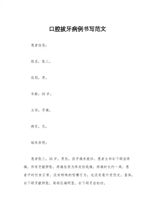
口腔拔牙病例书写范文患者信息:姓名,张三。
性别,男。
年龄,35岁。
主诉,牙痛。
病史,无。
临床表现:患者张三,35岁,男性,因牙痛来就诊。
患者主诉右下颌齿疼痛,伴有牙龈肿胀,疼痛性质为阵发性钝痛,疼痛时长约一周。
患者平时饮食正常,没有特殊的咀嚼行为,也没有意外受伤史。
查体,右下颌牙龈肿胀,局部压痛明显,右下颌牙齿松动。
辅助检查:口腔X线片显示右下颌第七号牙齿牙髓腔内有明显龋坏,周围组织有不同程度的炎症反应。
根管治疗后牙齿仍有疼痛感。
诊断:根尖周炎。
右下颌第七号牙齿龋齿。
右下颌第七号牙齿松动。
治疗方案:拔除右下颌第七号牙齿。
口腔抗炎消炎处理。
治疗过程:患者张三接受了根管治疗,但牙齿仍然有疼痛感,考虑到牙齿周围组织已经发生了炎症反应,为了消除疼痛并防止感染扩散,决定拔除右下颌第七号牙齿。
在局部麻醉后,进行了拔牙手术,手术过程顺利,术后给予口腔抗炎消炎处理,患者术后恢复良好,疼痛症状得到缓解。
随访观察:术后一周,患者口腔疼痛明显减轻,牙龈肿胀明显减退,局部无明显压痛,伤口愈合良好。
患者术后遵医嘱进行了口腔护理和消炎处理,术后恢复良好,未出现感染及其他并发症。
总结:口腔拔牙是一种常见的口腔手术,适用于牙齿严重龋齿、根尖周炎、牙周病等疾病。
对于患者张三来说,拔牙是有效的治疗方法,能够缓解患者的疼痛症状,并防止感染扩散。
在术后的护理和随访中,患者也积极配合医生的治疗建议,取得了良好的疗效。
口腔拔牙手术需要在专业医生的指导下进行,患者在术后也需要加强口腔护理,避免感染及其他并发症的发生。
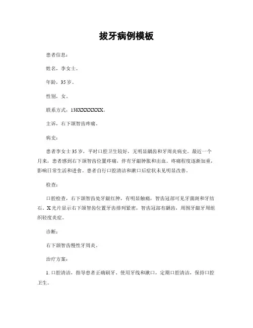
拔牙病例模板患者信息:姓名,李女士。
年龄,35岁。
性别,女。
联系方式,138XXXXXXX。
主诉,右下颌智齿疼痛。
病史:患者李女士35岁,平时口腔卫生较好,无明显龋齿和牙周炎病史。
最近一个月来,患者感到右下颌智齿位置疼痛,伴有牙龈肿胀和出血。
疼痛程度逐渐加重,影响日常生活和进食。
患者自行口腔清洁和漱口后症状未见明显改善。
检查:口腔检查,右下颌智齿处牙龈红肿,有明显触痛,智齿冠部可见牙菌斑和牙结石。
X光片显示右下颌智齿位置牙齿排列紧密,智齿冠部有龋齿,周围牙龈牙周组织轻度炎症。
诊断:右下颌智齿慢性牙周炎。
治疗方案:1. 口腔清洁,指导患者正确刷牙、使用牙线和漱口,定期口腔清洁,保持口腔卫生。
2. 牙周治疗,对右下颌智齿周围牙龈进行局部清洗和消炎,去除牙菌斑和牙结石,改善牙周组织炎症。
3. 智齿拔除,考虑右下颌智齿位置牙齿排列紧密,智齿冠部有龋齿,建议拔除右下颌智齿,以减轻患者疼痛和症状。
处理过程:患者接受口腔清洁和牙周治疗后,牙龈红肿和触痛明显减轻,牙周组织炎症得到改善。
随后进行右下颌智齿拔除手术,手术过程顺利,术后给予局部冷敷和口腔抗炎药物治疗。
患者术后恢复良好,疼痛症状明显减轻,牙龈红肿消退。
随访及效果评估:术后1周,患者口腔恢复良好,无明显疼痛和不适感。
牙龈红肿消退,牙周组织炎症减轻。
患者对治疗效果满意,口腔功能恢复正常。
结语:通过口腔清洁、牙周治疗和智齿拔除,成功治疗了患者右下颌智齿慢性牙周炎,取得了良好的治疗效果。
建议患者定期口腔检查和口腔清洁,保持口腔卫生,预防口腔疾病的发生。
拔牙病例书写案例
一、拔牙病例书写案例
一、病人资料
王先生,男,28岁,汉族,江苏人,未婚。
二、主诉
患者于2018年7月15日到本院牙科就诊,主诉右上颌牙九疼痛不适,痛经历持续1个月。
三、发病史
患者2017年5月就诊于本院牙科,提出右上颌牙九痛痛不适,X 线片显示右上颌牙九有炎症,给予关节联合止痛治疗,症状没有缓解,最近1个月症状加重。
四、发病前检查
周围牙状况良好,口腔检查无异常,X线片显示右上颌九牙根尖部骨破坏,前牙拇风角改变。
五、拔牙过程
根据检查,本院给予右上颌九拔牙治疗,施行无抗拔和无局部麻醉,使用钳器夹住牙根尖,缓慢旋转后,使牙根尖断开,完成拔牙治疗,见效。
六、出院指导
1.三天内准备止血牙膏
2.抗菌洁牙
3.口腔护理
4.定期复诊
七、出院
2018年7月20日出院,症状明显好转。
拔牙病历模板
患者姓名:性别:年龄:职业:住址:
主诉,患者因牙痛、牙松或其他口腔不适到我院就诊。
现病史,患者病程1天/1周/1月,疼痛性质为阵发性/持续性,疼痛部位为右上/右下/左上/左下/前牙/后牙,伴随症状为牙龈肿胀/牙齿松动/口臭等。
既往史,患者有无过往牙病史、手术史、药物过敏史等。
个人史,患者有无吸烟、饮酒等不良嗜好,饮食习惯等。
家族史,患者家族中有无牙病史、遗传疾病等。
体格检查,患者一般情况、身体发育、营养状况等。
辅助检查,口腔X线片、口腔CT等检查结果。
诊断,根据患者的病史、体格检查和辅助检查结果,做出初步诊断。
治疗方案,根据患者的病情,制定治疗方案,包括手术、药物治疗等。
随访观察,手术后患者的恢复情况、疼痛缓解情况等。
预后评估,根据患者的病情和治疗情况,对患者的预后进行评估。
医生签名,日期,年月日。
以上为患者的拔牙病历模板,仅供参考。
希望医生们在使用时能够根据实际情况进行填写,确保病历的准确性和完整性。
同时,希望患者在就诊时能够如实提供个人病史和症状,配合医生的治疗和检查,以便尽快康复。
感谢您的配合!。
口腔残根拔除病历书写范文英文回答:Patient Name: [Name]Age: [Age]Date of Procedure: [Date]Chief Complaint: The patient presented with a fractured tooth and severe pain in the lower left quadrant of the mouth.History of Present Illness: The patient reported experiencing pain and discomfort in the lower left quadrant of the mouth for the past few weeks. The pain worsened when chewing or applying pressure to the area. Upon examination, it was revealed that the pain was originating from a fractured root of a tooth that required extraction.Past Medical History: The patient has a history of hypertension and seasonal allergies. No known drugallergies were reported.Procedure: The patient was scheduled for a tooth extraction due to the fractured root. Local anesthesia was administered to ensure a painless procedure. The tooth was carefully extracted, and the area was thoroughly cleaned to prevent infection.Post-Procedure Instructions: The patient was advised to avoid chewing on the extraction site and to follow a soft diet for the next few days. Pain medication and antibiotics were prescribed to manage any discomfort and prevent infection. The patient was also instructed to maintain good oral hygiene and to gently rinse the mouth with salt water.Follow-Up: The patient was scheduled for a follow-up appointment in one week to monitor the healing process and remove any sutures if necessary.Complications: No immediate complications were notedduring the procedure. The patient was informed about the possibility of mild swelling, bleeding, or discomfort in the days following the extraction.Pathology Report: The extracted tooth and root fragments were sent for histopathological examination to rule out any underlying pathology.中文回答:患者姓名,[姓名]年龄,[年龄]手术日期,[日期]主诉,患者因下颌左侧四分之一处牙齿断裂并且疼痛就诊。
拔牙病历模板患者基本信息姓名:____________________________性别:____________________________年龄:____________________________就诊日期:____________________________主诉:____________________________现病史:____________________________ 11 牙齿情况111 患牙位置描述112 牙齿外观检查结果113 牙齿功能状态114 牙齿疼痛或其他不适症状记录115 影像学检查结果116 牙齿松动程度评估1111 X光片检查结果1112 CT扫描发现1113 其他影像学检查发现1121 牙齿颜色变化描述1122 腐烂程度1123 牙釉质完整性检查结果1124 牙龈健康状况1125 龋齿位置及大小描述1131 咀嚼功能受损程度1132 发音影响程度1133 外观影响程度1134 清洁困难程度1135 疼痛频率及强度描述1136 对日常生活影响评估1141 疼痛持续时间1142 疼痛性质描述1143 疼痛缓解因素1144 其他不适症状记录1151 X光片异常发现1152 CT扫描异常发现1153 其他影像学检查异常发现1161 松动程度分级1162 松动原因分析1163 是否存在感染迹象1164 预计牙齿保留可能性评估12 医疗历史121 过往口腔疾病治疗经历122 全身性疾病记录123 药物过敏史124 手术史125 家族病史126 吸烟饮酒习惯1211 既往口腔手术详情1212 上次看牙时间1213 上次治疗效果1214 使用药物名称及剂量1215 治疗后注意事项遵守情况1216 治疗过程中遇到的问题1221 患有慢性疾病情况1222 糖尿病控制情况1223 心脏疾病治疗经历1224 血液系统疾病情况1225 其他全身性疾病说明1231 已知药物过敏列表1232 最近一次过敏反应时间1233 过敏反应处理措施1234 过敏源避免措施1241 手术类型1242 手术时间1243 手术后恢复情况1244 是否需要特殊护理1245 手术并发症记录1251 家族遗传病1252 家族中类似病例1253 遗传病预防措施1261 吸烟频率1262 饮酒量1263 对牙齿影响评估1264 是否计划改变习惯13 拔牙方案131 拔牙指征132 拔牙方式选择133 局部麻醉剂使用134 拔牙器械准备135 拔牙步骤描述136 拔牙后止血措施137 患者术后护理指导138 预期恢复时间139 随访计划1311 拔牙必要性解释1312 拔牙对整体健康益处1313 不拔牙可能导致后果1314 患者同意拔牙记录1321 开放式拔牙适应症1322 闭合式拔牙适应症1323 患牙具体情况分析1324 预计拔牙难度评估1331 麻醉剂种类1332 麻醉剂量1333 麻醉剂过敏测试1334 麻醉后反应观察1335 麻醉效果评估1341 主要拔牙工具清单1342 辅助器械准备1343 消毒流程1344 应急设备准备1351 拔牙前准备工作1352 拔牙过程详细步骤1353 拔牙技巧要点1354 拔牙过程中可能出现问题及应对策略1361 止血材料使用1362 止血时间1363 嘴巴保持姿势1364 止血效果评估1365 止血后注意事项1371 术后饮食建议1372 口腔清洁方法1373 疼痛管理指南1374 活动限制说明1375 异常情况处理办法1381 初步愈合时间1382 完全恢复时间1383 影响恢复速度因素1384 加速恢复建议1391 术后首次复查时间1392 随访频率1393 随访内容1394 随访目标1395 随访记录保存1396 随访过程中可能遇到问题及解决方案患者签名:____________________________日期:____________________________医生签名:____________________________日期:____________________________。
典型病例分析之拔牙病例
对于拔除前磨牙的病例,由于隐形矫治器不能有效控制牙齿的整体移动和转矩,关闭间隙时容易发生牙齿倾斜,特别是需要后牙前移关闭间隙的病例,由于后牙冠根比较小,易近中倾斜。
作为支抗牙的后牙区附件的使用非常重要,要尽早粘接,避免支抗牙早起发生倾斜。
由于低角骨密度大,咀嚼力强,且面下1/3较短,一般不建议拔牙,效果可能不佳,间隙可能关不上,矫治周期也长,难度也大。
拔牙病例:拔牙不出血了带保持器,全部牙齿拔完了,不疼了,带矫治器。
THE ROYAL COLLEGE OF SURGEONS OF EDINBURGHMEMBERSHIP IN ORTHODONTICSFULL Y DOCUMENTED CASE HISTORYDATE OF EXAMINATION: Nov 2010CANDIDATE NUMBER: 109CASE NUMBER : 2PATIENT’S INITIALS : C.L.Q.COLOUR CODE :CASE SUMMARYCLQ, a 19-year-old Chinese schoolboy, presented with a Class III malocclusion on a skeletal Class III base. He had increased lower facial height, prominent lower lip, acute naso-labial angle, shallow labiomental fold and incompetent lips. He was in the permanent dentition stage with moderate crowding, crossbite in labial and left buccal segments, both upper and lower midlines shifted left, proclined upper incisors. Patient didn’t consent to surgical approach, so a camouflage treatment approach of extraction was commenced.SECTION 1. PRE-TREA TMENT ASSESSMENTPATIENT DETAILSInitials: C.L.Q.Sex: MaleDate of birth: 1st Dec 1989Age at start of treatment: 19 years oldPATIENTS COMPLAINTSPatient disliked his irregular teeth.RELEVANT MEDICAL HISTORYTonsillitis.CLINICAL EXAMINATION: EXTRA-ORAL FEATURESPatient had prominent lower lip, acute naso-labial angle, shallow labiomental fold, incompetent lips. There was no apparent facial asymmetry. The temporomandibular joints were normal.CLINICAL EXAMINATION: INTRA-ORAL FEATURESSoft tissues: GingivitisOral hygiene: InadequateGeneral dental condition: #15 was restored.CROWDING / SPACINGMaxillary arch 8 mm crowdingMandibular arch 2 mm crowdingOCCLUSAL FEATURESIncisor relationship: Class IIIOverjet (mm): 1 mm (on #11)Overbite: 1 mm (crossbite on #12,#21 and #22)Centrelines: Upper midline was 1.5 mm to left of facial midlineLower midline was 2 mm to left of facial midline1/4 unit Class IIILeft buccal segmentrelationship:1/2 unit Class IIIRight buccal segmentrelationship:Crossbites: #12,#21,#22,#24,#25,#26 Displacements: #23 blocked outOther occlusal features: NonePRE-TREATMENT PHOTOGRAPHS: EXTRA-ORALPRE-TREATMENT PHOTOGRAPHS: INTRA-ORALGENERAL RADIOGRAPHIC EXAMINATIONPre-treatment radiographs taken:Lateral Cephalogram 14th Oct 2008Dental panaromic tomogram 14th Oct 2008BitewingUnerupted teeth:Teeth absent:Other relevant radiographic findings:Dental panaromic tomogram Full permanent dentition was present. Roots’morphologywas normal. No other obvious pathology was noted. BitewingPRE-TREATMENT RADIOGRAPHSCEPHALOMETRIC TRACING: PRE-TREATMENT1CEPHALOMETRIC ANALYSISVARIABLE PRETREATMENT NORMAL SNA 81.5°82° ± 3.5 SNB 83°79° ± 3.0 ANB -1.5° 3.0° ± 2.0 Wits appraisal-4.5 mm-4.5 mm ± 3.0 Upper incisor to maxillary plane angle 128°118° ± 6 Lower incisor to mandibular plane angle 96°97° ± 7 Interincisal angle 113.5115° ± 8 Maxillary mandibular planes angle 15.5°26° ± 5 Upper anterior face height 58 mm 54.0 mm Lower anterior face height 78 mm64.0 mm Face height ratio57.4%55%± 1% Lower incisor to APo line 6.5 mm 5.5 mm ± 2.5 Lower lip to Ricketts E Plane 1 mm 4.0 mm ± 2.5Source of normal values for Chinese: Cooke MS and Wei SHY (1988)NormalOne standard deviationTwo standard deviationsINTERPRETATIONThe SNB value was increased and ANB value was negative which suggested a skeletal Class III pattern. Although lower facial height was increased, maxillary mandibular angle were decreased, indicating smaller ratio of posterior to anterior facial height.The upper incisors were proclined.DIAGNOSTIC SUMMARY19-year-old patient presenting with Class III malocclusion on a skeletal Class III base. He had increased facial height with increased lower facial height ratio, prominent lower lip, reduced upper facial height ratio, protrusive upper lip, acute naso-labial angle, shallow labiomental fold and incompetent lips. He was in the permanent dentition stage with moderate crowding, crossbite in labial and left buccal segments. both upper and lower midlines shifted left,proclined upper incisors.PROBLEM LIST1. Acute naso-labial angle, protrusive upper lip, shallow labiomental fold andincompetent lips.2. Class III malocclusion with crossbite in labial and left buccal segments.3. Crowding of both arches.4. Both midlines shifted left.RATIONALE FOR TREATMENT1. There was moderate crowding in the upper arch and mild in the lower, significant midlineshifts, proclined upper incisors and crossbite in labial segment. Extraction of upper second premolars and lower first premolars followed by fixed appliance treatment with maximum anchorage provides space for alignment, increasing overbite and overjet, correction of midlines.2. Retraction of lower frontal teeth can eliminate crossbite and improve lip competency.3. Placement of removable Hawley retainers.AIMS AND OBJECTIVES OF TREATMENT1. Relieve crowding, level and align teeth and harmonize arches2. Correct crossbite3. Correct both midlines4. Improve lip competency5. Retention and review third molar eruptionTREATMENT PLANExtractions: #15,#25,#34,#44Appliances: Fixed applianceSpecial anchorage requirements: NoneMinor adjunctive surgery: NoneMajor adjunctive surgery: NoneAdditional dental treatment:Proposed retention strategy: Hawley retainers.Additional notes on treatment plan: Refer to hygienist to improve the oral hygiene. Prognosis for stability: The prognosis for stability is good.SECTION 2. TREA TMENTTREATMENT PROGRESSStart of active treatment: Dec 2008 Age at start of active treatment: 19 years old End of active treatment: Oct 2010 Age at end of active treatment: 21 years old End of retention: OngoingKEY STAGES IN TREATMENT PROGRESSDATE STAGE1. Dec 2008 Referral to hygienist. Placement of fixed appliances with 0.022 X0.028” slot and initial levelling and alignment archwires on upperand lower teeth.TIME INTOSTAGETREATMENT1. 2 months Leveling in 0.012” NiTi archwireLower arch changed from0.018”NiTi to 0.016” x 0.022”NiTiarchwire2. 4 months Placement of broaden 0.016” x 0.022” NiTi archwire on upperarch and 0.017” x 0.025” NiTi archwire on lower arch.Class IIIelastics at night with light force to correct molar relationship3. 5 months Placement of 0.018” x 0.025” NiTi archwire on both upper andlower arch4. 6 months Placement of 0.019” x 0.025” NiTi archwire on both upper andlower arch5. 8 months #17 buccal tube repositioned, placement of 0.016” x 0.022” NiTiarchwire on upper arch and 0.016” x 0.022” NiTi reversed curveof spee archwire on the lower arch for further levelling6. 10 months Placement of 0.017” x 0.025” SS archwire on both upper andlower arch,oblique elastics in labial segment to correct shiftedmidlines7. 15 months Placement of 0.018” x 0.025” SS archwire on both upper andlower arch,asymmetric elastics to correct shifted midlines8. 17 months #35,#36,#45,#46 brackets repositioned, placement of 0.016”x 0.022” NiTi archwire on lower arch. -20° torque in upperarchwire at the place of #2210. 22 months Debonding and placement of Hawley retainers.MID-TREATMENT PHOTOGRAPHS:5mPlacement of 0.018” x 0.025” NiTi archwire on both upper and lower arch10mPlacement of 0.017” x 0.025” SS archwire on both upper and lower arch,oblique elastics in labial segment to correct shifted midlinesMID-TREATMENT PHOTOGRAPHS:12mPower chain to distalize #43 for coordination of midlines, #22 bracket occlusally positioned to slightly intrude #22, upper archwire back to 0.018” NiTi15mPlacement of 0.018” x 0.025” SS archwire on both upper and lower arch,asymmetric elastics to correct shifted midlines17m#35,#36,#45,#46 brackets repositioned, placement of 0.016” x 0.022” NiTi archwire on lower arch. -20° torque in upper archwire at the place of #22.SECTION 3. POST-TREA TMENT ASSESSMENTOCCLUSAL FEATURESIncisor relationship: Class IOverjet (mm): 2 mmOverbite: 2 mmCentrelines: Coincident with facial midlineLeft buccal segmentClass Irelationship:Class IRight buccal segmentrelationship:Crossbites: NoneDisplacements: NoneFunctional occlusal features Canine guidance was present on the left and rightduring lateral excursions and incisal guidance waspresent on protrusion. There were no non-workingside interferences during function.Other occlusal features: Good buccal interdigitation was achieved.COMPLICATIONS ENCOUNTERED DURING TREATMENTOverall, the patient was motivated, compliant with oral hygiene and tolerant with all aspects of treatment.OCCLUSAL INDICESINDEX PARAMETER VALUEIndex of Treatment NeedDental Health Component Start 5iFinish 1 Aesthetic Component Start 8Finish 1Start 56Peer Assessment Rating(PAR)Finish 1Change 55% Change 98RADIOGRAPHS TAKEN TOWARDS / AT END OF TREATMENTRadiographs taken:Lateral Cephalogram 21st Oct 2010Dental panaromic tomogram 21st Oct 2010Relevant findings:Dental panaromic tomogramRoot parallelism could have been better but clinical aesthetics did not support prolonging of treatment time. No other pathology was noted.POST-TREATMENT RADIOGRAPHSCEPHALOMETRIC TRACING: POST-TREATMENTCEPHALOMETRIC ASSESSMENTVARIABLE POSTTREATMENT CHANGE SNA 81 -0.5°SNB 81.5°-1.5°ANB -0.5°1°Wits appraisal -3 mm 1.5 mm Upper incisor to maxillary plane angle 122.5°-5.5°Lower incisor to mandibular plane angle 81.5°-14.5°Interincisal angle 135°22.5°MM angle 20°-2.5°Upper anterior face height 58 mm 0 mm Lower anterior face height 80 mm 2 mmFace height ratio 58%+0.6% Lower incisor to APo line 2 mm -4.5 mm Lower lip to Ricketts E Plane -0.5 mm -1.5 mmSource of normal values for Chinese: Cooke MS and Wei SHY (1988)NormalOne standard deviationTwo standard deviationsINTERPRETATIONMaxillary superimposition on maxillary stable structures indicates that there has been retroclination, some buccal root torque and extrution of vertical position of upper incisors. The upper molars have been mesialized.Mandibular superimposition indicates that there has been lower incisor retroclination over normal limits with reference to A-Po line and maintenance of vertical position. There was a small extrusion and mesialization of lower molars.C E P H A L O M E T R I C S U P E R I M P O S I T I O NOverall superimposition of pre- and post-treatment cephalometric radiographs registered on anterior cranial base.Pre-treatmentPost-treatmentCEPHALOMETRIC SUPERIMPOSITION (where appropriate)Maxillary superimposition and pre- and post-treatment cephalometric radiographs registered on the anterior border of zygomatic processPre-treatmentPost-treatmentMandibular superimposition and pre- and post-treatment cephalometric radiographs registered on Björk’s stable mandibular structuresPre-treatmentPost-treatmentPOST-TREATMENT PHOTOGRAPHS: EXTRA-ORALPOST-TREATMENT PHOTOGRAPHS: INTRA-ORALSECTION 4. CRITICAL APPRAISALThe patient was treated in 22 months.Dental Cast AnalysisClass I molar relationship was achieved. Upper and lower dental midlines are coincident. Arches are coordinated. Overjet and overbite are within normal limits. Buccal interdigitation has been achieved.Soft TissueDental midlines are also coincident with facial midline. Nasolabial angle is no longer acute after treatment. Lower lip is less protrusive and is in good relationship with upper lip and nose. Lip competency was possible without obvious mentalis muscle strain. Labiomental angle is more aesthetic but chin is still anteriorly positioned. The gingival condition is satisfactory.Radiographic AnalysisThe growth pattern of this patient is stable. After active treatment, the maxilla is almost in initial position, but the mandible presents a little growth vertically. Rretroclination of lower incisors and Class III elastics contribute to reduce the prominence of the lower lip. Facial height has almost been kept in initial proportion. Upper incisors have been retroclined to normal values while the lower incisors have been retroclined excessively so as to correct crossbite.Long term PrognosisThe patient has little growth potential at his age. The position of the developing #18 and #28 needs to be reviewed. As interincisal angle is increased, long-term retention of overbite and overjet involving removable retainers is suggested.。