Ex vivo gene transfer for improved adoptive immunotherapy of cancer
- 格式:pdf
- 大小:178.20 KB
- 文档页数:7

[4]J un Hiraki,Takafumi Ichika wa,Shin-ichi Ninomi ya,et al .Use of AD ME studi es to confirm the s afety of -poly-L-l ysine as a preservative in food [J].Regulatory T oxicology and Pharmacology ,2003,37(2):328-340.[5]Ki to M,Takimoto R,Yoshida T,et al .Purification and characteriz ationof -poly-L-lysine-degrading enzyme from an -pol y-L-lysine-produc ing s train Stre ptomyce s albulus [J].Arch Microbiol ,2002,178:325-330.[6]Nishi kawa M,Ogawa K.Distri bution of microbes producing anti microbi al -poly-L-Lysine polymers in s oil microflora determined by a novel method[J].Applied and Environmental Microbiology ,2002,68(7):3575-3581.[7]G Szokan,M Almas,K Krizsan,e t al .Structure determination and s ynthesis of l ysi ne isopeptides i nfluencing on cell proli ferati on[J].B iopolymers ,1997,42(3):305-318.[8]Kahar P,T Iwata,J Hiraki,et al .Enhance ment of -polylys ine production by Stre pto myce s albulus s train 410usi ng pH control[J ].J.Biosci.B ioeng ,2001,91(2):190-194.[9]Kahar P,K Kobayashi,T Iwata,et al .Production of -polylysine i n an air lift bioreactor (ABR)[J].J.B iosci.Bioeng ,2002,93(3):274-280.[10]Ki to M,Onji Y,Yoshida T,et al .Occurrence of -poly-L-lysine-de gradi ng enz yme in -poly-L-l ysine-tolerant Sphingobacte rium multivorum OJ10:purification and charac terization [J ].FEMS Microbiology Letters ,2002,207(2):147-151.[11]Kito M,Rika T,Onji Y,et al .Puri fication and characterization of an -poly-L-l ysi ne-Degrading Enz yme from the -poly-L-lysine-tolerant Chryseobacte rium sp .057[J].J.B iosci.Bioeng ,2003,96(1):92-94.[12]Hi raki J,Hatakeyama M ,M ori ta H,et al .Improved -poly-L-lysine production of an S-(2-aminoe thyl)-L-cysteine resis tant mutant of Stre pto myc es alblus [J].Seibu tsu Kogaku K ais hi ,1998,76:487-493.[13]Takagi H,Hoshino Y,Nakamori S,e t al .Isolation and sequence analysis of plasmid pNO33in the -pol y-L-lysineproduci ng antino myce te Stre ptomyces albulus IFO14147[J].J.Bios ci.Bioeng ,2000,89(1):94-96.[14]Kawai T,Kubota T,Hiraki J,et al .Bi os ynthesis of -poly-L-lysine i n a cell-free system of Stre ptomyces albulus [J ].Biochemical and B iophys ical Research Communications ,2003,311(3):635-640.[15]Yu-Ting H,Shoichi ro I,Munehiko T Improving emulsi fying activi ty of -polylysine by conjugation w i th dextran through the Maillard reac tion[J].Food Chemistry ,2000,68(4):449-455.[16]Keiji I,Kyos uke Y,Atsus hi H,et al .Polyi on comple x micelles from plas mid DNA and pol y (e thylene gl ycol)-poly(L -lysine)bl ock copol ymer as serum-tolerable pol yple x system:physicochemical properties of micelles rele vant to gene trans fection efficiency[J ].Biomaterials ,2003,24(24):4495-4506.[17]W Cui,G Barr,K M Faucher,et al .A me mbrane-mimetic barrier for islet encaps ulation[J].Transplantation Proceedings ,2004,36(4):1206-1208.[18]M aureen D B,Alexander I G,Laurence T,et al .In vi tro and in vi vo gene transfer with poly(amino aci d )vesicles [J].Journal of Controlled Release ,2003,93(2):193-211.[19]Tris tan M ,Pascal D,Thierry B,e t al .Efficient gene trans fer into human epithelial cell lines using gl ycosylated cationic carriers and neutral gl ycosylated co-lipids [J ].Blood Cells,Molecules,an d Diseases ,2004,32(2):271-282.[20]Hana H,David F,Petr K.Growth and elec trochemical ac tivity of the poly-L-lysine-poly-L-glutamic acid thin layer fi lms:an EQCM and electro chemical study[J].Journal of Electroanalytical Chemistry ,2004,562(1):261-265.[21]孙宇峰,王连超,吴仲城,等.石英晶体振荡器海洛因快速测定生物合成酶膜制备及反应机理研究[J].传感器世界,2003,9(12):2-5.[22]Kunioka M ,Choi H J.Properties of biodegradable hydrogels prepared by -irradiation of microbial poly( -l ysi ne)aqueous sol utions[J].J Appl Polym Sci ,1995,58:801-806.苯乙烯微生物降解机理的研究进展吴献花1,2,孙石2,邵丹1,李海涛1,章新1,林洪1(1.玉溪师范学院化学与环境科学系,云南玉溪653100;2.昆明理工大学环境科学与工程学院,云南昆明650093)摘要:综述苯乙烯微生物降解的机理。
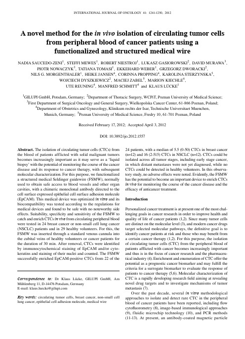
INTERNATIONAL JOURNAL OF ONCOLOGY 41: 1241-1250, 2012Abstract. The isolation of circulating tumor cells (CTCs) from the blood of patients afflicted with solid malignant tumors becomes increasingly important as it may serve as a ‘liquid biopsy’ with the potential of monitoring the course of the cancer disease and its response to cancer therapy, with subsequent molecular characterization. For this purpose, we functionalized a structured medical Seldinger guidewire (FSMW), normally used to obtain safe access to blood vessels and other organ cavities, with a chimeric monoclonal antibody directed to the cell surface expressed epithelial cell surface adhesion molecule (EpCAM). This medical device was optimized in vitro and its biocompatibility was tested according to the regulations for medical devices and found to be safe with no noteworthy side effects. Suitability, specificity and sensitivity of the FSMW to catch and enrich CTCs in vivo from circulating peripheral blood were tested in 24 breast cancer or non-small cell lung cancer (NSCLC) patients and in 29 healthy volunteers. For this, the FSMW was inserted through a standard venous cannula into the cubital veins of healthy volunteers or cancer patients for the duration of 30 min. After removal, CTCs were identified by immuno c ytochemical staining of EpCAM and/or cyto-keratins and staining of their nuclei and counted. The FSMW successfully enriched EpCAM-positive CTCs from 22 of the 24 patients, with a median of 5.5 (0-50) CTCs in breast cancer (n=12) and 16 (2-515) CTCs in NSCLC (n=12). CTCs could be isolated across all tumor stages, including early stage cancer, in which distant metastases were not yet diagnosed, while no CTCs could be detected in healthy volunteers. In this observa-tory study, no adverse effects were noted. Evidently, the FSMW has the potential to become an important device to enrich CTCs in vivo for monitoring the course of the cancer disease and the efficacy of anticancer treatment.IntroductionPersonalized cancer treatment is at present one of the most chal-lenging goals in cancer research in order to improve health and quality of life of cancer patients (1,2). Since many tumor cells are distinct on the molecular level (3), and modern cancer drugs target selected molecular pathways, the definitive goal is to identify cancer patients at risk and those who may benefit from a certain cancer therapy (1,2). For this purpose, the isolation of circulating tumor cells (CTC) from the peripheral blood of patients afflicted with cancer becomes increasingly important and thus is in the focus of cancer research and the pharmaceu-tical industry (4). Enrichment and enumeration of CTC offer the potential as a prognostic cancer biomarker and may fulfill the criteria for a surrogate biomarker to evaluate the response of patients to cancer therapy (5,6). Molecular characterization of CTC is a rapidly developing research field aiming at revealing novel drug targets and to investigate mechanisms of tumor metastasis (7).Over the past decade, several in vitro methodological approaches to isolate and detect rare CTC in the peripheral blood of cancer patients have been reported, including flow cytofluorometry (8), image-based immunological approaches (9), fluidic microchip technology (10), and PCR methods (11-13). At present, an antibody-coated magnetic particleA novel method for the in vivo isolation of circulating tumor cellsfrom peripheral blood of cancer patients using afunctionalized and structured medical wireNADIA SAUCEDO-ZENI1, STEFFI MEWES1, ROBERT NIESTROJ1, LUKASZ GASIOROWSKI2, DA VID MURAWA3, PIOTR NOWACZYK3, TATIANA TOMASI1, EKKEHARD WEBER1, GRZEGORZ DWORACKI2,NILS G. MORGENTHALER1, HEIKE JANSEN4, CORINNA PROPPING4, KAROLINA STERZYNSKA5,WOJCIECH DYSZKIEWICZ2, MACIEJ ZABEL5, MARION KIECHLE4,UTE REUNING4, MANFRED SCHMITT4 and KLAUS LÜCKE11GILUPI GmbH, Potsdam, Germany; 2Department of Thoracic Surgery, WCPiT, Poznan University of Medical Science;3First Department of Surgical Oncology and General Surgery, Wielkopolska Cancer Center, 61-866 Poznan, Poland;4Department of Obstetrics and Gynecology, Klinikum rechts der Isar, Technische Universitaet Muenchen, Munich, Germany; 5Poznan University of Medical Science, Fredry 10, 61-701 Poznan, PolandReceived February 17, 2012; Accepted April 3, 2012DOI: 10.3892/ijo.2012.1557Correspondence to: Dr Klaus Lücke, GILUPI GmbH, AmMühlenberg 11, D-14476 Potsdam, GermanyE-mail: klaus.luecke@Key words: circulating tumor cells, breast cancer, non-small celllung cancer, epithelial cell adhesion molecule, medical wireSAUCEDO-ZENI et al: In vivo ISOLATION OF CIRCULATING TUMOR CELLS 1242isolation system targeting the epithelial cell surface EpCAM is used in most studies designed for ex vivo quantification of CTC in the blood of patients with advanced breast, colon, or prostate cancer (14-16). Patients with metastasized cancer diseases exhibit detectable numbers of CTC in their blood (17), however, since all ex vivo detection systems are limited by the blood volume that can be obtained from the patients or handled by the detection system (18), these technologies are of relatively low sensitivity.To overcome the limitations of small blood sample volumes of the ex vivo CTC isolation techniques, new approaches are needed for screening large blood volumes in vivo using for example the principles of photoacoustic flow cytofluorometry (19). We developed an alternative medical device: a structured and functionalized medical wire (FSMW) based on a Seldinger guidewire (20) that offers the opportunity of capturing CTC from the circulating blood of cancer patients. Like in other ex vivo CTC-detection technologies, identification of CTC captured by the FSMW is performed by phenotyping CTC with antibodies directed to cytokeratins and/or epithelial cell markers. Occasionally trapped hematologic cells are identified by anti-bodies directed to respective hematologic cell surface markers. Here, we describe a novel in vivo CTC-catching medical device, the FSMW, its biocompatibility and first results of its applica-tion for in vivo enrichment of CTC from the peripheral blood of patients presenting with breast cancer or non-small cell lung cancer (NSCLC).Materials and methodsFunctionalized structured medical wire. The FSMW (Fig. 1) is based on a stainless steel medical wire (Seldinger guidewire) of 0.5 mm in diameter and 160 mm in length (EPflex, Dettingen, Germany). The first 20 mm are plated with a 2 µm thick gold layer deposited on the device by galvanization (OTEK, Brieselang, Germany). Subsequently, a hydrogel layer composed of a linear, synthetic polycarboxylate is attached to the gold layer (Xantec Bioanalytics, Düsseldorf, Germany). The carboxyl groups present in the hydrogel are then activated with EDC (1-ethyl-3-[3-dimethylaminopropyl] carbodiimide hydrochloride) and NHS (N-hydroxysuccinimide) (Sigma-Aldrich GmbH, Seelze, Germany) allowing for functionalization via covalent coupling of a chimeric antibody directed to the epithelial cell adhesion molecule CD326 (EpCAM; HEA 125, kindly provided by Dr G. Moldenhauer, German Cancer Research Center, DKFZ, Heidelberg, Germany) present on the surface of most CTC. Functionalization of the FSMW is carried out under clean room conditions. The FSMW is a sterile medical device and intended for single in vivo use only. For the in vivo application, the sterile FSMW is inserted into a standard 20G (pink color code) intra-venous cannula (Fig. 2).In vitro experiments. Prior to FSMW application in patients, a hemodynamic flow system was applied (Fig. 3). Within this system, blood was routed via flexible tubes through a flow chamber into which up to two FSMW are inserted. The flow rate, velocity, and flow direction can be regulated by a peristaltic pump. A flow rate of 20 ml/min was applied to reflect in vivo flow conditions within cubital veins (21). In this in vitro flow system the FSMW was tested for binding of cultured EpCAM-positive SK-BR-3 breast cancer cells (CLS, Eppelheim, Germany), which were grown in cell culture flasks until cell monolayers reached a confluency of ~80%. Adherent SK-BR-3 cells were detached with trypsin/EDTA (ethylene diamine tetraacetate) (Biochrom AG, Berlin, Germany), centrifuged, and suspended in phosphate-buffered saline (PBS). In addition, EDTA-anti-coagulated blood samples obtained from healthy donors were spiked with those cells and tested for binding to the FSMW. Furthermore, blood samples from breast or lung cancer patients were also assessed in the in vitro flow system to test for CTC binding to the FSMW.Cytotoxicity tests. To examine potential cytotoxic effects of the FSMW, we performed direct FSMW cell contact and material elution tests in vitro, based on the requirements for a class IIa medical device, as outlined in the ISO Guideline 10993-5 (). For the elution test, eluates of one or three FSMW devices (three to identify any variation in the produc-tion process), and of reference materials known to be toxic (copper wire, Goodfellow GmbH, Bad Nauheim, Germany) or non-toxic (Teflon wire, PTFE, Goodfellow GmbH) for normal human dermal fibroblasts (NHDF, C-12302, PromoCell GmbH, Heidelberg, Germany) were generated (0.095 m2 FSMW surface area eluted with 5 ml of RPMI-medium, FG 1235, Biochrom AG, at 37˚C for 24 h), and their influence on cell morphology and viability of NHDF examined. Evaluation of potential effects of these eluates on cell morphology was done qualita-tively by microscopic analysis for changes in cell morphology, cell adhesive capacity, and cell disintegration. Viability of cells was tested by use of the colorimetric TTC assay (triphenyltetra-zolium chloride test).In brief, for the microscopical inspection, NHDF were exposed to FSMW eluates for 48 h at 37˚C. Eluates of reference materials known to be toxic (copper wire, Goodfellow GmbH) or non-toxic (Teflon wire, PTFE, Goodfellow GmbH) for NHDF were tested in parallel. After 48 h of incubation, cells were stained with the dye CFSE [5-(and-6)-carboxyfluorescein diacetate succinimidyl ester; Invitrogen, Carlsbad, USA] and inspected under the fluorescence microscope. Impairment of cell morphology was scored according to a common reaction index (none versus slight, moderate or severe).To test for viability of NHDF, a colorimetric assay (TTC assay) was performed, which allows for the quantitative assessment of cell viability in the presence of material-derived eluates. After 48 h of incubation of NHDF with the eluates, a yellow tetrazolium compound (EZ4U, Cell Proliferation and Cytotoxicity Assay, Biomedica Medizinprodukte GmbH & Co. KG, Vienna, Austria) was added (3 h, 37˚C) which was converted due to metabolic mitochondrial cellular activity to a brick-red formazan. Change in color was recorded at 450 nm by use of a microtiter plate reader (SPECTROstar-Omega, BMG Labtech, Ortenberg, Germany). For direct contact tests, the FSMW device was placed on a layer of adherent NHDF, followed by a 24 h incubation of this set-up at 37˚C. Reference wires made of Teflon or copper were tested in parallel.Acute systemic toxicity. To assess potential hazardous effects of the FSMW in humans, possibly occurring, acute systemic toxicity of the FSMW was tested. In a parallel clinical study, aiming at the in vivo enrichment of trophoblasts from pregnantINTERNATIONAL JOURNAL OF ONCOLOGY 41: 1241-1250, 20121243women, potential acute systemic toxicity of the eluates had already been monitored. According to these tests, no adverse effects were caused by the FSMW (22). For evaluation of the acute systemic toxicity of the FSMW, four test groups of five mice each [Charles River, Sulzfeld, Germany; NMRI (Han) mice, female non-pregnant, nulliparous, 19-24 g body weight, 4-5 weeks old) were injected intravenously or intraperitoneally with a single dose of four different extracts of the FSMW. The amount of eluates administered were adjusted to body weight at a volume of 50 ml/kg for 0.9% (w/v) NaCl, 5% (v/v) ethanol, and cotton seed oil, and at 10 g/kg for polyethylene glycol-400. Four control groups of five mice each were treated in the same manner with the corresponding extraction vehicle not previ-ously exposed to the FSMW.This dosing regime supplied an about 100 times higher dose of the FSMW than the expected dose in humans. The animals were followed up immediately after injection, and at 4, 24, 48 and 72 h intervals for body weight and toxic effects. Cage side observations included spontaneous activity, lethargy, recumbent position, convulsions, tremors, apnoea, asphyxia, vocalization, diarrhea, obvious changes in the skin and fur, eyes and mucous membranes (salivation, discharge). At the end of the observation period the animals were sacrificed. All animals were subjected to gross necropsy. Any gross pathological changes were recorded. The studies were approved by the Ethics Committee of the Medical University of Poznan, Poland.Hemocompatibility test. Objectives of the hemocompatibility test were assessment of the patient's risk to develop thrombosis, coagulation, and hemolysis after in vivo exposure to the FSMW. The test method described in the ISO Guideline 10993-4:2002 (E) and according to Xu and coworkers (23) was employed. To assess any influence of the FSMW on the blood coagula-tion cascade, the plasma recalcification time was determined. The FSMW was incubated for 10 min at 37˚C in citrate anti-coagulated platelet-poor plasma, which was then treated with CaCl2 to induce blood coagulation. Clotting time of plasma, which had not been exposed to the FSMW, served as a control. Fibrin formation was determined during a rigid up and down movement of the FSMW in a plasma sample or in a plasma sample which was not exposed to the FSMW. Time intervals until fibrin deposits were formed on the FSMW were recorded. For examination of any potential hemolytic risk exerted by the FSMW, erythrocytes were isolated from EDTA anti-coagulated blood of healthy donors and eluates of the FSMW added. After incubation at room temperature (RT), the erythrocyte-eluate-suspension was centrifuged and the hemolysis rate (% increase in cell-free hemoglobin concentration) determined photo-me t rically. Products that have no hemolytic potential should exhibit a hemolytic rate of less than 5%.To assess non-specific blood cell adhesion to the FSMW device, the antibody-functionalized part of the FSMW was put into an in vitro hemodynamic flow system (Fig. 3) which simulates the in vivo blood flow situation, including FSMW contact period, body temperature, and flow rate. After removal, the FSMW was inspected microscopically for blood cells deposited on the device. Then, in order to detect any fibrin deposits on the FSMW, the device was stained with antibodies directed to fibrin(ogen) [mouse anti-human fibrin antibody, IgM, 1:100 (2 µg/ml) in PBS; Santa Cruz Biotechnology Inc., Heidelberg, Germany] for 30 min, followed by the addition of secondary FITC-conjugated anti-IgM (Santa Cruz Biotechnology Inc.), 1:200 (2 µg/ml), for 30 min at RT. For detection of adherent platelets, a fluorescence-labeled antibody directed to CD41 was applied [anti-CD41a-PE, 1:100 (2 µg/ml) in PBS; Santa Cruz Biotechnology Inc.].Study population. Patients were recruited at the Wielkopolska Cancer Center, Department of Surgical Oncology and General Surgery, and at the Poznan University of Medical Sciences, both in Poznan, Poland. The healthy donor population was recruited at the Department of Obstetrics and Gynecology, Klinikum rechts der Isar, Technische Universitaet Muenchen, Munich, Germany. The studies were approved by the Institutional Ethics Committees and written informed consents of the patients and volunteers were obtained.For the in vitro functionality test of the FSMW, we included 17 patients afflicted with breast cancer and 7 patients with NSCLC. For the in vivo functionality test of the FSMW, we included 12 breast cancer patients and 12 NSCLC patients (Tables I and II). Patients of both cancer types had different tumor stages and had not undergone surgery or received chemotherapy at the time of enrolment. Principal inclusion criteria for breast cancer and NSCLC patients were >18 years of age, histopathologically confirmed diagnosis of breast cancer or potentially resectable NSCLC with eligibility for radical surgery. Exclusion criteria: history of psychiatric disease, participation in other clinical trials, history of allergy, anaphylactic reactions, prior immunological diseases (anti-phospholipid antibody syndrome, Goodpasture's syndrome, lupus erythematosus, polychondritis, rheumatoid arthritis, sarcoidosis, scleroderma, Sjogren's syndrome, ANCA positive states), immunodeficiencies, prior infections with hepatitis viruses, the cytomegalovirus (CMV), or infectious diseases such as tuberculosis, syphilis, or toxoplasmosis. Principal exclusion criteria for the 29 healthy volunteers (premenopausal 24, postmenopausal 5) were >18 years of age, pregnancy or breastfeeding, any kind of oncological or allergic disease including asthma and thromboembolic complications. Median age of the volunteers was 27 (range 22-67).For in vitro studies of the FSMW in an in vitro flow system, peripheral venous blood from patients afflicted with breast cancer or NSCLC was harvested into EDTA-tubes (Sarstedt AG & Co., Nuembrecht, Germany). Blood samples were kept at RT and processed in the flow system at RT within 72 h after drawing of the blood.In vivo application of the FSMW. The FSMW was designed to fit into a standard 20G intravenous cannula, which is placed into the cubital vein of a cancer patient or a healthy donor (Fig. 2). An IN-Stopper (Sarstedt AG & Co.) allows secure fixation to the intravenous cannula. The FSMW is slowly pushed forward into the cannula until the EpCAM-antibody-functionalized FSMW surface of 2 cm in length is exposed to the blood flow within the lumen of the vein. The correct insertion is indicated by a mark on the distal part of the FSMW, which is not inserted into the cannula. The FSMW remains in the cubital vein for 30 min. In the present study, the insertion of the FSMW was done before the respective patient underwent surgery of the primary tumor. During the procedure of FSMW application, the patient remained in a flat or supine position. The total volume of bloodSAUCEDO-ZENI et al: In vivo ISOLATION OF CIRCULATING TUMOR CELLS 1244coming into contact with the FSMW during the 30 min applica-tion period is estimated at 1.5-3 liters (24).Inspection of the FSMW for bound CTC. After removal of the FSMW from the cubital vein, the FSMW was briefly and gently washed in PBS, followed by incubation in PBS containing 2% (w/v) bovine serum albumin (BSA, Carl Roth GmbH, Karlsruhe, Germany, purity grade ≥98%), for 30 min at RT. Characterization of CTC captured by the FSMW was done by immunocytochemical staining for EpCAM or cytokeratins 4, 5, 8, 9, and 18. Cells attached to the FSMW were incubated with an FITC-conjugated mouse monoclonal antibody directed to EpCAM [1:100 in PBS (10 µg/ml); Acris Antibodies GmbH, Herford, Germany] and a phycoerythrin (PE)-conjugated rabbit antibody raised against CD45 [1:25 in PBS (2 µg/ml); Life Technologies GmbH, Darmstadt, Germany]. Cells were counter-stained with the nuclear dye 4,6-diamidino-2-phenylindole (DAPI; 1 µg/ml PBS; Life Technologies GmbH). Intensity of the immunocytochemical staining of CTC was evaluated using an Axio Imager.A1m microscope (Zeiss, Jena, Germany) equipped with an AxioCam digital camera system and the AxioVision 4.6 software (Zeiss). EpCAM- or cytokeratin-positive cells included in the count had to disclose additional features such as a large cell body (diameter 10-50 µm), an irregular cell shape, a large irregularly shaped nucleus, and a high nuclear to cytoplasmic ratio (17,25,26). Cells were counted on each FSMW by an oper-ator blinded to the clinical background of the patients. Results are given as number of CTC immobilized on the surface of the EpCAM-antibody-functionalized FSMW. In some cases, before inspection, cells were fixed with 4% (w/v) buffered paraformal-dehyde.ResultsIsolation and subsequent molecular characterization of CTC from the blood of cancer patients becomes increasingly impor-tant as it may serve as a ‘liquid biopsy’ with the potential of monitoring the course of the cancer disease and response to cancer therapy. For this purpose, but different than currently employed ex vivo CTC enrichment protocols, we applied a structured medical Seldinger guidewire (FSMW), functional-ized with a chimeric monoclonal antibody directed to EpCAM, to be used in vivo to catch and enrich CTC from the peripheral blood pool (Fig. 1). The FSMW was first optimized in vitro for its CTC catching ability and then tested for biocompat-ibility according to the ISO guidelines for medical devices.Table I. Clinical characteristics of breast cancer patients assessed for CTC enrichment in vivo and histomorphological features of the primary tumor.Classification No. of Patients Patientspatients with 1-4 with ≥5CTC (%) CTC (%) All patients 12 3 (25) 9 (75) ER-/PR-statusPositive for either 10 3 (30) 7 (70) Negative for both 2 0 2 (100) HER2-statusPositive 3 0 3 (100) Negative 9 3 (33.3) 6 (66.7) Histological classificationInvasive-ductal 7 1 (14.3) 6 (85.7) Invasive-lobular 3 1 (33.3) 2 (66.7) Invasive-ductal/ 1 1 (100) 0invasive lobularInvasive-ductal/ 1 0 1 (100) bifocal cancerAdjuvant therapyChemotherapy 3 1 (33.3) 2 (66.7) Endocrine therapy 1 0 1 (100) No adjuvant therapy 8 2 (25) 6 (75) Tumor stageT1N1M0 2 1 (50) 1 (50) T1N3M0 1 0 1 (100) T2N1M0 3 1 (33.3) 2 (66.7) T2N3M0 1 0 1 (100) T4N0M0 1 0 1 (100) T4N2M0 1 0 1 (100) T4N+N/A 1 0 1 (100) T1N1M1 1 1 (100) 0T3N+N/A 1 0 1 (100)Table II. Clinical characteristics of NSCLC patients assessed for CTC enrichment in vivo and histomorphological features of the primary tumor.Classification No. of Patients Patientspatients with 1-4 with ≥5CTC (%) CTC (%) All patients 12 2 (16.7) 8 (66.7) Histological classificationSquamous cell lung 8 1 (12.5) 5 (62.5) carcinomaAdenocarcinoma 3 1 (33.3) 2 (66.7) Large cell lung carcinoma 1 0 1 (100) Histological gradeG2 9 2 (22.2) 6 (66.7) G3 2 0 1 (50) Unknown 1 0 1 (100) Tumor stageT2N0M0 4 1 (25) 2 (50) T2N1M0 3 0 3 (100) T2N2M0 2 1 (50) 1 (50) T3N0M0 1 0 1 (100) T3N1M0 2 0 1 (50)INTERNATIONAL JOURNAL OF ONCOLOGY 41: 1241-1250, 20121245Subsequently, suitability of the FSMW to catch and enrich CTC in vivo from circulating peripheral blood (Fig. 2) was tested in breast cancer and NSCLC patients in the framework of clinical trials, in comparison to healthy volunteers.In vitro evaluation of the CTC capture capability of the FSMW. Functionality of the FSMW in regard to its antibody-mediated CTC enrichment capability was tested in an in vitro dynamic flow system (Fig. 3) by capturing SK-BR-3 breast cancer cells simulating human CTC which had been added to20 ml of anti-coagulated blood of healthy volunteers or blood obtained from breast cancer or NSCLC patients. The intention of these experiments was to test: a) whether EpCAM-antibody mediated tumor cell immobilization on the FSMW occurs under conditions similar to venous blood flow and b) whether non-malignant blood cells are interfering with the capture capability of the FSMW. Representative microscopic images of FSMW-immobilized fluorescence-labeled SK-BR-3 cells from the in vitro flow system experiments are shown in Fig. 4. The functionality of the FSMW in the in vitro flow system wasalso tested with blood samples from breast cancer and NSCLCFigure 1. Schematic drawing of the functionalized tip of the FSMW. Antibodies to the epithelial cell surface antigen EpCAM are attached to a polycarboxylate hydrogel (1-5 µm) which is coated on a gold-plated (200 nm) Seldinger guidewire. Then the hydrogel is functionalized with antibodies to the EpCAM. This FSMW interacts with target cells expressing EpCAM antigen on their surface, e.g., CTC of breast and lung cancer patients.Figure 3. Schematic drawing of an in vitro flow system for repeated interaction of CTC with the FSMW. This in vitro flow system allows the simulation of in vivo venous blood flow conditions. A bidirectional flow is maintained by a peristaltic pump to enable repeated interaction of blood with the FSMW. This flow system is used to test interaction of CTC present in anti-coagulated blood or anti-coagulated blood spiked with cultured tumor cells with up to two FSMW simultaneously. The blood is not passing through the peristaltic pump to avoid mechanical impairment of cells. The flow system contains 16 ml of blood. Flow conditions are 20 ml/min, 44 cycles per 30 min, at RT.Figure 2. Insertion of the FSMW into the cubital vein through a conventional cannula. Above: arm bend showing the place where the FSMW is inserted into the cubital vein. Below: the FSMW is slowly pushed forward into the can-nula until the anti-EpCAM-antibody-functionalized FSMW surface of 2 cm in length is exposed to the blood flow within the lumen of the vein. An IN-Stopper allows its secure fixation to the intravenous cannula. The correct length of insertion is indicated by a mark on the distal part of the FSMW, which is not inserted into the cannula. The FSMW remains in the cubital vein for 30 min.SAUCEDO-ZENI et al : In vivo ISOLATION OF CIRCULATING TUMOR CELLS1246patients. Under these conditions, the FSMW captured CTC in 7 out of 17 (41%) anti-coagulated blood samples of breast cancer patients (range 1-44, median 5). Also, all anti-coagulated blood samples of NSCLC patients (n=7) turned out to be positive for CTC (range 1-8, median 7).Biocompatibility of the FSMW device. To demonstrate the biocompatibility and safety of the FSMW for cancer patients or healthy volunteers during its in vivo application, tests were performed according to ISO guidelines recommended for class IIa medical devices. Eluates prepared from the FSMW as described in Materials and methods were tested for theirpotential effects on the viability of cultured NHDF. Microscopic inspection of NHDF after treatment with FSMW or Teflon wire eluates did not disclose any changes in cell morphology. In contrast, NHDF subjected to copper wire extract, detached from the cell culture dish substratum and assumed a spindle-shaped cell morphology (Fig. 5). Cellular mitochondrial activity (as a surrogate marker for cell viability) assessed by the TTC test remained unchanged when NHDF were exposed to the FSMW eluate, compared to that observed in NHDF treated with copper wire eluate, resulting in a more than 80% reduction of cell viability (Fig. 6). Direct contact of NHDF with the FSMW gave similar results. Even after a 24-h incubation period of the FSMW on cultured NHDF, no perturbation of cell membrane integrity was observed. In contrast, the copper wire as a positive control had a negative influence on NHDF since they detached from the substrate and showed a reduced proliferation rate.Tests for in vivo biocompatibility of the FSMW did not reveal any signs of acute toxicity when FSMW eluates (n=4) were injected intravenously or intraperitoneally into NMRI mice (n=5; for details see Materials and methods). No FSMW-related mortalities were recorded. All mice survived the test period of 72 h independent of whether a negative control or a FSMW eluate was applied and the mice showed normal food intake and unchanged body weight.Hemocompatibility tests did not indicate any hemolytic effects of FSMW eluates. The re-calcification time of platelet-poor plasma of citrate anti-coagulated blood from healthy donors in the presence of the FSMW (n=5) was comparable to the recalcification time of citrate anti-coagulated blood which had not been exposed to the FSMW (n=5).In vivo application of the FSMW in healthy volunteers and in breast cancer and NSCLC patients. The FSMW was inserted into the cubital veins of 29 healthy volunteers, 12 patientsafflicted with breast cancer and 12 patients presenting withFigure 6. Test for potential adverse effects of eluates obtained from different wire materials by TTC assay according to ISO Guideline 10993-5. The ability of vital cells to convert a tetrazolium salt into a red formazan derivative was employed to determine the influence of different wire eluates on the viability of cultured NHDF. Reference eluates prepared from known non-cytotoxic (Teflon) and cytotoxic (copper) wire materials and from eluates prepared from different FSMW were added to cultured NHDF and formazan production recorded photometrically at 450 nm after 3 h of incubation at RT. Obtained optical density values are expressed as relative cell viability in percent. Eluates were prepared from indicated numbers of test materials. Formazan production by NHDF in the absence of any test wire eluates was set to 100%.Figure 5. Test for potential adverse effects of eluates obtained from different wire materials on cell integrity by assessment of cell morphology; according to ISO guideline 10993-5. Cultured CFSE-stained NHDF were incubated with different wire eluates for 24 h at 37˚C to demonstrate any influence of the eluates on cell morphology. (A), Reference Teflon wire eluate with known non-cytotoxic effect on NHDF: no apparent changes in cell morphology. (B), Reference copper wire eluate with known cytotoxic effect on NHDF: severe changes in cell morphology. (C), FSMW eluate prepared from a single FSMW: no apparent change in cell morphology. (D), FSMW eluate prepared from three FSMW: no apparent change in cell morphology. Staining of NHDF with CFSE was performed to improve microscopic inspection.Figure 4. Representative image of cultured SK-BR-3 breast cancer cells cap-tured by the FSMW. Cultured SK-BR-3 breast cancer cells were captured by the FSMW, fixed with buffered 4% (w/v) paraformaldehyde and then stained with FITC-labeled antibodies raised against EpCAM. The white lines denote the borders of the FSMW. Green fluorescence indicates SK-BR-3 cells. Note: view of the FSMW from the top focal plane, thus only cells attached here are in focus, cells in lower planes are not. The upper part of the FSMW appears in red due to fluorescence illumination of the gold layer.。

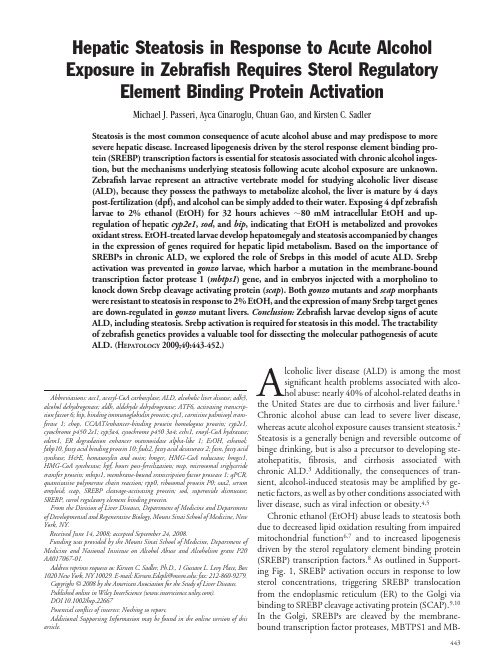
Hepatic Steatosis in Response to Acute Alcohol Exposure in Zebrafish Requires Sterol RegulatoryElement Binding Protein ActivationMichael J.Passeri,Ayca Cinaroglu,Chuan Gao,and Kirsten C.SadlerSteatosis is the most common consequence of acute alcohol abuse and may predispose to more severe hepatic disease.Increased lipogenesis driven by the sterol response element binding pro-tein (SREBP)transcription factors is essential for steatosis associated with chronic alcohol inges-tion,but the mechanisms underlying steatosis following acute alcohol exposure are unknown.Zebrafish larvae represent an attractive vertebrate model for studying alcoholic liver disease (ALD),because they possess the pathways to metabolize alcohol,the liver is mature by 4days post-fertilization (dpf),and alcohol can be simply added to their water.Exposing 4dpf zebrafish larvae to 2%ethanol (EtOH)for 32hours achieves ϳ80mM intracellular EtOH and up-regulation of hepatic cyp2e1,sod ,and bip ,indicating that EtOH is metabolized and provokes oxidant stress.EtOH-treated larvae develop hepatomegaly and steatosis accompanied by changes in the expression of genes required for hepatic lipid metabolism.Based on the importance of SREBPs in chronic ALD,we explored the role of Srebps in this model of acute ALD.Srebp activation was prevented in gonzo larvae,which harbor a mutation in the membrane-bound transcription factor protease 1(mbtps1)gene,and in embryos injected with a morpholino to knock down Srebp cleavage activating protein (scap ).Both gonzo mutants and scap morphants were resistant to steatosis in response to 2%EtOH,and the expression of many Srebp target genes are down-regulated in gonzo mutant livers.Conclusion:Zebrafish larvae develop signs of acute ALD,including steatosis.Srebp activation is required for steatosis in this model.The tractability of zebrafish genetics provides a valuable tool for dissecting the molecular pathogenesis of acute ALD.(H EPATOLOGY 2009;49:443-452.)Alcoholic liver disease (ALD)is among the most significant health problems associated with alco-hol abuse:nearly 40%of alcohol-related deaths in the United States are due to cirrhosis and liver failure.1Chronic alcohol abuse can lead to severe liver disease,whereas acute alcohol exposure causes transient steatosis.2Steatosis is a generally benign and reversible outcome of binge drinking,but is also a precursor to developing ste-atohepatitis,fibrosis,and cirrhosis associated with chronic ALD.3Additionally,the consequences of tran-sient,alcohol-induced steatosis may be amplified by ge-netic factors,as well as by other conditions associated with liver disease,such as viral infection or obesity.4,5Chronic ethanol (EtOH)abuse leads to steatosis both due to decreased lipid oxidation resulting from impaired mitochondrial function 6,7and to increased lipogenesis driven by the sterol regulatory element binding protein (SREBP)transcription factors.8As outlined in Support-ing Fig.1,SREBP activation occurs in response to low sterol concentrations,triggering SREBP translocation from the endoplasmic reticulum (ER)to the Golgi via binding to SREBP cleavage activating protein (SCAP).9,10In the Golgi,SREBPs are cleaved by the membrane-bound transcription factor proteases,MBTPS1and MB-Abbreviations:acc1,acetyl-CoA carboxylase;ALD,alcoholic liver disease;adh3,alcohol dehydrogenase;aldh,aldehyde dehydrogenase;ATF6,activating transcrip-tion factor 6;bip,binding immunoglobulin protein;cpt1,carnitine palmitoyl trans-ferase 1;chop,CCAAT/enhancer-binding protein homologous protein;cyp2e1,cytochrome p4502e1;cyp3a4,cytochrome p4503a4;echs1,enoyl-CoA hydratase;edem1,ER degradation enhancer mannosidase alpha-like 1;EtOH,ethanol;fabp10,fatty acid binding protein 10;fads2,fatty acid desaturase 2;fasn,fatty acid synthase;H&E,hematoxylin and eosin;hmgcr,HMG-CoA reductase;hmgcs1,HMG-CoA synthetase;hpf,hours post-fertilization;mtp,microsomal triglyceride transfer protein;mbtps1,membrane-bound transcription factor protease 1;qPCR,quantitative polymerase chain reaction;rpp0,ribosomal protein P0;saa2,serum amyloid;scap,SREBP cleavage-activating protein;sod,superoxide dismutase;SREBP,sterol regulatory element binding protein.From the Division of Liver Diseases,Department of Medicine and Department of Developmental and Regenerative Biology,Mount Sinai School of Medicine,New York,NY.Received June 14,2008;accepted September 24,2008.Funding was provided by the Mount Sinai School of Medicine,Department of Medicine and National Institute on Alcohol Abuse and Alcoholism grant P20AA017067-01.Address reprints requests to:Kirsten C.Sadler,Ph.D.,1Gustave L.Levy Place,Box 1020New York,NY 10029.E-mail:Kirsten.Edepli@:fax:212-860-9279.Copyright ©2008by the American Association for the Study of Liver Diseases.Published online in Wiley InterScience ().DOI 10.1002/hep.22667Potential conflict of interest:Nothing to report.Additional Supporting Information may be found in the online version of this article.443TPS2(also called site 1and site 2proteases).11This releases the active transcription factors to the nucleus where SREBP1regulates genes essential for lipogenesis acetyl-coenzyme A (i.e.carboxylase (Acc1)and fatty acid synthase (Fasn ))and SREBP2activates genes required for cholesterol biogenesis,including HMG CoA reductase (Hmgcr )and HMG CoA synthase (Hmgcs1).Although SCAP is specific for the SREBPs,MBTPS1and MBTPS2are also required for activating the unfolded protein re-sponse and the hepatic acute phase response via cleavage of activating transcription factor 6(ATF6)12and cAMP responsive element binding protein H (CREBH)13tran-scription factors,respectively (Supporting Fig.1).This suggests a possible link between hepatic lipogenesis,ER stress,and inflammation,each of which are central fea-tures of chronic ALD.The pathophysiology of steatohepatitis caused by chronic alcoholism is well understood,whereas the mech-anisms underlying steatosis following acute EtOH expo-sure are less clearly defined.The dearth of suitable animal models is a major obstacle to studying the genetic and cellular factors that contribute to hepatic pathology fol-lowing acute EtOH exposure.Rodent models of ALD are informative for identifying key biochemical and cellular abnormalities after chronic EtOH,however,these models are time-consuming and costly 14and,with the exception of chronic intragastric alcohol feeding,14rodents are rela-tively resistant to ALD.This highlights the need for new approaches.Among vertebrates,zebrafish have emerged as a pow-erful system because of its genetic tractability,optical clar-ity,and simple culture system.15The large clutch size (number of offspring),rapid generation time,and the ability to manipulate the externally developing zebrafish embryo allows for forward genetic screening,creation of transgenic fish,and transient knockdown of geneexpres-Fig.1.Acute EtOH exposure re-duces viability of zebrafish larvae.(A)Outline of the EtOH treatment protocol.EtOH exposure began at 96hpf and continued for up to 32hours (128hpf).(B)Larvae treated with 0%-4%EtOH were scored for mortality,and the average of three clutches is plotted during 32hours of exposure;error bars show stan-dard deviation.(C)Tissue EtOH con-centration was determined using homogenates of whole larvae treated with 0,171,and 343mM EtOH (i.e.,0%,1%,and 2%)as a substrate for an alcohol dehydroge-nase activity,and the measure-ments from untreated larvae was subtracted as background from measurements obtained from treated animals.Values indicate the average millimolar concentrations of internal EtOH in at least three sep-arate clutches.*indicates P ϭ0.046.(D-F)qPCR analysis of gene expression in whole larvae treated for 24or 32hours with 2%EtOH (D)or for 32hours with either 1%or 2%EtOH (E).Values indicate the aver-age fold change in gene expression compared to untreated controls in three clutches.(F)Livers were dis-sected from four to eight clutches of larvae that were treated with 0%or 2%EtOH for 32hours,qPCR analy-sis demonstrates a significant in-crease in the expression of cyp2e1,bip and sod in response to EtOH (*indicates P value Ͻ0.03).444PASSERI ET AL.HEPATOLOGY,February 2009sion via morpholino injection.16Zebrafish possess key enzymes that metabolize EtOH,including alcohol dehy-drogenase(adh3),17aldehyde dehydrogenase(aldh2),18 and cytochrome oxidase p4502e1(cyp2e1).Behavioral studies with adult and larval zebrafish exposed to alco-hol19-22and the use of early zebrafish embryos to study fetal alcohol syndrome23-28demonstrate their utility for studying the effects of alcohol.No investigators have yet reported using zebrafish to study ALD.The ex vivo development of transparent zebrafish em-bryos allows detailed and dynamic analysis of embryonic lethal mutants.For example,homozygous deletion of Mbtps1in mice is embryonic lethal,29requiring the cre-ation of tissue-specific knockout mice to study the role of this important gene in liver metabolism and steatosis.29 The zebrafish mbtps1mutant line,gonzo(goz),also do not survive to adulthood and die at7days post-fertilization (dpf).30,31By4dpf,several measures of liver function are present,including the high expression of many hepato-cyte-specific genes,32bile secretion,33glycogen storage, and lipid metabolism.34,35Thus,the role of mbtps1in hepatic physiology can be studied in goz mutants. Here,we use zebrafish larvae to address the genetic basis of steatosis associated with acute ALD.Early ze-brafish embryos are sustained by yolk,and although larvae are capable of feeding by4dpf,36they can survive for several days without external nutrients.Thus,zebrafish larvae at4-5dpf present an ideal system to study the effects of alcohol on a mature liver without the metabolic changes introduced by nutrient metabolism.Exposing4 dpf larvae to EtOH results in increased mortality,mor-phologic and behavioral abnormalities,and -ing goz mutants and morpholino knockdown of scap,we establish that the SREBP pathway is required for steatosis in this system.Thesefindings provide mechanistic insight into the pathophysiology of acute ALD and present ze-brafish as a new model for identifying genes that contrib-ute to ALD.Materials and MethodsAnimals.Adult wild-type(WT)zebrafish and het-erozygous carriers of the hi1487allele30,37of mbtps1(herein referred to as goz hi1487)were maintained on a14:10hour light:dark cycle at28°C.Fertilized embryos collected fol-lowing natural spawning were cultured at28°C infish water(0.6g/L Crystal Sea Marinemix;Marine Enter-prises International,Baltimore,MD)containing methyl-ene blue(0.002g/L).The Mount Sinai School of Medicine Institutional Animal Care and Use Committee approved all protocols.Ethanol Treatment.At4dpf(between96-98hours post-fertilization[hpf]),20-50larvae were anesthetized with2.5g/L tricane and transferred to0%,0.5%,1%, 2%,3%,or4%EtOH infish water and cultured in sealed dishes for up to32hours.Each experiment was repeated on at least three clutches.Tissue Alcohol Concentration Assay.Approximately 50of the4dpf larvae were treated with0%,1%,or2% EtOH and collected at8,24,or32hours of EtOH vae were washed twice in phosphate-buffered sa-line(PBS),homogenized on ice in50L of PBS,and pelleted by centrifugation.The supernatant was adjusted with PBS to equal1L/larvae,and EtOH concentration was quantified using the Ethanol Assay(Diagnostic Chemicals Limited,Oxford,CT).Briefly,the superna-tant was incubated with alcohol dehydrogenase and nic-otinamide adenine dinucleotide at room temperature. The ultraviolet absorbance at340nm was measured,and the reading from untreated larvae at each time point was set as0mM.The concentration was calculated according to measurements of a standard containing20mM EtOH. Each sample was measured in triplicate and repeated on three clutches per time point.Oil Red O Staining.Whole larvae werefixed with 4%paraformaldehyde,washed with PBS,infiltrated with a graded series of propylene glycol,and stained with0.5% Oil Red O in100%propylene glycol overnight.Stained larvae were washed with decreasing concentrations of pro-pylene glycol followed by several rinses with PBS and transferred to80%glycerol.Steatosis was scored as three or more lipid droplets per liver.Images were captured on a Nikon SMZ1500with a Nikon Digital Sight color cam-era.Histologic rvae werefixed by embedding in HistoGel(Richard-Allan Scientific,Kalamazoo,MI) and paraffin according to standard procedures.Then,4m sections were stained with hematoxylin and eosin(H&E)and imaged on an Olympus BX41microscope using a Nikon Digital Sight-Ri1camera.Reverse Transcription and Quantitative PCR. Poly deoxythymidylate–primed complementary DNA was prepared from RNA isolated with the RNeasy kit (Qiagen)using Superscript II(Invitrogen,Carlsbad,CA). Real-time quantitative PCR(qPCR)was carried out on Light Cycler480(Roche,Basel,Switzerland)with one cycle of95°C for3minutes,followed by40cycles of95°C for15seconds,and60°C for1minute using1mol/L of gene-specific primers(Supporting Table1)and the iQ SYBR Green Supermix(Roche).rpp0was used as a refer-ence,and expression was calculated using the cycle thresh-old(C t)method(2-Ct(target)/2-Ct(rppo)).Typically,20fishHEPATOLOGY,Vol.49,No.2,2009PASSERI ET AL.445were pooled for each sample and3-10separate clutches were analyzed per experiment.Morpholino Injections.A splice-blocking morpholino targeting scap(MO;5Ј-cccgatactgcaagaagatt-3Јas de-scribed31)and a control morpholino(5Јaggttcctttggttttgca-cagcgc-3Ј;Gene Tools,Philomath,OR)were diluted with water to0.5mM,and approximately4nL was injected into over50one-cell to two-cell stage embryos using a Narishege IM-300microinjector.Abnormal embryos were removed at 6hpf.Statistical Analysis.All evaluations were performed with MS Excel software.P values less than0.05were considered statistically significant.ResultsEthanol Induces Abnormalities in Zebrafish Lar-vae.Zebrafish hepatogenesis occurs between1-3dpf,38 and larval feeding begins on4-5dpf when the yolk has been completely utilized.Fasting-induced steatosis occurs if larvae are not fed by6dpf(K.C.S.and C.G.,unpub-lished results).Thus,4-5dpf is the ideal time to assess the effects of alcohol without interference by the metabolic changes that accompany external nutrient metabolism or fasting.To optimize a protocol for exposing zebrafish larvae to EtOH,wefirst assessed EtOH toxicity.The4dpf larvae were exposed to EtOH concentrations ranging from 0.5%to4.0%for32hours(i.e.,until128hpf;Fig.1A) and were visually scored for viability,morphology,and behavioral changes.Exposure to4%EtOH causes rapid mortality,whereas nearly all larvae exposed to2%EtOH or less survived the entire exposure(Fig.1B).Thus,4dpf larvae are affected by EtOH in a dose-dependent and time-dependent manner,the median lethal dose(LD50)is 2.6%at32hours of treatment,and2%EtOH is the maximal tolerable dose.The cellular EtOH concentration was determined by an alcohol dehydrogenase assay using homogenate pre-pared from larvae exposed to0,171,and343mM EtOH (i.e.,0%,1%,and2%EtOH)for8,24,or32hours as a substrate.By8hours of exposure,tissue EtOH levels reached40mM in larvae treated with1%EtOH and63 mM in those treated with2%EtOH(Fig.1C).The level of internal alcohol remained constant at all time points, but at24hours of exposure,the internal EtOH concen-tration was higher in larvae treated with2%compared to 1%EtOH(Fig.1C).The values measured in our exper-iments are comparable to those measured in other ze-brafish studies22,39and are within an order of magnitude of the legal limit in humans.If zebrafish metabolize EtOH,then the genes which metabolize EtOH,such as cyp2e1,cyp3a4,and aldh3 should be induced,and the generation of free oxygen radicals should cause oxidative stress and induce superox-ide dismutase(sod)expression.40Neither extended time (Fig.1D)nor increased concentration(Fig.1E)of EtOH exposure caused changes in the expression of these genes in extracts prepared from whole embryos,but cyp2e1and sod expression increased significantly(P valuesϽ0.05), albeit modestly,in livers dissected from larvae treated with2%EtOH for32hours.Strikingly,bip expression was induced more than30-fold in the liver(Fig.1F), indicating significant ER stress.Based on these data,we conclude that zebrafish can ingest and metabolize EtOH, and this causes hepatic oxidative stress,signifying the suit-ability of this model for studying acute ALD. Exposing early zebrafish embryos to EtOH disrupts patterning and causes morphologic abnormalities.25,28To avoid these effects on early development,we began our treatment protocol at4dpf,yet we found that EtOH concentrations of2%or greater induced reproducible morphologic abnormalities(Fig.2A).Fish with edema, hepatomegaly,and body curvature were considered“se-verely affected”,whereas those which only failed to inflate their swim bladder were scored as“moderately affected”(Fig.2B).The number of affectedfish increased as either time of exposure or EtOH concentration increased,with higher concentrations(3%and4%EtOH)inducing more severe abnormalities(Fig.2B).Alcohol induces pro-found behavioral effects in mammals as well as in adult 19,21,41and larval22zebrafish.We also observed irregular and nondirectional swimming,lethargy,and immobility in zebrafish larvae treated with EtOH,and scored the number of animals exhibiting any of these behaviors as “affected”(Fig.2C).These results demonstrate that EtOH ingestion by4dpf zebrafish larvae increases mor-tality and causes changes in morphology and behavior. Acute Ethanol Exposure Induces Steatosis.Acute ethanol exposure in mammals causes steatosis.2To deter-mine whether this also occurs in zebrafish larvae,we treated4dpf larvae with1%or2%EtOH for24or32 hours and visualized neutral lipids by whole-mount oil red O staining(Fig.3A).Untreated larvae and those treated with1%EtOH(not shown)rarely developed ste-atosis by5.5dpf,whereas2%EtOH treatment resulted in 53%and64%steatosis at24and32hours of exposure, respectively(Fig.3B).Histological analysis revealed ma-crovesicular lipid droplets in the hepatocytes of larvae treated for32hours with2%EtOH(Fig.3C).Despite this marked steatosis,there was no change in the expres-sion of genes regulating several other hepatic functions446PASSERI ET AL.HEPATOLOGY,February2009(Supporting Fig.2),suggesting that acute alcohol expo-sure does not compromise liver function.Steatosis in chronic ALD is due to both decreased lipid utilization due to mitochondrial dysfunction 6,7and to lipogenesis driven by SREBP1-c.8The molecular basis of steatosis associated with acute EtOH exposure is not known.To assess the effects of EtOH on hepatic lipid metabolism in zebrafish,qPCR was used to detect the expression of genes involved in lipid and cholesterol syn-thesis,and lipid oxidation and transport.The expression of these genes was compared between the livers of 5.5dpf fish treated with 2%EtOH and those of untreated fish.In Fig.3D,the fold change in expression is plotted as a single point for each clutch,with the mean fold change from all experiments indicated by an “X”.We found significant changes in the expression of genes in each category,in-cluding the putative Srebp target genes fasn ,hmgcs1,and the hmgcra and hmgcrb genes,which resulted from the genome duplication that occurred during teleost evolu-tion.42Ethanol-Mediated Steatosis Requires mbtps1and scap:A Role for Srebps.To address whether acute alco-hol-induced steatosis requires Srebps,we used mbtps1mutants and scap morpholino knock-down to prevent Srebp activation (Supporting Fig.1).Both genes are ex-pressed in the 5dpf liver (Fig.4A),suggesting they func-tion at the time in which larvae develop steatosis in response to EtOH.The hi 1487allele of goz mutants (i.e.,goz hi1487)has a viral insertion in the intron preceding the initiator ATG codon of the mbtps1gene,resulting in a loss of function and embryonic lethality.30,37However,unlike the Mbtps1knockout mice,which die in utero 43preclud-ing examination of hepatic function,goz hi1487mutants survive until 7dpf with fully mature livers.Additionally,injecting embryos with a morpholino to prevent scap splicing 31should specifically prevent Srebp activation.We found no difference in the internal EtOH concentra-tion of WT larvae and goz hi1487mutants (Fig.4B)or scap morphants (not shown)or the morphological changes induced by 32hours of treatment with 2%EtOH (Fig.4C and not shown),indicating that these models do not alter the ability of zebrafish larvae to ingest and respond to alcohol.There was,however,a marked inhibition of ste-atosis in response to 32hours exposure to 2%EtOH in goz hi1487mutants (Fig.4D,E),indicating a requirement for mbtps1in steatosis resulting from acute EtOH exposure.The most straightforward interpretation of this data is that acute alcohol exposure causes hepatic Srebp activa-tion,thereby inducing the expression of genes required for lipid and cholesterol synthesis (Fig.3D),and that mutation of mbtps1blocks this pathway.Alternatively,because ATF6is critical to the unfolded protein response (UPR),and this pathway has been implicated in ALD 44the role of mbtps1in activating the ATF612may also be important (Supporting Fig.1).However,with the excep-tion of bip up-regulation (Fig.1F),we found that other Atf6targets (Supporting Fig.3A)and or xbp1splicing (Supporting Fig.3B),were not induced the livers of EtOH-treated larvae.Serum amyloid (saa2)is regulated by CREBH 13and was substantially up-regulated inre-Fig.2.Zebrafish exposed to alcohol de-velop morphological and behavioral abnor-malities.(A)The 5.5dpf larvae treated with 0%or 2%EtOH for 32hours develop mor-phologic abnormalities including upward curvature of the trunk and tail,hepatomeg-aly (liver is outlined by a dotted line),peri-cardial edema (arrow),and failure to inflate the swim bladder (sb).Scale bar represents 250m.(B)Morphologic abnormalities induced by 0%,1%,2%,and 3%EtOH become more severe with duration of expo-sure.Failure to inflate the swim bladder was scored as a moderate abnormality,and occurrence of one additional phenotype (i.e.,edema,body curvature or hepatomeg-aly)was scored as severe.(C)Behavioral abnormalities induced by EtOH,including nondirectional swimming and lethargy,was scored for animals treated with 0%,1%,2%,and 3%EtOH.All experiments were repeated on three clutches,and error bars indicate standard deviation.HEPATOLOGY,Vol.49,No.2,2009PASSERI ET AL.447sponse to EtOH (Supporting Fig.3C),but the degree of up-regulation varied from clutch to clutch.These findings raised the possibility that failure to ac-tivate Atf6and/or Crebh may contribute to the striking inhibition of steatosis in goz hi1487mutants.Transient knock-down of scap expression in WT fish by injecting embryos with a morpholino that blocks splicing of scap message is predicted to specifically prevent Srebp activa-tion without affecting Atf6or Crebh.scap morpholino injection did not adversely affect embryonic development or mortality,and the same internal EtOH concentration was measured measured in scap morphants,larvae in-jected with a control morpholino and uninjected controls treated with 2%EtOH for 24hours (not shown).In both sets of control larvae,i.e.,uninjected and those injected with a nontargeting morpholino,more than 60%of the larvae developed steatosis in response to 2%EtOH,in contrast to only 25.7%of larvae developed steatosis in scap morphants (Fig.4F).If Srebp activation does not occur in goz hi1487mutants in response to acute alcohol exposure,then Srebp target genes should not be up-regulated in these fish.The qPCR analysis of gene expression in the livers from three clutches of goz hi1487larvae and their nonmutant siblings confirms this hypothesis.The expression of acc1,fasn ,hmgcra ,and hmgcs1is significantly lower in untreated goz hi1487livers compared to WT livers,signifying that ex-pression of these genes is regulated by Srebps during nor-mal liver homeostasis (Fig.5A).The hepatic expression of each of these genes is increased after 2%EtOH exposure in WT livers (Fig.3D),whereas acc1,fasn ,hmgcra ,and hmgcs1expression was markedly reduced in goz hi1487(Fig.5B).Interestingly,the hmgcrb expression is the same in WT and goz hi1487livers,regardless of EtOH exposure,suggesting that it is independent of Srebps.This is con-sistent with the finding that hmgcrb expression is not liver-specific and that it primarily functions in the zebrafish heart,not in the liver.45Importantly,changes in echs1,cyp2e1,and mtp expres-sion in response to EtOH are similar in WT and goz hi1487livers,indicating the specificity of the effect on Srebp target genes.The Atf6target gene,bip ,is stronglyinducedFig.3.EtOH exposure results in steatosis and alteration of lipid metabolism.(A)Whole-mount oil red O staining of 5.5dpf larvae after exposure to 0%or 2%EtOH for 32hours reveals steatosis.Dotted line outlines the liver.(B)The 4dpf larvae were treated with 0%or 2%EtOH,collected at 24or 32hours of expo-sure and stained with oil red O.The percent of larvae with steatosis was scored in at least four clutches with an average n ϭ22fish per group;*P ϭ0.07;**P ϭ0.0002.(C)H&E staining of sections through the liver of 5.5dpf larvae treated with 0%,1%,or 2%EtOH for rge,clear cytoplasmic lipid droplets (arrows)are seen in livers from larvae treated with 2%EtOH.(D)Livers were dissected from 5.5dpf larvae treated with 0%or 2%for 32hours and the expression of genes involved in lipid metabolism was analyzed by qPCR.Each gene was analyzed in up to eight clutches and each point represents the fold change of each gene in livers treated with 2%EtOH relative to their untreated siblings from a single clutch.The “X”marks the mean fold change,which is indicated numerically.*,P Ͻ0.02;**,P ϭ0.07.448PASSERI ET AL.HEPATOLOGY,February 2009in the liver following EtOH treatment,similar to what is observed in adult zebrafish brains following chronic alco-hol exposure.46There is not a significant decrease in bip expression,however,in goz hi1487livers (Fig.5C),suggest-ing that Atf6may not be the primary regulator of bip in response to EtOH.We thus conclude that Srebp activa-tion occurs in response to acute alcohol exposure,and is required for steatosis.DiscussionWe demonstrate that 32hours of continuous exposure to 2%EtOH causes zebrafish larvae to develop character-istic signs of acute ALD,including hepatomegaly,steato-sis,and changes in hepatic gene expression,suggesting that alcohol is metabolized and causes oxidative stress.47Symptomatic fish were generated rapidly and in large numbers via a relatively simple treatment protocol,which provides distinct advantages over mammalian models.Genetic tractability is among the most useful attributes ofzebrafish.We present two examples,using goz hi1487mu-tants and scap morphants,to demonstrate that zebrafish genetics can be used to quickly and efficiently identify genes required for alcohol-mediated steatosis.Several studies established early zebrafish embryos as a model for fetal alcohol syndrome 23,25-27,48and adult ze-brafish are used to study the effects of alcohol on behav-ior.19-21,41,46Although we also found that EtOH induces morphological and behavioral abnormalities in zebrafish larvae,the effects we report are less likely to be attributable to disrupting developmental processes,because we ex-posed larvae to alcohol once major developmental mile-stones are complete.Alcohol exposure in both early-stage and late-stage embryos does,however,appear to affect cholesterol metabolism.Early embryos exposed to EtOH display growth retardation and cyclopia attributed to de-creased cholesterol modification of sonic hedgehog,27whereas we found that alcohol increases the expression of genes required for cholesterol biogenesis.It ispossibleFig.4.goz hi1487mutants and scap morphants are resistant to EtOH-induced steatosis.(A)PCR analysis of mbtps1and scap expression in 5dpf liver and whole embryo extracts.rpp0is used as a control.(B)Tissue EtOH concentration was determined by spectrophotometric analysis of alcohol dehydrogenase activity using homogenates of whole 5.5dpf larvae treated for 24hours with 2%EtOH as a substrate.Values on each bar indicate the average millimolar concentration of internal EtOH in four separate clutches of nonmutant siblings (WT)and goz hi1487larvae.Error bars indicate the standard error of the mean.(C)Live images of 5.5dpf larvae treated with 0%or 2%EtOH for 32hours indicate that EtOH induces the same morphological changes in WT and goz hi1487larvae.Additionally,goz hi1487mutants have defective craniofacial development 30illustrated by the small jaw (arrow).Scale bar represents 250m.(D)goz hi1487larvae and their nonmutant siblings were stained with oil red O on 5.5dpf following 32hours exposure to 2%EtOH.Neutral lipid accumulation is seen in the liver (dotted line)of WT fish (top)but not in goz hi1487mutants (bottom).The percentage of 5.5dpf (E)goz hi1487mutants or (F)scap morphants that develop steatosis with and without 32hours of 2%EtOH exposure was scored in four goz hi148clutches (n Ն14fish per clutch;total n ϭ230larvae)and five clutches injected with scap or control morpholinos (MO;n Ն8fish per clutch,average clutch size of 21,and total n ϭ502larvae)and averaged.Control larvae were nonmutant siblings in (E)and the sum of uninjected and control morpholino-injected larvae in (F).Error bars indicate standard error of the mean and P values are indicated.HEPATOLOGY,Vol.49,No.2,2009PASSERI ET AL.449。
structural capacities of smart biomaterials,we add intelligence by introducing selected proteins by ex vivo gene transfer technologies.Due to high availability we currently purify lipoaspirate derived adult progenitor cells as susbtrate and carriers for growth factor release into full thickness defects.In this overview we focus on current promising strategies in soft tissue reconstruction using this‘composite’smart autologous tissue engineering concept.Effects of in vitro pre-culture on the biomechanical properties and further in vivo development of humannasal engineered cartilage graftsJ.Farhadi a,b,I.Fulco a,b,S.Miot b,M.Haug a,b,G.Pierer a,b,M.Heberer b,I.Martin ba Department of Plastic,Reconstructive and Aesthetic Surgery, University Hospital Basel,Switzerlandb Institute for Surgical Research and Hospital Management, University Hospital Basel,SwitzerlandEngineered nasal cartilage grafts need to have sufficient mechanical integrity at the time of implantation and have to reach sufficient mechanical stability in a time frame of about2 weeks,whenfixation is typically removed.The goal of this study was thus to determine suture retention strength and tensile stiffness of engineered human nasal cartilage,respec-tively,at different times of pre-culture and following2weeks in vivo implantation.The same mechanical properties were also measured in native human nasal cartilage,as a reference. Chondrocytes from nasal septum cartilage from four patients were isolated by collagenase.The cells were expanded for two passages and seeded on hyaluronan scaffolds(Hyalograft-C, FAB,Italy).The constructs were implanted for2weeks in the back of nude mice either directly(freshly seeded scaffold),or after2and4weeks of pre-culture.Biomechanical(i.e.,suture pull out tests,tensile tests,three point bending tests)and histological(i.e.,Safranin O stain)assessments were performed on native nasal cartilage and on engineered cartilage,immedi-ately after seeding,after pre-culture,and after explantation. The results showed that the intensity of Safranin O stain progressively increased during pre-culture and in vivo develop-ment.The suture pull out force normalised to the specimen’s thickness was3.6-fold higher in2week-pre-cultured constructs than in freshly seeded scaffolds,did not further increase with longer pre-culture time and reached levels about4.3-fold lower than native nasal cartilage.The approximate modulus of elasticity in tension following in vivo implantation was21% higher if constructs were pre-cultured for2weeks than if they were directly implanted,and reached levels which were2.7-fold lower than in native nasal cartilage.The approximate modulus of elasticity in bending following in vivo implantation was2.1-fold higher if constructs were pre-cultured for2weeks than if they were directly implanted,and reached levels which were7.5-fold lower than in native nasal cartilage.Based on the increased suture pull out force at the time of implantation and the increased tensile and bending stiffness2weeks after implantation,engineered cartilage tissues pre-cultured for2 weeks are more suitable for clinical use than freshly seeded scaffolds.Demineralised bovine spongiosa as three-dimensionalmatrix in Adipose Tissue EngineeringK.Koellensperger,M.Markowicz,N.PalluaDepartment of Plastic Surgery and Hand Surgery,Burns Center, University Hospital,Aachen University of Technology, Aachen,Germany Background:Adipose Tissue Engineering represents a promising perspective for soft tissue reconstruction.A three-dimensional defect-adapted matrix is used as basis onto which autologous preadipocytes are seeded.So far an ideal material as scaffold structure for the transplanted cells has not been found.Fur-thermore vascularisation of such a cell e matrix compound con-tinues to pose problems and remains a main obstacle for survival of the transplanted cells and thus long-term tissue re-placement.Consequently,a suited material has to guarantee (1)a three-dimensional mechanical stability,allow(2)prolifer-ation and differentiation of preadipocytes,and,finally,support (3)growth of microvascular endothelial cells as basis for neoan-giogenesis and mature vascularisation.Methods:Excised adipose tissue taken from healthy donors was digested enzymatically to isolate primary human preadipocytes and microvascular endothelial cells.Cells were seeded in dem-ineralised bovine spongiosa sterilised using y-rays as both mono-and co-cultures.Subsequent in vitro and in vivo experiments, including a mouse model of NOD-SCID mice,proliferation and differentiation were assessed.Results:In vitro both cell types displayed good proliferation and migration characteristics.Capability of adipogenic differ-entiation in preadipocytes remained intact.In vivo apart from mechanical three-dimensional stability of the cell-matrix com-pounds the formation of mature adipose tissue with a sustained cellular composition was achieved.Conclusion:In vitro and in vivo demineralised bovine spongiosa displays ideal characteristics for culture of human preadipo-cytes and endothelial cells.Among the advantages for Adipose Tissue Engineering are ready availability and high mechanical stability of the three-dimensional structure.Demineralised bo-vine spongiosa might be a biotechnological option for plastic and reconstructive surgery of soft tissue defects.Matrigel-induced adipogenesis is host rather than graft derived in the murine tissue engineering chamberF.B.J.L.Stillaert a,b,M.Findlay b,R.Idrizi b,J.Palmer b, S.Cheang b,A.Messina b,K.Abberton b,P.Blondeel a,W.A.Morrison b,E.W.Thompson ba Ghent University,Belgiumb Bernard O’Brien Institute of Microsurgery,Melbourne, AustraliaWe have previously shown that Matrigel-filled chambers placed around the epigastric bundle in the mouse groin are highly adipogenic,so long as contact with a source of adipose tissue is ensured(Cronin et al.,2004).Here we used human adipose tissue biopsy autografts(w3mg)in chambers implanted in Se-vere Combined Immuno Compromised(SCID)mice together with species specific probes to determine the graft and/or host origins of the resultant adipose tissue.The adipose tissue which formed in Matrigel-filled chambers seeded with human fat from three anatomical sources(mammary,axillary,and ab-dominal)lacked human-specific staining for the intermediate filament protein vimentin,indicating that it was of mouse ori-gin.The murine origin was confirmed by the preponderance of mouse-specific Cot-1DNA hybridisation byfluorescent in situ hybridisation,and the absence of human Cot-1labelling.Con-sistent with these data,Matrigel-containing chambers seeded with cultured human stromal-vascular fraction isolated from different adipose tissue beds(preadipocytes)showed promi-nent human vimentin staining only of cells which did not adopt an adipocyte phenotype,and the abundant,newly formed adi-pocytes lacked this staining.These results are surprising,given the strict requirement we have observed for close contact with pre-existing adipose tissue,which we anticipated would provide precursor cells.ECSAPS2005Abstracts S9。
骨 科 基 因 治 疗王爱民 李起鸿 随着分子生物学等学科的飞速发展,基因治疗应运而生,发展也十分迅速。
所谓基因治疗,就是向靶细胞(target cell)引入外源性基因,以纠正或补偿其基因缺陷,从而达到治疗的目的。
在众多动物实验取得成功的基础上,临床实践开始起步。
Anderson和Blase于1990年9月为1例腺苷脱氨酶(adenosine deaminase ADA)基因缺陷患儿进行基因治疗,取得了令人鼓舞的结果,开创了临床基因治疗的新篇章[1]。
目前,美国DNA重组委员会(N IH,Recombinant DNA Advisory Committee)已经批准80余种临床基因治疗,并有15种基因治疗取得一定疗效。
尽管骨科临床基因治疗尚未开始,但是导致肌肉骨骼系统严重并发症的高歇氏病(gaucher disease)和类风湿性关节炎的临床基因治疗已获DNA重组委员会的批准。
因此,骨科临床基因治疗不久也会很快开展。
一、基因治疗的概况本世纪60年代后期,针对由于遗传和单基因突变引起的典型基因缺陷性疾病,提出了基因治疗的构想[2]。
80年代初,Anderson首先阐明了基因治疗的前景及其发展方向。
以后Joyner(1983)等最先在体外成功地通过逆转录病毒载体,把细菌新霉素磷酸转移酶基因(neo r)转入造血干细胞。
neo r的表达可以使靶细胞获得耐G148(一种新霉素同类物)的能力。
实验结果发现0.3%的粒细胞-巨噬细胞集落形成单位(CFU-GM)具有G148耐受性。
1984年,Willams等人把经如上改造的细胞输入受致死量照射的小鼠,在20%的脾细胞集落形成单位(CFU-S)中检测到neo r的存在。
随后, K ohn等人在非人灵长类长目动物中把载有腺苷酸脱氨酶(ADA)基因的逆转录病毒载体转入体内,该基因可在动物体内持续达数日,表达水平为正常水平的0. 5%[2]。
ADA缺陷病基因治疗在动物体内获得成功后, Ferrari等(1991)将转导有人ADA基因的淋巴细胞输入免疫缺陷的小鼠内,转导细胞在小鼠体内能长期存活,表达ADA分子,重建免疫功能[2]。
AIM-V® Medium CTS™Therapeutic Grade serum free cell expansion mediumGIBCO® AIM-V Medium CTS™ (Therapeutic Grade) is the first commercially available defined, serum-free formulation for proliferation and/or manipulation of T-cells and dendritic cells and manufactured in compliance with cGMP. AIM-V Medium CTS™ is an FDA 510(k) cleared device which is intended for human ex-vivo tissue & cell culture processing applications.Description Cat. No. SizeAIM-V® Medium CTS™, Liquid 0870112DK 1000mL AIM-V® Medium CTS™, Liquid 0870112BK 10L(Bag)IntendedUseFor human ex-vivo tissue & cell culture processing applications. CAUTION: When used as a medical device, Federal Law restricts this device to sale by or on the order of a physician. StorageStore medium at 2 to 8°C. Protect from light.Shelf Life14 monthsCulture Procedure:The procedure below serves as a general guideline for static T- cell and dendritic cell culture, regardless of vessel. For high-density culture in bioreactors, optimal procedures should be determined empirically by the investigator.T Cells Culture:1. Prepare fresh peripheral blood mononuclear cells (PBMCs)or rapidly thaw (< 1 minute) frozen vials of PBMCs cells in a 37°C water bath according to standard PBMC thawing protocols.2. Wash cells with DPBS CTS™ without calcium andmagnesium (Cat. No A12856), with 2-5% heat-inactivated human pooled Type AB serum according to the applications, if desired or required.3. Count cells using either electronic (i.e. Coulter Counter, Vi-Cell) or manual (i.e. hemocytometer) methods.4. Centrifuge cells and remove wash buffer.5. Resuspend PBMC at roughly 0.5-1x10⁶ CD3+ T cells/mL inmedium supplemented with cytokines (e.g. IL-2), if used at culture initiation. Transfer the desired number of cells to the desired tissue culture vessel. A variety of protocols may be used for activating T-cells for subsequent expansion, including adding stimulatory antibodies or antigen presenting cells. Similarly, for either small or the large scale T-cell expansion, cells can be isolated, activated and expanded with Dynabeads® ClinExVivo™ CD3/CD28 or Dynabeads® CD3/CD28 CTS TM(Cat. No. 402-03D) according to instructions in the product insert. 6. Incubate the culture vessel at 37°C in a humidifiedatmosphere with 5% CO2. Feed and maintain cells at desired concentrations while cells are in log phase growth.To maintain log phase growth, it may be preferable to split cells to achieve a density of 0.5-1x10⁶cells/mL whenever cell density gets above 1x10⁶cells/mL (e.g. 2x10⁶cells/mL, split 1:4 to continue culture at 0.5x10⁶cells/mL). For optimal gas exchange in static plate cultures it is recommended that medium depth not exceed 1 to 1.2cm.Monocyte Derived Dendritic Cell Culture:1. Prepare fresh peripheral blood mononuclear cells (PBMCs).2. Plate PBMC in culture flask with 25 mL RPMI 1640 (Cat.No 72400) or AIM-V® Medium CTS™ (Therapeutic Grade).3. Incubate for 2 to 3 hours at 36 to 38˚C in a humidified atmosphere of 5% CO2 in air.4. Discard medium containing non-adherent cells.5. Wash the adherent cells (mainly CD14+ monocytes) threetimes with DPBS without calcium and magnesium (Cat. No A12856).6. Add medium containing 50 to 100 ng/mL recombinanthuman IL-4 (Cat. No. CTP0043 1mg or Cat. No. CTP0041 100ug) and 50 ng/mL recombinant human GM-CSF (Cat.No. CTP2011 100ug or Cat. No. CTP2013 1mg). Cell density should be between 1 to 3x10⁵ cells/mL.7. Incubate cells at 36 to 38˚C in a humidified atmosphere of5% CO2 in air for 5 days. It is recommended to replace medium once after 3 days with fresh medium containing IL-4 and GM-CSF. Save all non-adherent or loosely adherentcells by centrifuging the removed culture medium 10 minutes at 200xg and adding the pellet to the fresh culture medium.8. After 6 days, the loosely adherent or non-adherent cellsshould display typical dendritic cell morphology and surface markers (CD1a, CD80, CD86, and HLA-DR).9. The maturation of dendritic cells is induced by the additionof either 1 µg/mL LPS or 50µl/mL TNF-α (cat. No. CTP3013 1mg or Cat. No. CTP3011 100ug) to the medium.Note: Alternatively to plastic adherence, monocytes can also be isolated by magnetic separation.Related ProductsDulbecco's Phosphate Buffered Saline CTS™ (DPBS) without calcium, magnesium (1X), liquid (A12856) L-Glutamine-200mM (100X), liquid (25030)Dynabeads ClinExVivo™ CD3/CD28 ®or Dynabeads CD3/CD28 ®CTS TM (402-03D) DynaMag™ CTS™ (121-02)IL-2 CTS™ REC HU (CTP0021 100ug or CTP0023 1mg) IL-7 CTS™ REC HU (CTP0071 100ug or CTP0073 1mg) IL-4 CTS™ REC HU (CTP0041 100ug or CTP0043 1mg) GM-CSF CTS™ REC HU (CTP2011 100ug or CTP2013 1mg) TNF-α CTS™ (CTP3011 100ug or CTP3013 1mg)Technical SupportFor additional product and technical information, such as Material Safety Data Sheets (MSDS), Certificate of Analysis,etc, please visit our website at /celltherapysupport/. For further assistance, please email our Technical Support team at celltherapysupport@The trademarks mentioned herein are the property of Life Technologies Corporation or their respective ownersReferences1.Rebecca J et al., (2010) Natural exposure to cutaneous anthrax gives long lasting T cell immunity encompassing infection-specific Epitopes. J. Immunol., 184: 3814 – 38212.Fabricius D et al., (2010) Prostaglandin E2 inhibits IFN-α secretion and Th1 costimulation by human plasmacytoid dendritic cells via E-prostanoid 2 and E-prostanoid 4 receptor engagement. J. Immunol., 184: 677 – 6843.Nesbit L et al., (2010) Polyfunctional T Lymphocytes Are in the Peripheral Blood of Donors Naturally Immune to Coccidioidomycosis and Are Not Induced by Dendritic Cells . Infect. Immun., 78: 309 - 3154. Jahrsdorfer B et al., (2010) Granzyme B produced by human plasmacytoid dendritic cells suppresses T-cell expansion . Blood, 115: 1156 – 11655.Csillag A et al., (2010) Pollen-Induced Oxidative Stress Influences Both Innate and Adaptive Immune Responses via Altering Dendritic Cell Functions. J. Immunol., 184: 2377 – 23856.Cornberg M et al., (2010) CD8 T Cell Cross-Reactivity Networks Mediate Heterologous Immunity in Human EBV and Murine Vaccinia Virus Infections. J. Immunol., 184: 2825 - 2838.7.Bellone S et al., (2009) Human Papillomavirus Type 16 (HPV-16) Virus-Like Particle L1-Specific CD8 Cytotoxic T Lymphocytes (CTLs) Are Equally Effective as E7-Specific CD8 CTLs in Killing Autologous HPV-16-Positive Tumor Cells in Cervical Cancer Patients: Implications for L1 Dendritic Cell-Based Therapeutic Vaccines ++. J. Virol., 83: 6779 - 67898. Sato K et al., (2009) Impact of culture medium on the expansion of T cells for immunotherapy. Cytotherapy 11: 4-119.Liu ZW et al., (2009) A CD26-Controlled Cell Surface Cascade for Regulation of T Cell Motility and Chemokine Signals . J. Immunol ., 183: 3616 - 3624.10. Megyeri M et al., (2009) Complement Protease MASP-1 Activates HumanEndothelial Cells: PAR4 Activation Is a Link between Complement and Endothelial Function. J. Immunol., 183: 3409 - 3416. 11.Manfred L et al., (2005) Functional characterization of monocyte-derived dendritic cells generated under serum free culture conditions. Immunology letters 99: 209-21612. Nagorsen D et al., (2003) Biased epitope selection by recombinant vaccinia-virus (rVV)-infected mature or immature dendritic cells. Gene Therapy 10: 1754-1765 13. Lotem M et al., (2006) Presentation of tumor antigens by dendritic cells genetically modified with viral and nonviral vectors. J immunotherapy 29: 616-62714.Dietze B et al. (2008) An improved method to generate equine dendritic cells from peripheral blood mononuclear cells: divergent maturation programs by IL-4 and LPS. Immunobiology 213:751–758.15.Meehan KR et al. (2008) Development of a clinical model for ex vivo expansion of multiple populations of effector cells for adoptive cellular therapy. Cytotherapy 10: 30–37.16. Ye Z et al. (2006) Human dendritic cells engineered to express alpha tumornecrosis factor maintain cellular maturation and T-cell stimulation capacity. Cancer Biother Radiopharm 21:613–622.17. Choi BH et al. (2006) Optimization of the concentration of autologous serum forgeneration of leukemic dendritic cells from acute myeloid leukemic cells for clinical immunotherapy. J Clin Apher 21:233–240. 18.Imataki O et al. (2006) Efficient ex vivo expansion of alpha24+ NKT cells derived from G-CSF-mobilized blood cells. J Immunother 29:320–327.19. Peng JC et al. (2005) Generation and maturation of dendritic cells for clinicalapplication under serum-free conditions. J Immunother 28:599–609. 20. Trickett AE et al. (2002) Ex vivo expansion of functional T lymphocytes from HIV infected individuals. J Immunol Methods 262:71–83.21.Carlens S et al. (2000) Ex vivo T lymphocyte expansion for retroviral transduction: influence of serum-free media on variations in cell expansion rates and lymphocyte subset distribution. Exp Hematol 28:1137–1146.22.Kambe N et al. (2000) An improved procedure for the development of human mast cells from dispersed fetal liver cells in serum-free culture medium. J Immunol Methods 240:101–110.23. Gerin PA et al. (1999) Production of retroviral vectors for gene therapy with thehuman packaging cell line FLYRD18. Biotechnol Prog 15:941–948. 24. Slunt JB et al. (1997) Human T-cell responses to Trichophyton tonsurans:inhibition using the serum free medium Aim-V. Clin Exp Allergy 27:1184–1192. 25. Kreuzfelder E (1996) Assessment of peripheral blood mononuclear cellproliferation by [2-3H]adenine uptake in the woodchuck model. Clin Immunol Immunopathol 78:223–227. 26.Causey AL (1994) A serum-free medium for human primary T lymphocyte culture. J Immunol Methods 175:115–121.27. Freedman RS et al. (1994) Large-scale expansion in interleukin-2 oftumorinfiltrating lymphocytes from patients with ovarian carcinoma for adoptive immunotherapy. J Immunol Methods 167:145–160. 28.Nomura K et al. (1993) [Study of adoptive immunotherapy for metastatic renal cell carcinoma with lymphokine-activated killer (LAK) cells and interleukin-2. II. Clinical evaluation.] Nippon Hinyokika Gakkai Zasshi 84:831–840. Japanese.29.Kaldjian EP et al. (1992) Enhancement of lymphocyte proliferation assays by use of serum-free medium. J Immunol Methods 147:189–195.30. Hayakawa K et al. (1991) Study of tumor-infiltrating lymphocytes for adoptivetherapy of renal cell carcinoma (RCC) and metastatic melanoma: sequential proliferation of cytotoxic natural killer and noncytotoxic T cells in RCC. J Immunother 10:313–325. 31.McVicar DW et al. (1991) A comparison of serum-free media for the support of in vitro mitogen-induced blastogenic expansion of cytolytic lymphocytes. Cytotechnology 6:105–113.32. Burg S et al. (1991) [Effect of different media on long-term cultivation of humansynovial macrophages.] Z Rheumatol 50:142–150. German. 33. Helinski EH et al. (1988) Long-term cultivation of functional human macrophagesin Teflon dishes with serum-free media. J Leukoc Biol 44:111–121. 34. Robyn S et al. (2007) RA8, A human anti-CD25 antibody against human Tregcells. Hybridoma 26:119–130. 35.Chena X et al. (2006) Induction of primary anti-HIV CD4 and CD8 T cell responses by dendritic cells transduced with self-inactivating lentiviral vectors. Cell Immunol 243:10–18.36. Grant R et al. (2008) CCL2 increases X4-tropic HIV-1 entry into resting CD4+ Tcells. J Biol Chem 283:30745–30753. 37.Hagihara M et al. (2003) Increased frequency of CD3/8/56-positive umbilical cord blood T lymphocytes after allo-priming in vitro. Ann Hematol 82:166–170.38. Wang Z et al. (2006) Application of serum-free culture medium for preparation ofA-NK cells. Cell Mol Immunol 3:391–395. 39. Morecki S et al. (1991) Retrovirus-mediated gene transfer into CD4+ and CD8+human T cell subsets derived from tumor-infiltrating lymphocytes and peripheral blood mononuclear cells. Cancer Immunol Immunother 32:342–352. 40.Johansen P et al. (2003) CD4 T cells guarantee optimal competitive fitness of CD8 memory T cells. Eur J Immunol 34:91–97.June 2010Form No. 5047。
AIM-V® Medium CTS™Therapeutic Grade serum free cell expansion mediumGIBCO® AIM-V Medium CTS™ (Therapeutic Grade) is the first commercially available defined, serum-free formulation for proliferation and/or manipulation of T-cells and dendritic cells and manufactured in compliance with cGMP. AIM-V Medium CTS™ is an FDA 510(k) cleared device which is intended for human ex-vivo tissue & cell culture processing applications.Description Cat. No. SizeAIM-V® Medium CTS™, Liquid 0870112DK 1000mL AIM-V® Medium CTS™, Liquid 0870112BK 10L(Bag)IntendedUseFor human ex-vivo tissue & cell culture processing applications. CAUTION: When used as a medical device, Federal Law restricts this device to sale by or on the order of a physician. StorageStore medium at 2 to 8°C. Protect from light.Shelf Life14 monthsCulture Procedure:The procedure below serves as a general guideline for static T- cell and dendritic cell culture, regardless of vessel. For high-density culture in bioreactors, optimal procedures should be determined empirically by the investigator.T Cells Culture:1. Prepare fresh peripheral blood mononuclear cells (PBMCs)or rapidly thaw (< 1 minute) frozen vials of PBMCs cells in a 37°C water bath according to standard PBMC thawing protocols.2. Wash cells with DPBS CTS™ without calcium andmagnesium (Cat. No A12856), with 2-5% heat-inactivated human pooled Type AB serum according to the applications, if desired or required.3. Count cells using either electronic (i.e. Coulter Counter, Vi-Cell) or manual (i.e. hemocytometer) methods.4. Centrifuge cells and remove wash buffer.5. Resuspend PBMC at roughly 0.5-1x10⁶ CD3+ T cells/mL inmedium supplemented with cytokines (e.g. IL-2), if used at culture initiation. Transfer the desired number of cells to the desired tissue culture vessel. A variety of protocols may be used for activating T-cells for subsequent expansion, including adding stimulatory antibodies or antigen presenting cells. Similarly, for either small or the large scale T-cell expansion, cells can be isolated, activated and expanded with Dynabeads® ClinExVivo™ CD3/CD28 or Dynabeads® CD3/CD28 CTS TM(Cat. No. 402-03D) according to instructions in the product insert. 6. Incubate the culture vessel at 37°C in a humidifiedatmosphere with 5% CO2. Feed and maintain cells at desired concentrations while cells are in log phase growth.To maintain log phase growth, it may be preferable to split cells to achieve a density of 0.5-1x10⁶cells/mL whenever cell density gets above 1x10⁶cells/mL (e.g. 2x10⁶cells/mL, split 1:4 to continue culture at 0.5x10⁶cells/mL). For optimal gas exchange in static plate cultures it is recommended that medium depth not exceed 1 to 1.2cm.Monocyte Derived Dendritic Cell Culture:1. Prepare fresh peripheral blood mononuclear cells (PBMCs).2. Plate PBMC in culture flask with 25 mL RPMI 1640 (Cat.No 72400) or AIM-V® Medium CTS™ (Therapeutic Grade).3. Incubate for 2 to 3 hours at 36 to 38˚C in a humidified atmosphere of 5% CO2 in air.4. Discard medium containing non-adherent cells.5. Wash the adherent cells (mainly CD14+ monocytes) threetimes with DPBS without calcium and magnesium (Cat. No A12856).6. Add medium containing 50 to 100 ng/mL recombinanthuman IL-4 (Cat. No. CTP0043 1mg or Cat. No. CTP0041 100ug) and 50 ng/mL recombinant human GM-CSF (Cat.No. CTP2011 100ug or Cat. No. CTP2013 1mg). Cell density should be between 1 to 3x10⁵ cells/mL.7. Incubate cells at 36 to 38˚C in a humidified atmosphere of5% CO2 in air for 5 days. It is recommended to replace medium once after 3 days with fresh medium containing IL-4 and GM-CSF. Save all non-adherent or loosely adherentcells by centrifuging the removed culture medium 10 minutes at 200xg and adding the pellet to the fresh culture medium.8. After 6 days, the loosely adherent or non-adherent cellsshould display typical dendritic cell morphology and surface markers (CD1a, CD80, CD86, and HLA-DR).9. The maturation of dendritic cells is induced by the additionof either 1 µg/mL LPS or 50µl/mL TNF-α (cat. No. CTP3013 1mg or Cat. No. CTP3011 100ug) to the medium.Note: Alternatively to plastic adherence, monocytes can also be isolated by magnetic separation.Related ProductsDulbecco's Phosphate Buffered Saline CTS™ (DPBS) without calcium, magnesium (1X), liquid (A12856) L-Glutamine-200mM (100X), liquid (25030)Dynabeads ClinExVivo™ CD3/CD28 ®or Dynabeads CD3/CD28 ®CTS TM (402-03D) DynaMag™ CTS™ (121-02)IL-2 CTS™ REC HU (CTP0021 100ug or CTP0023 1mg) IL-7 CTS™ REC HU (CTP0071 100ug or CTP0073 1mg) IL-4 CTS™ REC HU (CTP0041 100ug or CTP0043 1mg) GM-CSF CTS™ REC HU (CTP2011 100ug or CTP2013 1mg) TNF-α CTS™ (CTP3011 100ug or CTP3013 1mg)Technical SupportFor additional product and technical information, such as Material Safety Data Sheets (MSDS), Certificate of Analysis,etc, please visit our website at /celltherapysupport/. For further assistance, please email our Technical Support team at celltherapysupport@The trademarks mentioned herein are the property of Life Technologies Corporation or their respective ownersReferences1.Rebecca J et al., (2010) Natural exposure to cutaneous anthrax gives long lasting T cell immunity encompassing infection-specific Epitopes. J. Immunol., 184: 3814 – 38212.Fabricius D et al., (2010) Prostaglandin E2 inhibits IFN-α secretion and Th1 costimulation by human plasmacytoid dendritic cells via E-prostanoid 2 and E-prostanoid 4 receptor engagement. J. Immunol., 184: 677 – 6843.Nesbit L et al., (2010) Polyfunctional T Lymphocytes Are in the Peripheral Blood of Donors Naturally Immune to Coccidioidomycosis and Are Not Induced by Dendritic Cells . Infect. Immun., 78: 309 - 3154. Jahrsdorfer B et al., (2010) Granzyme B produced by human plasmacytoid dendritic cells suppresses T-cell expansion . Blood, 115: 1156 – 11655.Csillag A et al., (2010) Pollen-Induced Oxidative Stress Influences Both Innate and Adaptive Immune Responses via Altering Dendritic Cell Functions. J. Immunol., 184: 2377 – 23856.Cornberg M et al., (2010) CD8 T Cell Cross-Reactivity Networks Mediate Heterologous Immunity in Human EBV and Murine Vaccinia Virus Infections. J. Immunol., 184: 2825 - 2838.7.Bellone S et al., (2009) Human Papillomavirus Type 16 (HPV-16) Virus-Like Particle L1-Specific CD8 Cytotoxic T Lymphocytes (CTLs) Are Equally Effective as E7-Specific CD8 CTLs in Killing Autologous HPV-16-Positive Tumor Cells in Cervical Cancer Patients: Implications for L1 Dendritic Cell-Based Therapeutic Vaccines ++. J. Virol., 83: 6779 - 67898. Sato K et al., (2009) Impact of culture medium on the expansion of T cells for immunotherapy. Cytotherapy 11: 4-119.Liu ZW et al., (2009) A CD26-Controlled Cell Surface Cascade for Regulation of T Cell Motility and Chemokine Signals . J. Immunol ., 183: 3616 - 3624.10. Megyeri M et al., (2009) Complement Protease MASP-1 Activates HumanEndothelial Cells: PAR4 Activation Is a Link between Complement and Endothelial Function. J. Immunol., 183: 3409 - 3416. 11.Manfred L et al., (2005) Functional characterization of monocyte-derived dendritic cells generated under serum free culture conditions. Immunology letters 99: 209-21612. Nagorsen D et al., (2003) Biased epitope selection by recombinant vaccinia-virus (rVV)-infected mature or immature dendritic cells. Gene Therapy 10: 1754-1765 13. Lotem M et al., (2006) Presentation of tumor antigens by dendritic cells genetically modified with viral and nonviral vectors. J immunotherapy 29: 616-62714.Dietze B et al. (2008) An improved method to generate equine dendritic cells from peripheral blood mononuclear cells: divergent maturation programs by IL-4 and LPS. Immunobiology 213:751–758.15.Meehan KR et al. (2008) Development of a clinical model for ex vivo expansion of multiple populations of effector cells for adoptive cellular therapy. Cytotherapy 10: 30–37.16. Ye Z et al. (2006) Human dendritic cells engineered to express alpha tumornecrosis factor maintain cellular maturation and T-cell stimulation capacity. Cancer Biother Radiopharm 21:613–622.17. Choi BH et al. (2006) Optimization of the concentration of autologous serum forgeneration of leukemic dendritic cells from acute myeloid leukemic cells for clinical immunotherapy. J Clin Apher 21:233–240. 18.Imataki O et al. (2006) Efficient ex vivo expansion of alpha24+ NKT cells derived from G-CSF-mobilized blood cells. J Immunother 29:320–327.19. Peng JC et al. (2005) Generation and maturation of dendritic cells for clinicalapplication under serum-free conditions. J Immunother 28:599–609. 20. Trickett AE et al. (2002) Ex vivo expansion of functional T lymphocytes from HIV infected individuals. J Immunol Methods 262:71–83.21.Carlens S et al. (2000) Ex vivo T lymphocyte expansion for retroviral transduction: influence of serum-free media on variations in cell expansion rates and lymphocyte subset distribution. Exp Hematol 28:1137–1146.22.Kambe N et al. (2000) An improved procedure for the development of human mast cells from dispersed fetal liver cells in serum-free culture medium. J Immunol Methods 240:101–110.23. Gerin PA et al. (1999) Production of retroviral vectors for gene therapy with thehuman packaging cell line FLYRD18. Biotechnol Prog 15:941–948. 24. Slunt JB et al. (1997) Human T-cell responses to Trichophyton tonsurans:inhibition using the serum free medium Aim-V. Clin Exp Allergy 27:1184–1192. 25. Kreuzfelder E (1996) Assessment of peripheral blood mononuclear cellproliferation by [2-3H]adenine uptake in the woodchuck model. Clin Immunol Immunopathol 78:223–227. 26.Causey AL (1994) A serum-free medium for human primary T lymphocyte culture. J Immunol Methods 175:115–121.27. Freedman RS et al. (1994) Large-scale expansion in interleukin-2 oftumorinfiltrating lymphocytes from patients with ovarian carcinoma for adoptive immunotherapy. J Immunol Methods 167:145–160. 28.Nomura K et al. (1993) [Study of adoptive immunotherapy for metastatic renal cell carcinoma with lymphokine-activated killer (LAK) cells and interleukin-2. II. Clinical evaluation.] Nippon Hinyokika Gakkai Zasshi 84:831–840. Japanese.29.Kaldjian EP et al. (1992) Enhancement of lymphocyte proliferation assays by use of serum-free medium. J Immunol Methods 147:189–195.30. Hayakawa K et al. (1991) Study of tumor-infiltrating lymphocytes for adoptivetherapy of renal cell carcinoma (RCC) and metastatic melanoma: sequential proliferation of cytotoxic natural killer and noncytotoxic T cells in RCC. J Immunother 10:313–325. 31.McVicar DW et al. (1991) A comparison of serum-free media for the support of in vitro mitogen-induced blastogenic expansion of cytolytic lymphocytes. Cytotechnology 6:105–113.32. Burg S et al. (1991) [Effect of different media on long-term cultivation of humansynovial macrophages.] Z Rheumatol 50:142–150. German. 33. Helinski EH et al. (1988) Long-term cultivation of functional human macrophagesin Teflon dishes with serum-free media. J Leukoc Biol 44:111–121. 34. Robyn S et al. (2007) RA8, A human anti-CD25 antibody against human Tregcells. Hybridoma 26:119–130. 35.Chena X et al. (2006) Induction of primary anti-HIV CD4 and CD8 T cell responses by dendritic cells transduced with self-inactivating lentiviral vectors. Cell Immunol 243:10–18.36. Grant R et al. (2008) CCL2 increases X4-tropic HIV-1 entry into resting CD4+ Tcells. J Biol Chem 283:30745–30753. 37.Hagihara M et al. (2003) Increased frequency of CD3/8/56-positive umbilical cord blood T lymphocytes after allo-priming in vitro. Ann Hematol 82:166–170.38. Wang Z et al. (2006) Application of serum-free culture medium for preparation ofA-NK cells. Cell Mol Immunol 3:391–395. 39. Morecki S et al. (1991) Retrovirus-mediated gene transfer into CD4+ and CD8+human T cell subsets derived from tumor-infiltrating lymphocytes and peripheral blood mononuclear cells. Cancer Immunol Immunother 32:342–352. 40.Johansen P et al. (2003) CD4 T cells guarantee optimal competitive fitness of CD8 memory T cells. Eur J Immunol 34:91–97.June 2010Form No. 5047。
Ex vivo gene transfer for improved adoptive immunotherapy of cancerMinhtran C.Ngo,Cliona M.Rooney,Jeffrey M.Howard and Helen E.Heslop ∗Center for Cell and Gene Therapy,Baylor College of Medicine,The Methodist Hospital and Texas Children’s Hospital,Houston,TX,USAReceived January 18,2011;Revised February 24,2011;Accepted March 7,2011Adoptive immunotherapy is an appealing approach to cancer treatment,with the potential for more precise targeting and reduced toxicity.While early clinical trial data using adoptive T cells against post-transplant virus-associated hematologic malignancies,lymphoma and melanoma have been promising,treating other solid tumors has proven to be more challenging.Adoptive lymphocytes have been genetically modified in many ways to improve activity and circumvent tumor evasion,including transfer of transgenic T-cell recep-tors and chimeric antigen receptors to redirect T cell and natural killer cell antigen specificity.Gene transfer may also allow expression of homeostatic cytokines or their receptors to overcome the lack of stimulatory signals or expression of dominant-negative receptors for inhibitory cytokines to compensate for an immuno-suppressive tumor milieu.In addition,suicide genes can install a ‘safety switch’on adoptively transferred cells to allow ablation if necessary.Although further refinement and validation are necessary,these genetic modification strategies offer hope for significant improvements in cancer immunotherapy.INTRODUCTIONOver the past few decades,advances in cancer immunology have been increasingly translated into clinical testing of immune-based approaches to cancer treatment.By exploiting the exquisite specificity of the immune system,cancer immu-notherapy offers the potential for more precise targeting and reduced toxicity compared with traditional chemotherapy.Numerous immune-based therapies have been evaluated,including monoclonal antibody treatment,cell-and DNA-based vaccines and adoptive transfer of natural killer (NK)and T lymphocytes (1).Adoptive cell therapies have achieved promising results in clinical trials.Ex vivo -expanded autologous tumor-infiltrating lymphocytes (TILs)have induced regression in patients with metastatic melanoma (2),and virus-specific T cells have been shown to be effective in treatment of virus-associated hematologic malignancies (3).Unmanipulated donor lympho-cyte infusions have been widely used to treat patients with relapsed hematologic malignancies after allogeneic hemopoie-tic stem cell transplantation (4).Allogeneic NK cell therapies have also shown activity in patients with acute myeloid leuke-mia with little or no toxicity (5).Although these results arepromising,other studies,particularly in solid tumors,have shown less impressive responses,likely due to a combination of immune evasion by tumors and poor function of adoptively transferred cells.In this review,we discuss a variety of ex vivo genetic manipulations designed to overcome tumor evasion and improve function of adoptively transferred lymphocytes.GENETIC AUGMENTATION OF ADOPTIVE T CELLSThe development of T cell immunotherapy for cancer faces significant challenges.Most tumor-associated antigens that serve as targets for T cell therapies are self-proteins which are weakly antigenic,due to the development of tolerance.Therefore,endogenous antigen-specific T cells isolated from patients with cancer may be present in low frequency and will likely have low-affinity T-cell receptors (TCRs)due to negative selection during maturation,(6)limiting their cyto-toxic activity against tumor cells.In addition,tumors have evolved to evade the immune system through both passive and active mechanisms (7).Tumors can passively avoid T cell targeting by downregulating major histocompatibility∗To whom correspondence should be addressed at:Center for Cell and Gene Therapy,Baylor College of Medicine,The Methodist Hospital and Texas Children’s Hospital,1102Bates Street,Suite 1630,Houston,TX 77030,USA.Tel:+18328244662;Fax:+18328254668;Email:hheslop@#The Author 2011.Published by Oxford University Press.All rights reserved.For Permissions,please email:journals.permissions@Human Molecular Genetics,2011,Vol.20,Review Issue 1R93–R99doi:10.1093/hmg/ddr102Advance Access published on March 17,2011complex (MHC)or co-stimulatory molecules.Tumors actively antagonize the immune response by expressing inhibitory ligands and secreted factors.All of these evasion mechanisms can potentially be overcome by genetic modification of adoptively transferred T cells as summarized in Figure 1.Redirection of T cells to tumor antigens by expressing trans-genic TCRs or chimeric antigen receptors (CARs)can bypass negative selection and yield much higher levels of tumor-specific cells,with reduced dependence on co-stimulation and target cell MHC expression.Lymphocyte activity can be restored by transgenic expression of activating cytokines such as interleukin-2(IL-2)and IL-15,and T cells can also be made resistant to suppressive factors by overexpression of dominant-negative receptors (8,9).Homing of T cells to the tumor site can be improved by transgenic expression of receptors for tumor-secreted chemokines (10).T cells can also be provided with resistance to immunosuppressive drugs by genetic modification (11).Redirecting T cell specificity with transgenic TCRs Tumor-specific T cells are frequently negatively selected during development because tumor antigens are typically recognized as ‘self’.Those tumor-specific T cells that survive have low affinity TCRs,are often anergic and conse-quently have poor tumor-killing activity.However,tumor specificity can be conferred on non-tumor-specific T cells by expression of transgenic TCRs specific for antigens expressed on tumor cells.This approach permits large numbers of highly active,tumor-specific T cells to be generated in a short period of time.Transgenic TCRs are typically generated by cloning the a and b subunits of class I human leukocyte antigen (HLA)-restricted TCRs from tumor-reactive cytotoxic T cell clones.The cloned TCR is then transferred ex vivo into patient T cells using integrating retroviral or lentiviral vectors or,in some studies,plasmids.In one example of this strategy,Morgan et al.(12)treated metastatic melanoma patients with autologous T cells geneti-cally modified with a retroviral vector to express a transgenic TCR targeting melanoma antigen recognized by T cells (MART)-1.The infused cells persisted up to a year and objec-tive regression of tumor was seen in 4of 31patients.None experienced clinical signs of graft-versus-host disease (GvHD)or melanocyte toxicity (13).One of the advantages of transgenic TCR expression is the ability to optimize affinity between TCRs and their target anti-gens to improve activation.In follow-up studies,higher avidity TCRs recognizing MART-1and the human melano-cyte differentiation antigen gp100were generated.Objective cancer regression was observed in 19–30%of patients who received these higher affinity TCRs (14).However,in the study with the higher affinity TCR targeting MART,some patients also experienced toxicity to normal melanocytes in the skin,eye and ear,requiring administration of steroids to treat hearing loss and uveitis (14).Another example of the effects of highly avid TCRs on normal tissue was observed in a study in which autologous T cells expressing a murine TCR against human carcinoembryonic antigen (CEA)were administered to three patients with metastatic colorectal cancer (15).Although one patient had an objective tumor regression,all three developed an inflammatory colitis,likely due to recognition of low levels of CEA in normal colonic mucosa.Using a large pool of non-specific T cells as a source for adoptive transfer also has some potential drawbacks.Although these T cells gain new antigen specificity through the presence of a transgenic TCR,their original TCRs are still functional.Studies in murine models with TCR gene transferhaveFigure 1.(A )Tumor evasion mechanisms.(1)Downregulation of HLA;(2)lack of co-stimulation;(3)lack of homing signals for effector T cells;(4)recruitment of Treg and Th2subsets;(5)secretion of immunosupressive molecules;(6)expression of inhibitory ligands.(B )Genetic modifications of adoptive T cells to overcome tumor evasion.(1)Redirecting T cell antigen specificity with transgenic TCRs or bypassing the need for HLA with CARs;(2)intrinsic co-stimulatory signals;(3)expression of homing signals;(4)expression of transgenic cytokines or their receptors;(5)expression of dominant-negative receptors;(6)down-regulation of negative effectors’signaling cascade.R94Human Molecular Genetics,2011,Vol.20,Review Issue 1shown that mispairing of endogenous and introduced TCR chains can lead to off-target effects or GvHD(16). However,GvHD has not occurred in the.100human patients who have received autologous T cells transduced with human or mouse TCRs,illustrating that preclinical models do not always predict clinical effects in humans(17).While transgenic TCRs overcome the problems of low number and avidity of cancer-specific T cells,they are still HLA restricted,limiting the treatment scope of each trans-genic TCR to MHC-matched tumors and tumors whose HLA antigens have not been downregulated.In addition, they can dimerize with native TCR,leading to loss of function. An alternative approach that avoids some of these issues is the use of transgenic CARs instead of TCRs.Redirecting T cell specificity with CARsCARs are synthetic constructs that can confer target antigen specificity without HLA restriction and avoid development of hybrid TCRs.A CAR contains an extracellular antigen-binding domain,a transmembrane region and a signaling endodomain.The extracellular domain is typically a single-chain variable fragment(scFv)derived from a tumor-specific monoclonal antibody.The hinge/spacer region between the binding and transmembrane domains permitsflexibility and increases the CAR’s access to antigens by allowing it to pro-trude higher than other molecules on the plasma membrane (18).The endodomain consists of one or more intracellular signaling components of the TCR complex.There are several advantages to using an antibody-derived domain for antigen recognition.Antibodies are not dependent on MHC presentation,so CARs can recognize both protein-derived peptides and surface proteins with varying degrees of post-translational modification(19).In addition,antibodies bind antigens with much greater affinity than TCRs,permitting the formation of a more stable immunological synapse(20). Current CARs can be grouped into three generations,with progressively increasing co-stimulatory activity.These differ primarily in the structure of the signaling endodomain.First-generation CARs contain a single signaling unit derived from the CD3z chain or Fc1RI g IgG receptor(21).Overall, modest clinical responses have been achieved in studies wherefirst-generation CARs have been transferred to adop-tively transferred lymphocytes for treatment of lymphoma, neuroblastoma,ovarian and renal cancer as summarized in Table1.The results from these studies suggest that CAR sig-naling through the CD3z chain alone is not adequate to achieve full T cell activation.Pule et al.(22)attempted to overcome this limitation and enhance in vivo T cell persistence by expressing a tumor-specific CAR in Epstein–Barr virus (EBV)-specific T cells.Unlike non-specific T cells,EBV-specific T cells experience additional co-stimulation when encountering EBV antigens in vivo.These investigators expressed distinguishable CARs targeting the neuroblastoma antigen GD2in EBV-specific T cells and T cells non-specifically activated with CD3antibody.They found better persistence of the EBV-specific cytotoxic T lympho-cytes(CTLs)and an encouraging response rate,with tumor regression or necrosis in four out of eight patients with active disease.Because full activation and proliferation of T cells require signaling through the CD28receptor,in second-generation CARs the CD28intracellular domain is inserted proximal to the CD3z endodomain to enhance the stimulatory effects of the CAR(23).The synergistic effect of combining the two sig-naling domains results in increased proliferation,decreased activation-induced apoptosis and increased cytokine secretion in response to antigen(19).The improvement in T cell function with addition of the CD28endodomain to the CAR encouraged further addition of other signaling sequences such as CD137(4-1BB)and CD134(OX40)to third-generation CARs(24,25).In preclini-cal studies,these second-and third-generation CARs showed superior ability to eliminate tumor xenograft models(26) and are currently under evaluation in the clinic(27).In one recent report,a complete response was seen in a patient with high-grade progressive follicular lymphoma who received T cells transduced with an anti-CD19CAR(FMC63 antibody-CD28-CD3z)(28).A summary of other active clini-cal trials involving all three generations of CARs as of2010is listed by Cooper and colleagues(29)in a recent review. While second-and third-generation CARs showed superior ability to target tumor xenografts(26),their potentially supra-physiological signal is also a source of concern.By lowering the activation threshold,later generation CARs have a higher risk for low-avidity off-target binding(30)and may also produce an overly vigorous activation signal,with on-target binding resulting in adverse effects from cytokine release.Morgan et al.(31)recently reported an adverse event when a patient with metastatic colon cancer receiving autologous T cells transduced with an ERBB2-specific CAR (herceptin-CD28-CD137-CD3z)rapidly developed acute res-piratory distress syndrome within15min after infusion,and died5days after treatment despite intensive medical interven-tion.This event was associated with very high cytokine levels attributed to cross-reactivity with normal tissues expressing HER2.In a second report,a patient with bulky chronic lym-phocytic leukemia developed fever and hypotension with elev-ated cytokine levels within24h of receiving autologous T cells transduced with a CD19-28z CAR(32).In this case, low-grade sepsis was considered the most likely cause,but elevated cytokine levels may also have enhanced the in vivo activation of modified T cells.It is worth noting that similar ‘on-target,off-organ’adverse events have been observed in other studies using native T cells(33),suggesting that this is not solely due to genetic modifications.Modification to reduce T cell dose,as well as splitting infusions across multiple days,has been suggested to reduce the risk of such serious adverse events(31,32).Supplying homeostatic cytokinesAdministration of cytokines such as IL-2and IL-15can over-come the lack of stimulatory signals within the tumor micro-environment and enhance antitumor effects of adoptively transferred T cells(9).However,systemic toxicity and expan-sion of regulatory T cells limit the use of these cytokines when administered systemically.An alternative approach is to modify T cells to express cytokine or cytokine receptor genes that recapitulate the milieu found during lymphoidR96Human Molecular Genetics,2011,Vol.20,Review Issue1regeneration and restoration of homeostasis.Transgenic expression of IL-2and IL-15has been shown to increase antigen-specific T cell expansion in vivo and enhance antitu-mor activity without systemic toxicity in preclinical models (34).For cytokines like IL-7,whose receptors are downregu-lated upon exposure,T cells can be modified to stably express IL-7R a(35).Antigen-specific T cells transduced with transgenic cytokines or cytokine receptors showed improved antitumor effects in animal models(36,37). However,in a clinical trial of adoptive transfer of TILs to treat metastatic melanoma,overexpression of IL-2did not increase in vivo persistence or overall clinical effectiveness compared with unmodified T cells(38).Enhancing homing signalsIt is well established that tumors secrete chemokines that selectively recruit only Th2and regulatory subsets of T cells.In murine studies,transgenic expression of CCR4on CD8(+)T cells that are specific for the Hodgkin’s lymphoma marker CD30helped to direct them to the tumor site while retaining their cytotoxic function and cytokine secretion in vivo(10).Similar enhancements in antitumor effects were obtained when T cells were co-transduced with anti-GD2 CAR and the receptor for CCL2,a chemokine which is highly secreted by neuroblastoma cells(39).At present,strat-egies involving genetic modification of T cells to enhance homing signals are still in the preclinical phase and need to consider chemokines produced by tumor stromal cells as well as tumor cells.Resisting hostile tumor environmentEven after migration to tumor tissues,T cells continue to encounter challenges such as suppressive cytokines and inhibi-tory ligands produced by tumors and their stroma.One of the most potent inhibitory cytokines is transforming growth factor (TGF)b,and expression of a dominant-negative TGF b type II receptor in T cells rendered them resistant to TGF b-secreting EBV-positive lymphoma in in vitro studies and murine models (40).This approach is currently being evaluated in a phase I clinical trial.Similar strategies to confer resistance to other negative signals such as IL-10are also under evaluation (41).T cells can also be protected from apoptosis by knock-down of inhibitory ligands such as Fas using retrovirally encoded small interfering RNAs(siRNAs)(42).Resisting concomitant immunosuppressive therapy Adoptive transfer of EBV-specific T cells has been shown to be safe and efficacious in preventing post-transplantation lym-phoproliferative diseases(43,44).However,most solid organ transplant recipients require continuous administration of immunosuppressive drugs to prevent graft rejection,which can limit the efficacy and long-term persistence of adoptively transferred T cells(45).By knocking down FK506-binding protein(FKBP12)with a stably expressed siRNA,De Angelis et al.(11)enabled EBV-specific T cells to continue to expand and maintain their cytotoxicity in the presence of FK506in a xenogenic mouse model.Installing a safety switch on adoptively transferred T cells Along with enhancing potency,sustainability and resistance to suppressive signals,genetic modification can also install a ‘safety switch’so that genetically modified T cells can be ablated if adverse effects occur.One of the most well-studied suicide strategies is the herpes simplex viral thymidine kinase (TK)gene.In the presence of ganciclovir or acyclovir pro-drugs,TK will phosphorylate the substrate to produce a toxic product that can interfere with the infused T cell’s DNA synthesis.Thus,prodrugs can be administered when transferred cells have deleterious effects.T cells genetically modified with TK have been infused in clinical trials to .120patients after allogeneic stem cell transplants(13)and have controlled GvHD in every occurrence,confirming the ability of this approach to control alloreactivity.The strategy is now being evaluated in phase III clinical trials in preventing GvHD when donor lymphocytes are infused following stem cell transplantation(46).There are,however,several shortcomings to using TK as a suicide gene.One is its immunogenicity,which might lead to premature clearance of infused cells.Second is the removal of a therapeutically valuable drug as an option in treating viral infection post-transplant.Another concern is the time required to ablate infused cells,usually days to weeks.This is accepta-ble in treatment of GvHD,but would be inadequate in cases where infused cells cause acute on-or off-target toxicity. An attractive alternative suicide strategy is the inducible Caspase9transgene(iCaspase9),which is non-immunogenic and rapidly produces apoptosis even in non-dividing cells (47).iCaspase9is trigged upon administration of a small molecule dimerizer,AP20187,yields.90%apoptosis and is currently being tested in a phase I study.GENETIC MODIFICATIONS OF NK CELLSIn recent years,interest in using NK cells for cancer immu-notherapy has been increasing.In a study in which acute myeloid leukemia patients were infused with freshly isolated and expanded haploidentical,related-donor NK cells,com-plete hematologic remissions were seen in5out of19patients (5).Unlike T cells,NK cells are not antigen-specific,and their cytotoxicity is directed at a number of targets on cells expres-sing low levels of MHC class I(48).Genetic modification with CARs can retarget NK cells specifically to tumor antigens,as demonstrated by reports of improved killing of a Her-2/neu breast cancer cell line and CD20-positive lymphoma(49,50).A recent comparison between the classical CAR endodomain of CD28-CD3z versus2B4(CD244),an important regulator of NK cell activation,showed that2B4signaling on its own can only trigger a degranulation response but not full NK cell activation(51).However,addition of the2B4endodomain proximally to CD3z significantly enhances NK cell activation as well as cytokine secretion in a tumor-specific manner(51). As with T cells,genetic modifications to produce cytokines (IL-2,IL-12and IL-15)can increase NK cell in vivo survival and antitumor activities(52–54).NK cells may be an attrac-tive platform for redirecting antigen specificity modification after allogeneic transplant due to their lower potential to induce GvHD compared with T cells.However,NK cellHuman Molecular Genetics,2011,Vol.20,Review Issue1R97trials are at an earlier stage compared with T cells,mostly due to technical difficulties in NK cell in vitro expansion(29).This limitation may be overcome by development of an efficient ex vivo culturing system using gas-permeable cultureware (55)and artificial antigen-presenting cells(56). CONCLUSIONPotentially immunogenic tumors have evolved many different immune evasion strategies,including reduced antigen presen-tation and inhibition of effector lymphocyte function.Genetic modifications of adoptively transferred cells may be able to overcome these challenges and improve clinical outcomes. Early clinical reports have been promising;however,there have also been increased rates of both on-and off-target toxi-cities,some of which may be amenable to dose optimization. Inclusion of a safety switch with other genetic modifications can further ensure long-term safety of adoptively transferred lymphocytes.With these novel approaches,the results of on-going and future clinical trials with genetically modified NK and T cells promise to be exciting. ACKNOWLEDGEMENTWe would like to thank Juan F Vera for his assistance in designing thefigure.FUNDINGThis work was supported in part by CA094237and P50CA126752from the NCI,U54HL081007from NHLBI and a Leukemia and Lymphoma Society Specialized Center of Research award.J.M.H.was supported by T32HL092332. REFERENCES1.Perez-Gracia,J.L.,Berraondo,P.,Martinez-Forero,I.,Alfaro,C.,Suarez,N.,Gurpide,A.,Sangro,B.,Hervas-Stubbs,S.,Ochoa,C.,Melero,J.A.and Melero,I.(2009)Clinical development of combination strategies in immunotherapy:are we ready for more than one investigational product in an early clinical trial?Immunotherapy,1,845–853.2.Rosenberg,S.A.,Restifo,N.P.,Yang,J.C.,Morgan,R.A.and Dudley,M.E.(2008)Adoptive cell transfer:a clinical path to effective cancer immunotherapy.Nat.Rev.Cancer,8,299–308.3.Leen,A.M.,Rooney,C.M.and Foster,A.E.(2007)Improving T celltherapy for cancer.Annu.Rev.Immunol.,25,243–265.4.Moosmann,A.,Bigalke,I.,Tischer,J.,Schirrmann,L.,Kasten,J.,Tippmer,S.,Leeping,M.,Prevalsek,D.,Jaeger,G.,Ledderose,G.et al.(2010)Effective and long-term control of EBV PTLD after transfer of peptide-selected T cells.Blood,115,2960–2970.ler,J.S.,Soignier,Y.,Panoskaltsis-Mortari,A.,McNearney,S.A.,Yun,G.H.,Fautsch,S.K.,McKenna,D.,Le,C.,Defor,T.E.,Burns,L.J.et al.(2005)Successful adoptive transfer and in vivo expansion of human haploidentical NK cells in patients with cancer.Blood,105,3051–3057.6.Brenner,M.K.and Heslop,H.E.(2010)Adoptive T cell therapy of cancer.Curr.Opin.Immunol.,22,251–257.7.Stewart,T.J.and Abrams,S.I.(2008)How tumours escape massdestruction.Oncogene,27,5894–5903.8.Bollard,C.M.,Ro¨ssig,C.,Calonge,M.J.,Huls,M.H.,Wagner,H.,Massague,J.,Brenner,M.K.,Heslop,H.E.and Rooney,C.M.(2002)Adapting a transforming growth factor beta-related tumor protectionstrategy to enhance antitumor immunity.Blood,99,3179–3187.9.Westwood,J.A.and Kershaw,M.H.(2010)Genetic redirection of T cellsfor cancer therapy.J.Leukoc.Biol.,87,791–803.10.Di Stasi,A.,De Angelis,B.,Rooney,C.M.,Zhang,L.,Mahendravada,A.,Foster,A.E.,Heslop,H.E.,Brenner,M.K.,Dotti,G.and Savoldo,B.(2009)T lymphocytes coexpressing CCR4and a chimeric antigenreceptor targeting CD30have improved homing and antitumor activity ina Hodgkin tumor model.Blood,113,6392–6402.11.De Angelis,B.,Dotti,G.,Quintarelli,C.,Huye,L.E.,Zhang,L.,Zhang,M.,Pane,F.,Heslop,H.E.,Brenner,M.K.,Rooney,C.M.and Savoldo,B.(2009)Generation of Epstein-Barr virus-specific cytotoxic T lymphocytes resistant to the immunosuppressive drug tacrolimus(FK506).Blood,114, 4784–4791.12.Morgan,R.A.,Dudley,M.E.,Wunderlich,J.R.,Hughes,M.S.,Yang,J.C.,Sherry,R.M.,Royal,R.E.,Topalian,S.L.,Kammula,U.S.,Restifo,N.P.et al.(2006)Cancer regression in patients after transfer of genetically engineered lymphocytes.Science,314,126–129.13.Bonini,C.,Brenner,M.K.,Heslop,H.E.and Morgan,R.A.(2011)Geneticmodification of T cells.Biol.Blood Marrow Transplant.J.Am.Soc.Blood Marrow Transplant.,17,S15–S20.14.Johnson,L.A.,Morgan,R.A.,Dudley,M.E.,Cassard,L.,Yang,J.C.,Hughes,M.S.,Kammula,U.S.,Royal,R.E.,Sherry,R.M.,Wunderlich, J.R.et al.(2009)Gene therapy with human and mouse T-cell receptors mediates cancer regression and targets normal tissues expressing cognate antigen.Blood,114,535–546.15.Parkhurst,M.R.,Yang,J.C.,Langan,R.C.,Dudley,M.E.,Nathan,D.N.,Feldman,S.A.,Davis,J.L.,Morgan,R.A.,Merino,M.J.,Sherry,R.M.et al.(2010)T cells targeting carcinoembryonic antigen can mediateregression of metastatic colorectal cancer but induce severe transientcolitis.Mol.Ther.,doi:10.1038/mt.2010.272.16.Bendle,G.M.,Linnemann,C.,Hooijkaas,A.I.,Bies,L.,de Witte,M.A.,Jorritsma,A.,Kaiser,A.D.M.,Pouw,N.,Debets,R.,Kieback,E.et al.(2010)Lethal graft-versus-host disease in mouse models of T cell receptor gene therapy.Nat.Med.,16,565–570(1p following570).17.Rosenberg,S.A.(2010)Of mice,not men:no evidence forgraft-versus-host disease in humans receiving T-cell receptor-transduced autologous T cells.Mol.Ther.,18,1744–1745.18.Moritz,D.and Groner,B.(1995)A spacer region between the singlechain antibody-and the CD3zeta-chain domain of chimeric T cellreceptor components is required for efficient ligand binding and signaling activity.Gene Ther.,2,539–546.19.Sadelain,M.,Brentjens,R.and Rivie`re,I.(2009)The promise andpotential pitfalls of chimeric antigen receptors.Curr.Opin.Immunol., 21,215–223.20.Beckman,R.A.,Weiner,L.M.and Davis,H.M.(2007)Antibodyconstructs in cancer therapy:protein engineering strategies to improve exposure in solid tumors.Cancer,109,170–179.21.Weijtens,M.E.,Willemsen,R.A.,Valerio,D.,Stam,K.and Bolhuis,R.L.(1996)Single chain Ig/gamma gene-redirected human T lymphocytes produce cytokines,specifically lyse tumor cells,and recycle lyticcapacity.J.Immunol.,157,836–843.22.Pule,M.A.,Savoldo,B.,Myers,G.D.,Rossig,C.,Russell,H.V.,Dotti,G.,Huls,M.H.,Liu,E.,Gee,A.P.,Mei,Z.et al.(2008)Virus-specific T cells engineered to coexpress tumor-specific receptors:persistence andantitumor activity in individuals with neuroblastoma.Nat.Med.,14,1264–1270.23.Geiger,T.L.,Nguyen,P.,Leitenberg,D.and Flavell,R.A.(2001)Integrated src kinase and costimulatory activity enhances signaltransduction through single-chain chimeric receptors in T lymphocytes.Blood,98,2364–2371.24.Pule`,M.A.,Straathof,K.C.,Dotti,G.,Heslop,H.E.,Rooney,C.M.andBrenner,M.K.(2005)A chimeric T cell antigen receptor that augments cytokine release and supports clonal expansion of primary human T cells.Mol.Ther.,12,933–941.25.Brentjens,R.J.,Santos,E.,Nikhamin,Y.,Yeh,R.,Matsushita,M.,LaPerle,K.,Quinta´s-Cardama,A.,Larson,S.M.and Sadelain,M.(2007) Genetically targeted T cells eradicate systemic acute lymphoblasticleukemia xenografts.Clin.Cancer Res.,13,5426–5435.26.Ahmed,N.,Salsman,V.S.,Yvon,E.,Louis,C.U.,Perlaky,L.,Wels,W.S.,Dishop,M.K.,Kleinerman,E.E.,Pule,M.,Rooney,C.M.,Heslop,H.E.and Gottschalk,S.(2009)Immunotherapy for osteosarcoma:geneticmodification of T cells overcomes low levels of tumor antigen expression.Mol.Ther.,17,1779–1787.27.Kohn,D.B.,Dotti,G.,Brentjens,R.,Savoldo,B.,Jensen,M.,Cooper,L.J.,June,C.H.,Rosenberg,S.,Sadelain,M.and Heslop,H.E.(2011) CARs on track in the clinic.Mol.Ther.J.Am.Soc.Gene Ther.,19,432–438.R98Human Molecular Genetics,2011,Vol.20,Review Issue1。