双肺弥漫性病变的诊断与鉴别诊断
- 格式:ppt
- 大小:3.19 MB
- 文档页数:49
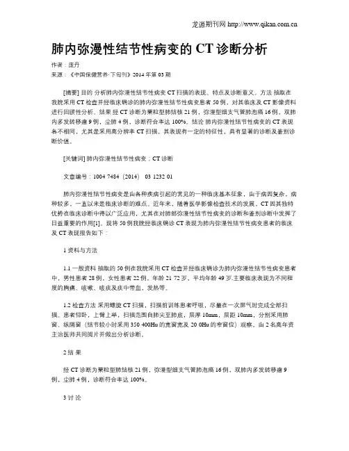
肺内弥漫性结节性病变的CT诊断分析作者:庞丹来源:《中国保健营养·下旬刊》2014年第03期[摘要] 目的分析肺内弥漫性结节性病变CT扫描的表现、特点及诊断意义。
方法抽取在我院采用CT检查并经临床确诊的肺内弥漫性结节性病变患者50例,对其临床及CT影像资料进行回顾性分析。
结果经CT诊断为粟粒型肺结核21例,弥漫型细支气管肺泡癌16例,双肺内多发转移瘤9例,尘肺4例,诊断符合率达100%。
结论肺内弥漫性结节性病变的CT表现各不相同,尤其是采用高分辨率CT扫描,其表现有一定的特征性,具有显著的诊断及鉴别诊断价值。
[关键词] 肺内弥漫性结节性病变;CT诊断文章编号:1004-7484(2014)-03-1232-01肺内弥漫性结节性病变是由各种疾病引起的常见的一种临床基本征象,由于病因复杂,病种较多,一直以来是临床诊断的难点。
近年来,随着医学影像检查技术的发展,CT因其独特优势在临床诊断中得以广泛应用,尤其在对肺部弥漫性结节性病变的诊断和鉴别诊断中发挥了日益重要的作用[1]。
现将50例我院经临床确诊CT表现为肺内弥漫性结节性病变患者的临床及CT表现报告如下:1 资料与方法1.1 一般资料抽取的50例在我院采用CT检查并经临床确诊为肺内弥漫性结节性病变患者中,男性患者28例,女性患者22例,年龄21-72岁,平均年龄49岁.主要临床表现为不同程度的胸痛、咳嗽、咳痰及痰中带血,发热等。
1.2 检查方法采用螺旋CT扫描,扫描前训练患者呼吸,尽量在一次屏气时完成全部扫描。
患者仰卧,上臂上举,扫描范围自肺尖至肺底,层厚10mm,层距10mm,分别采用肺窗、纵隔窗(结节较小时采用350-400Hu的宽窗宽及-20-0Hu的窄窗位)观察。
由2名高年资主治医师共同阅片并做出分析诊断。
2 结果经CT诊断为粟粒型肺结核21例,弥漫型细支气管肺泡癌16例,双肺内多发转移瘤9例,尘肺4例,诊断符合率达100%。
3 讨论3.1 弥漫性细支气管肺泡癌的CT影像表现可见两肺多发弥漫分布的结节状、斑片状阴影或肺叶及肺段多发的实变阴影[2],部分可融合成大片状的实变影。


双肺弥漫性病变1例周磊;卢进昌;吴波;杜春玲【期刊名称】《临床肺科杂志》【年(卷),期】2016(000)001【总页数】3页(P177-178,183)【作者】周磊;卢进昌;吴波;杜春玲【作者单位】201700 上海,复旦大学附属中山医院青浦分院呼吸内科;201700 上海,复旦大学附属中山医院青浦分院呼吸内科;201700 上海,复旦大学附属中山医院青浦分院呼吸内科;201700 上海,复旦大学附属中山医院青浦分院呼吸内科【正文语种】中文作者单位:201700 上海,复旦大学附属中山医院青浦分院呼吸内科患者70岁女性,咳嗽、咳痰伴纳差2周余入院。
患者咳少量白色粘痰,纳差,当地医院查胸部CT示慢性支气管炎伴左肺下叶少许感染可能,两肺弥漫结节;纵隔、右侧心膈角及两肺门淋巴结肿大;胃镜示慢性浅表性胃炎;腹部B超示血吸虫肝病声像。
给予“头孢丙烯”口服抗感染治疗效果不佳。
至我院求治,门诊拟“两肺弥漫性病变待查”收入我科。
病程中精神一般,胃纳欠佳,无发热,无呼吸困难,无咯血胸痛,无体重进行性下降。
既往有高血压病史,口服氯沙坦钾氢氯噻嗪,珍菊降压片降压治疗,血压控制情况不详。
有2型糖尿病病史,口服格列美脲降糖治疗,血糖可控制。
有血吸虫病史,否认病毒性肝炎、结核病史及接触史,否认饲养家禽、宠物、鸽子史;近期无外出旅游史;长期务农。
体格检查:T:36.7℃ P:90次/min R:18次/min BP:130/85 mmHg;神志清晰,呼吸平稳。
全身皮肤黏膜无黄染,全身浅表淋巴结未及肿大,口唇无紫绀,颈软,甲状腺未及肿大,气管居中,胸廓无畸形,双肺叩诊呈清音,双肺呼吸音粗,未及明显干湿啰音。
心界不大,心率90次/min,律齐,各瓣膜听诊区未及病理性杂音。
腹部平坦,触诊腹软,全腹无压痛,无反跳痛,肝脾肋下未及,未及腹块,肝肾区无叩击痛,四肢肌力、肌张力均正常,双下肢不浮肿,生理反射存在,病理反射未引出。
门诊辅助检查:血常规:白细胞总数 4.9*109/L;中性粒细胞70.3%;淋巴细胞18.1%;血沉 33 mm/h;C-反应蛋白 1.1 mg/L;血气分析(未吸氧):pH 7.381;二氧化碳分压 42.3 mmHg;氧分压 68.8 mmHg;SO2% 92.9%;红细胞压积 41%;碳酸氢根 25.3 mmol/l;类风湿因子、抗“O”:正常;肿瘤标记物:NSE、CYFRA21-1、CEA、AFP、SCC等均正常范围;降钙素原 0.073 ng/ml;T-SPOT:阴性。


[作者简介]雷志丹(1971-),男,四川丹棱人,在读硕士,主治医师。
研究方向:胸腹部影像学。
E -mail :leizhidan @ [通讯作者]葛英辉,河南省人民医院放射科,郑州,450003。
E -mail :cjr.geyinghui @ [收稿日期]2007-01-09 [修回日期]2007-06-05CT diagnosis and differential diagnosis of pulmonarydiffuse ground -glass opacityLEI Zhi -dan ,GE Y ing -hui*,S H I Da -peng(De partment of Radiology ,Henan Provincial Peop le 's Hospital ,Z hengz hou 450003,China )[Abstract ] Objective T o study the CT features of pulmo na ry disease s w ith diffuse g ro und -glass o pacity (DG GO ),and to improv e diagno stic and differential diag no stic accuracy of theses diseases.Methods T he clinical ,CT and high -re so lutio n CT (H RCT )ma te rials of 121cases 'DG GO which wer e pr oved by pa tho log y o r clinical comprehensive diagnosis w ere ret rospec -tively analy zed ,and its CT features wer e summarized.T he valuable diag no stic and differ ential diagno stic sig ns w ere investi -g ated.Results In these 121ca ses ,23cases w ere the diseases w ith abnor mity of inherent pulmo nary structur es ,including 18ca ses 'interstitial pneumo nia a nd 5cases 'co nnective tissue disease.T he DGG O had pe ripherally distributed with inter stitialthickening ,fibro sis ,t ractive bro nchiectasis o r honey comb shadow.T w enty cases were the disea ses with abno rmity of pul -mo na ry air -space 's filling state ,including 10ca ses 'aller gic pneumonia ,5ca ses 'pulmo nar y hemo r rhage ,3cases 'metastaic ne -o plasm and 2cases 'alveo lar cell carcino ma.T he DGG O had cent rally dist ributed w ith lobular centra l node ,acino se node o r mass.26ca ses we re the diseases w ith abno rmity of ex trav ascula r fluid ove rlo ad ,including 21cases 'pulmo nar y edema and 5ca ses 'co ntusio n of lung.T he DG GO had dist ributed pulmo na ry pendulous region or r elativ e pendulo us reg io n.15case s we re the diseases w ith abno rmity o f pulmo na ry blood 's no n -homo genous per fusio n ,including 11cases 'pulmonary embo lism and 4ca ses 'Behce t 's syndrome.T he DG GO had presented mo saic -fa shio n 's shado w or the DGG O had distributed throug ho ut one later al lung.37case s w ere the disea ses w ith two o r super -tw o king s o f abno rmity ,including 25cases 'chr onic o bstr uctive pulmo na ry disease ,7ca ses 'pulmo na ry alveo lar pr oteino sis and 5cases 'viral pneumonia.T he DGG O had pre sented mosaic -fashio n 's shado w o r g eog raphic change o r crazing paving pat te rn ,or the DGG O had centrally dist ributed with lobular central no de ,acinose node and inter stitial fibr osis.C onclusion DGG O may be seen in v ario us disea ses.T o analy ze the mor pho log y ,distribution ,co nco mitant sig ns and dy namic chang es of DGG O can preliminarily comprehend its pathological basis and patho -g enic mechanism.In combinatio n w ith clinical materials ,the r ang e of diag no stic po ssibilities can be shrinked.[Key words ] L ung disease ;G ro und -g lass o pacity ;T omog raphy ,X -ray computed肺部弥漫性磨玻璃阴影的CT 诊断与鉴别诊断雷志丹,葛英辉*,史大鹏(1.河南省人民医院放射科,河南郑州 450003)[摘 要] 目的 探讨以弥漫性磨玻璃阴影为主要表现肺部疾病的CT 特征,提高对本类型肺部疾病的诊断以及鉴别诊断水平。