Brodmann分区系统
- 格式:docx
- 大小:13.26 KB
- 文档页数:8

大脑的解剖结构和功能——xxxx分区系统xxxx分区是一个根据将划分为一系列解剖区域的系统。
中所谓细胞结构(Cytoarchitecture),是指在染色的脑组织中观察到的的组织方式。
xxxx分区1909年由医生(Korbinian Brodmann)提出。
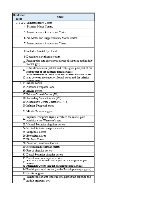
根据皮质细胞的类型及纤维的疏密把大脑皮质分为52个区,并用数字给予表示。
Brodmann Area 1, BA1Brodmann Area 2, BA2Brodmann Area 3, BA3位置:位于中央后回 (postcentral gyrus) 和前顶叶区。
功能:分别为体感皮层内侧、末尾和前端区,BA1、BA2、BA3共同组成体感皮层;具备基本体感功能(first somatic sensory area)接受对侧肢体的感觉传入。
Brodmann Area 4, BA4位置:位于中央前回(precentral gyrus),中央沟(central sulcus)的内侧面功能:初级运动皮层(first somatic motor area),包含“运动小人”(motor homunculus )。
控制行为运动,与BA6 (前)和BA3 、BA2 、BA1、(后)相连,同时与丘脑腹外侧核相连。
体感小人(Somatosensory Homunculus )传入体感信息较多的身体区域获得的皮层代表区域较大。
比如手部在初级体感皮层中的代表区域比背部的大。
体感皮质定位可用“体感小人”(Somatosensory homunculus)来表示。
Brodmann Area 5, BA5位置:位于顶叶前梨状皮质区(梨状皮质piriform cortex为下边缘皮质的组成部分)。
功能:与BA7形成体感联合皮层。
Brodmann Area 7, BA7位置:位于顶叶皮质顶部,体感皮层后方,视觉皮层(visual area)上方。
功能:将视觉和运动信息联合起来;与BA5形成体感联合皮层;视觉-运动协调功能。

Brodmann分区系统Brodmann分区是一个根据细胞结构将大脑皮层划分为一系列解剖区域的系统。
神经解剖学中所谓细胞结构(Cytoarchitecture),是指在染色的脑组织中观察到的神经元的组织方式。
Brodmann分区最早由德国神经科医生科比尼安·布洛德曼(Korbinian Brodmann)提出。
他的分区系统包括每个半球的52个区域。
其中一些区域今天已经被细分,例如23区被分为23a和23b区等。
从物种间差异来讲,同一分区号码在不同的物种间并不一定代表相似的区域。
人类大脑皮层的Brodmann分区:1,2和3区:中央后回(体感皮层,习惯上常称为“3,1和2区”)4区- 中央前回(初级运动皮层)5区- 体感联合皮层6区- 前运动皮层7区- 体感联合皮层8区- 包括额叶眼动区(Frontal eye field)9区- 后外侧前额叶皮层10区- 额极区(上额回和中额回最前侧的部分)11区- 额眶区(眶回,直回和上额回前侧的一部分)12区- 额眶区(上额回和下前回之间的区域)13区- 岛皮层(脑岛,岛叶)17区- 初级视皮层18区- 视觉联合皮层19区- 视觉联合皮层20区- 颞下回21区- 颞中回22区- 颞上回,其前侧部分属于韦尼克区23区- 下后扣带皮层24区- 下前扣带皮层25区- 膝下皮层26区- 压外区(Ectosplenial area)28区- 后内嗅皮层29区- 压后扣带皮层30区- 扣带皮层的一部分31区- 上后扣带皮层32区- 上前扣带皮层34区- 前扣带皮层,位于海马旁回35区- 旁嗅皮层,位于海马旁回36区- 海马旁皮层37区- 梭状回38区- 颞极区39区- 角回,威尔尼克区的一部分40区- 缘上回,威尔尼克区的一部分41,42区- 初级听皮层和听觉联合皮层43区- 中央下区(Subcentral area)44区- 三角部,布洛卡区的一部分45区- 岛盖部,布洛卡区的一部分46区- 上外额叶皮层47区- 下额叶皮层48区- 下脚后区,颞叶内侧的一小部分49区- 岛旁区,位于颞叶和岛叶的交界处。

brodmann分区染色法Brodmann分区染色法Brodmann分区染色法是一种神经学研究中常用的方法,用于确定大脑皮层的不同功能区域。
这种方法是由德国神经学家科尔特·布罗德曼(Korbinian Brodmann)于20世纪初提出的,他根据对大脑组织的观察和研究,将大脑皮层分为多个不同的区域,并为每个区域编制了编号。
布罗德曼的分区方法是基于对大脑组织的显微镜观察,他发现不同区域的细胞形态和结构特征存在差异。
通过染色技术,他能够清晰地观察到大脑皮层的细胞层次结构,并根据这些观察结果将其分为不同的区域。
根据布罗德曼的分类系统,大脑皮层被分为52个不同的区域,每个区域被赋予一个编号。
这些编号不仅仅是为了方便分类和研究,更重要的是为了揭示大脑皮层在不同区域中的功能差异。
布罗德曼提出的分区系统至今仍然被广泛应用于神经学研究中,成为了大脑皮层功能定位的重要工具。
布罗德曼的分区方法基于显微镜下对细胞形态的观察,他认为不同区域的细胞形态和结构特征反映了该区域的功能特点。
例如,一些区域主要包含神经元细胞体,而另一些区域则主要包含神经纤维。
这些不同的细胞结构可以帮助我们理解大脑在感知、运动、记忆等方面的功能。
布罗德曼分区法的主要优势在于其客观性和可重复性。
通过对大量大脑样本的观察和比较,布罗德曼能够确定不同区域的边界和特征,并将其编制为编号。
这使得不同研究者在使用布罗德曼分区法时可以得到一致的结果,从而方便了不同研究之间的比较和交流。
然而,布罗德曼分区法也存在一些局限性。
首先,该方法是基于对已知样本的观察,因此可能无法完全覆盖大脑皮层的所有功能区域。
其次,由于大脑皮层的结构和功能之间的复杂关系,仅仅通过细胞形态的观察可能无法全面揭示不同区域的功能特点。
因此,布罗德曼分区法在研究大脑功能时仅仅是一个起点,还需要结合其他方法和技术来进行深入研究。
布罗德曼分区染色法是一种常用的大脑皮层功能定位方法。
通过观察大脑组织的细胞形态和结构特征,布罗德曼将大脑皮层分为多个不同的区域,并为每个区域编制了编号。

Brodmann大脑皮层分区是德国神经解剖学家Korbinian Brodmann 于1909年提出的一种关于大脑皮层分区的理论。
这一分区方案被广泛应用于解剖学、神经科学和临床医学领域,并对了解大脑结构和功能起着重要作用。
本文将详细介绍Brodmann大脑皮层分区的背景、原理和应用,旨在帮助读者全面了解这一重要的神经科学理论。
一、背景Korbinian Brodmann是20世纪初期的神经解剖学家,他在研究大脑皮层时发现不同的脑区在细胞结构和组织类型上存在巨大的差异。
为了更好地描述和理解这些差异,Brodmann开始系统地研究大脑皮层的细胞组织结构,并最终提出了大脑皮层分区的理论。
这一理论基于对大量人类和动物大脑的显微镜观察和测量,被视为解剖学上关于大脑皮层最具权威的分类系统之一。
二、原理Brodmann大脑皮层分区的原理基于神经元的细胞结构和分布。
根据Brodmann的研究,大脑皮层中的神经元可以根据其形态、密度和分布分为不同的类型,不同类型的神经元聚集在一起形成具有特定功能的脑区。
Brodmann通过对大脑组织进行显微镜观察和测量,将大脑皮层分为了52个不同的区域,每个区域有其独特的细胞结构和功能特征。
这一分区方式为后续的大脑研究奠定了重要的基础,也为神经科学领域的发展做出了重要贡献。
三、应用Brodmann大脑皮层分区的理论在解剖学、神经科学和临床医学领域有着广泛的应用。
它为解剖学和神经科学研究提供了重要的工具和参考标准。
研究人员可以利用Brodmann的分区方式对大脑进行定量和定性的分析,揭示不同区域的功能和相互通联,从而深入了解大脑的结构和功能。
Brodmann的分区方案对于临床诊断和治疗也具有重要意义。
医生可以根据大脑分区的特定功能特征,判断患者的脑损伤位置和程度,为临床治疗提供重要参考。
现代的神经影像技术也常常结合Brodmann的分区方式,帮助医生观察和分析大脑结构,对各种疾病进行诊断和治疗。
1909年,布鲁德曼(Brodmann)根据皮质细胞的类型及纤维的疏密把大脑皮质分为52个区,并用数字给予表示。
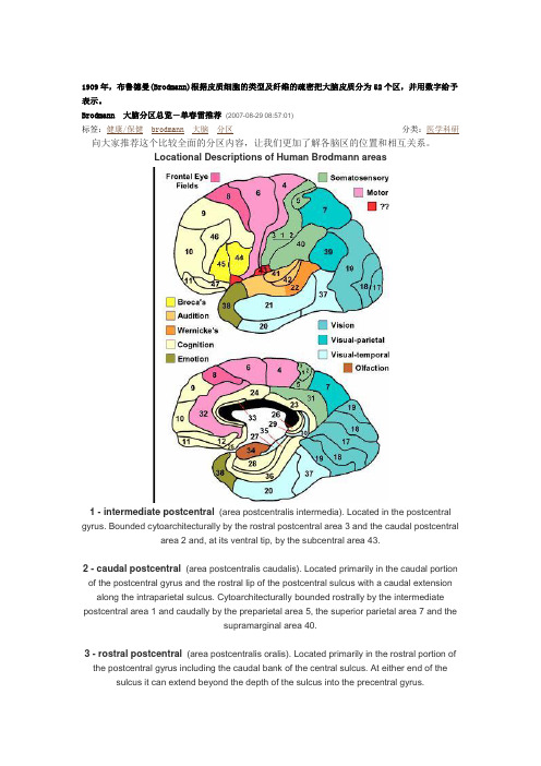
Brodmann 大脑分区总览-单春雷推荐(2007-08-29 08:57:01)标签:健康/保健brodmann大脑分区分类:医学科研向大家推荐这个比较全面的分区内容,让我们更加了解各脑区的位置和相互关系。
Locational Descriptions of Human Brodmann areas1 - intermediate postcentral(area postcentralis intermedia). Located in the postcentral gyrus. Bounded cytoarchitecturally by the rostral postcentral area 3 and the caudal postcentral area2 and, at its ventral tip, by the subcentral area 43.2 - caudal postcentral(area postcentralis caudalis). Located primarily in the caudal portion of the postcentral gyrus and the rostral lip of the postcentral sulcus with a caudal extension along the intraparietal sulcus. Cytoarchitecturally bounded rostrally by the intermediate postcentral area 1 and caudally by the preparietal area 5, the superior parietal area 7 and thesupramarginal area 40.3 - rostral postcentral(area postcentralis oralis). Located primarily in the rostral portion ofthe postcentral gyrus including the caudal bank of the central sulcus. At either end of the sulcus it can extend beyond the depth of the sulcus into the precentral gyrus.Cytoarchitecturally bounded rostrally by the gigantopyramidal area 4 and caudally by theintermediate postcentral area 1.4 - gigantopyramidal(area gigantopyramidalis). Located in the precentral gyrus. Cytoarchitecturally the caudal boundary with the rostral postcentral area 3 does not coincide precisely with the floor of the central sulcus but lies variably in the banks of the postcentral gyrus and the precentral gyrus. The area also does not extend in all cases to the cingulate sulcus medially or to the end of the central sulcus ventro-laterally. Bounded rostrally by thefrontal agranular area 6.5 - preparietal(area praeparietalis). Occupies the superior parietal lobule and a portion of thepostcentral gyrus, particularly on the medial aspect of the hemisphere. Bounded approximately by the cingulate sulcus on the medial aspect of the hemisphere and by the superior postcentral sulcus on the lateral aspect. Cytoarchitecturally bounded by the caudal postcentral area 2, the superior parietal area 7 and on the medial bank of the hemisphere by the gigantopyramidal area 4 and the dorsal posterior cingulate area 31.6 - agranular frontal(area frontalis agranularis). Located primarily in the caudal portions of the superior frontal gyrus and the middle frontal gyrus and the rostral portions of the precentral gyrus not occupied by the gigantopyramidal area 4. It extends from the cingulate sulcus on the medial aspect of the hemisphere to the lateral sulcus on the lateral aspect. Cytoarchitecturally bounded rostrally by the frontal region and caudally by the gigantopyramidal area 4.7 - superior parietal(area parietalis superior). Occupies much of the superior parietal lobuleand some of the precuneus. Bounded approximately by the superior postcentral sulcus rostrally, the intraparietal sulcus laterally, the parieto-occipital sulcus caudally and, on the medial bank of the hemisphere, the subparietal sulcus. Cytoarchitecturally bounded rostrally by the preparietal area 5 and the caudal postcentral area 2; caudally by the peristriate area 19;and medially by the dorsal posterior cingulate area 31.8 - intermediate frontal(area frontalis intermedia). Located primarily in the superior frontalgyrus extending from the cingulate sulcus on the medial surface over the margin of the hemisphere to the middle frontal gyrus. Cytoarchitecturally bounded caudally by the agranular frontal area 6 and ventrally by the granular frontal area 9.9 - granular frontal(area frontalis granularis). Occupies portions of the superior frontal gyrusand the middle frontal gyrus. Its approximate boundary on the medial aspect of the hemisphere is the cingulate sulcus and, on the lateral aspect, the inferior frontal sulcus. Cytoarchitecturally bounded dorsocaudally by the intermediate frontal area 8, caudally by the agranular frontal area 6, and ventrally by the frontopolar area 10, the middle frontal area 46and the opercular area 44.10 - frontopolar(area frontopolaris). Occupies the most rostral portions of the superiorfrontal gyrus and the middle frontal gyrus. On the medial aspect of the hemisphere it isbounded ventrally by the superior rostral sulcus. It does not extend as far as the cingulate sulcus. Cytoarchitecturally bounded dorsally by the granular frontal area 9, caudally by the middle frontal area 46, and ventrally by the orbital area 47 and by the frontopolar area 12.11 - prefrontal(area praefrontalis). Constitutes most of the orbital gyri, gyrus rectus and themost rostral portion of the superior frontal gyrus. Bounded medially by the inferior rostral sulcus and laterally approximately by the frontomarginal sulcus. Cytoarchitecturally bounded on the rostral and lateral aspects of the hemisphere by the frontopolar area 10, the orbital area 47, and the triangular area 45; on the medial surface it is bounded dorsally by the area 12 andcaudally by the subgenual area 25.12 - prefrontal(area praefrontalis). Occupies the area between the superior rostral sulcus and the inferior rostral sulcus. Cytoarchitecturally bounded dorsally by the frontopolar area 10 and the dorsal anterior cingulate area 32; caudally, ventrally and rostrally it is bounded by the prefrontal area 11. (Originally described as part of prefrontal area 11 but not shown in the map, subsequently it was labeled as an independent area 12.)17 - striate(area striata). Part of the occipital lobe of the cerebral cortex that is defined on thebasis of cyto- and myeloarchitecture, primarily by the band/stripe of Gennari.18 - parastriate(area parastriata). Located in parts of the cuneus, the lingual gyrus and the lateral occipital gyrus of the occipital lobe. Cytoarchitecturally bounded on one side by the striate area 17, from which it is distinguished by absence of a band/stripe of Gennari, and onthe other by the peristriate area 19.19 - peristriate(area peristriata). Located in parts of the lingual gyrus, the cuneus, the lateraloccipital gyrus and the superior occipital gyrus of the occipital lobe where it is bounded approximately by the parieto-occipital sulcus. Cytoarchitecturally bounded on one side by the parastriate area 18 which it surrounds. Rostrally it is bounded by the angular area 39 and theoccipitotemporal area 37.20 - inferior temporal(area temporalis inferior). Corresponds approximately to the inferior temporal gyrus. Cytoarchitecturally bounded medially by the ectorhinal area 36, laterally by the middle temporal area 21, rostrally by the temporopolar area 38 and caudally by theoccipitotemporal area 37.21 - middle temporal(area temporalis media). Corresponds approximately to the middletemporal gyrus. Bounded rostrally by the temporopolar area 38, ventrally by the inferior temporal area 20, caudally by the occipitotemporal area 37, and dorsally by the superiortemporal area 22.22 - superior temporal(area temporalis superior). Corresponds approximately to the lateral and caudal two thirds of the superior temporal gyrus. Bounded rostrally by the temporopolar area 38, medially by the posterior transverse temporal area 42, ventrocaudally by the middletemporal area 21 and dorsocaudally by the supramarginal area 39.23 - ventral posterior cingulate(area cingularis posterior ventralis). Occupies most of the posterior cingulate gyrus adjacent to the corpus callosum. At the caudal extreme it is bounded approximately by the parieto-occipital sulcus. Cytoarchitecturally bounded dorsally by the dorsal posterior cingulate area 31, rostrally by the ventral anterior cingulate area 24, and ventrorostrally in its caudal half by the retrosplenial region.24 - ventral anterior cingulate(area cingularis anterior ventralis). Occupies most of theanterior cingulate gyrus in an arc around the genu of corpus callosum. Its outer border corresponds approximately to the cingulate sulcus. Cytoarchitecturally bounded internally by the pregenual area 33, externally by the dorsal anterior cingulate area 32, and caudally by the ventral posterior cingulate area 23 and the dorsal posterior cingulate area 31.25 - subgenual(area subgenualis). A narrow band located in the caudal portion of the subcallosal area adjacent to the paraterminal gyrus from which it is separated by the posterior parolfactory sulcus. It is bounded by the prefrontal area 11rostrally and by the paraterminalgyrus caudally.26 - ectosplenial(area ectosplenialis). A narrow band located in the isthmus of cingulategyrus adjacent to the fasciolar gyrus internally. It is bounded externally by the granularretrolimbic area 29.28 - entorhinal(area entorhinalis). Located in the entorhinal area on the medial aspect of thetemporal lobe. It and the dorsal entorhinal area 34 together constitute approximately theentorhinal area.29 - granular retrolimbic(area retrolimbica granularis). A narrow band located in the isthmus of cingulate gyrus. Cytoarchitecturally bounded internally by the ectosplenial area 26 and externally by the agranular retrolimbic area 30.30 - agranular retrolimbic(area retrolimbica agranularis). Located in the isthmus of cingulate gyrus. Cytoarchitecturally bounded internally by the granular retrolimbic area 29, dorsally by the ventral posterior cingulate area 23 and ventrolaterally by the ectorhinal area 36.31 - dorsal posterior cingulate(area cingularis posterior dorsalis). Occupies portions of the posterior cingulate gyrus and medial aspect of the parietal lobe. Approximate boundaries are the cingulate sulcus dorsally and the parieto-occipital sulcus caudally. It partially surrounds the subparietal sulcus. Cytoarchitecturally bounded rostrally by the ventral anterior cingulate area 24, ventrally by the ventral posterior cingulate area 23, dorsally by the gigantopyramidal area 4 and preparietal area 5 and caudally by the superior parietal area 7.32 - dorsal anterior cingulate(area cingularis anterior dorsalis). Forms an outer arc around the anterior cingulate gyrus. The cingulate sulcus defines approximately its inner boundaryand the superior rostral sulcus its ventral boundary; rostrally it extends almost to the margin of the frontal lobe. Cytoarchitecturally bounded internally by the ventral anterior cingulate area 24, externally by medial margins of the agranular frontal area 6, intermediate frontal area 8, granular frontal area 9, frontopolar area 10, and prefrontal area 11.33 - pregenual(area praegenualis). A narrow band located in the anterior cingulate gyrusadjacent to the supracallosal gyrus in the depth of the callosal sulcus. Cytoarchitecturally bounded by the ventral anterior cingulate area 24 and the supracallosal gyrus.34 - dorsal entorhinal(area entorhinalis dorsalis). Located in the entorhinal area on themedial aspect of the temporal lobe. It and the entorhinal area 28 together constituteapproximately the entorhinal area.35 - perirhinal(area perirhinalis). Located along the rhinal sulcus. Cytoarchitectuallybounded medially by the entorhinal area 28 and laterally by the ectorhinal area 36.36 - ectorhinal(area ectorhinalis). Located primarily in the fusiform gyrus, with its medialboundary corresponding approximately to the rhinal sulcus. Cytoarchitecturally bounded laterally and caudally by the inferior temporal area 20, medially by the perirhinal area 35 androstrally by the temporopolar area 38.37 - occipitotemporal (area occipitotemporalis). Located primarily in the caudal portions of the fusiform gyrus and inferior temporal gyrus on the mediobasal and lateral surfaces at the caudal extreme of the temporal lobe. Cytoarch itecturally bounded caudally by the peristriate area 19, rostrally by the inferior temporal area 20 and middle tempor al area 21 and dorsally on the lateral aspect of the hemisphere by the angular area 39.38 - temporopolar (area temporopolaris). Located primarily in the most rostral portions of the superior temporal gy rus and the middle temporal gyrus. Cytoarchitecturally bounded caudally by the inferior temporal area 20, the mid dle temporal area 21, the superior temporal area 22 and the ectorhinal area 36.39 - angular (area angularis). Corresponds to the angular gyrus surrounding the caudal tip of the superior temporal sulcus. Dorsally it is bounded approximately by the intraparietal sulcus. Cytoarchitecturally bounded rostrally by t he supramarginal area 40, dorsally and caudally by the peristriate area 19, and ventrally by the occipitotemporal ar ea 37.40 - supramarginal (area supramarginalis). Located primarily in the supramarginal gyrus surrounding the posterior ascending limb of lateral sulcus. Bounded approximately by the intraparietal sulcus, the inferior postcentral sulcus the posterior subcentral sulcus and the lateral sulcus. Cytoarchitecturally bounded caudally by the angular area 39, rostrally and dorsally by the caudal postcentral area 2, and ventrally by the subcentral area 43 and the superior tem poral area 22.41 - anterior transverse temporal (area temporalis transversa anterior). Occupies the anterior transverse temporal g yrus in the bank of the lateral sulcus on the dorsal surface of the temporal lobe. Cytoarchitecturally bounded media lly by the parainsular area 52 and laterally by the posterior transverse temporal area 42.42 - posterior transverse temporal (area temporalis transversa posterior). Located in the bank of the lateral sulcus on the dorsal surface of the temporal lobe. Cytoarchitecturally bounded medially by the anterior transverse temporal area 41 and laterally by the superior temporal area 22.43 - subcentral (area subcentralis). Occupies the postcentral gyrus and the precentral gyrus between the ventrolater al extreme of the central sulcus and the depth of the lateral sulcus at the insula. Its rostral and caudal borders are ap proximated by the anterior subcentral sulcus and the posterior subcentral sulcus respectively. Cytoarchitecturally b ounded rostrally by the agranular frontal area 6 and caudally, for the most part, by the caudal postcentral area 2 and the supramarginal area 40.44 - opercular (area opercularis). Corresponds approximately to the opercular part of the inferior frontal gyrus. Bo unded caudally by the inferior precentral sulcus and rostrally by the anterior ascending limb of lateral sulcus. It sur rounds the diagonal sulcus. In the depth of the lateral sulcus it borders on the insula. Cytoarchitectonically bounde d caudally and dorsally by the agranular frontal area 6, dorsally by the granular frontal area 9 and rostrally by the tr iangular area 45.45 - triangular (area triangularis). Occupies the triangular part of the inferior frontal gyrus and, surrounding the ant erior horizontal limb of lateral sulcus, a portion of the orbital part of inferior frontal gyrus. Bounded caudally by th e anterior ascending limb of lateral sulcus, it borders on the insula in the depth of the lateral sulcus . Cytoarchitecto nically bounded caudally by the opercular area 44, rostrodorsally by the middle frontal area 46 and ventrally by the orbital area 47.46 - middle frontal (area frontalis media). Occupies approximately the middle third of the middle frontal gyrus and the most rostral portion of the inferior frontal gyrus. Cytoarchitecturally bounded dorsally by the granular frontal a rea 9, rostroventrally by the frontopolar area 10 and caudally by the triangular area 45.47 - orbital (area orbitalis). Surrounds the caudal portion of the orbital sulcus from which it extends laterally into t he orbital part of inferior frontal gyrus. Cytoarchitectonically bounded caudally by the triangular area 45, medially by the prefrontal area 11, and rostrally by the area frontopolaris 10.48 - retrosubicular (area retrosubicularis). Located on the medial surface of the temporal lobe. Cytoarchitectually b ounded rostrally by the perirhinal area 35 and medially by the presubiculum. (While described by Brodmann, it wa s not included in his areal maps of human cortex.)52 - parainsular (area parainsularis) Located in the bank of the lateral sulcus on the dorsal surface of the temporal l obe. Its medial boundary corresponds approximately to the junction between the temporal lobe and the insula. Cyt oarchitecturally bounded laterally by the anterior transverse temporal area 41.Brodmann分区系统Brodmann分区系统Brodmann分区是一个根据细胞结构将大脑皮层划分为一系列解剖区域的系统。
broadmann大脑分区法
Broadmann大脑分区法是一种将大脑皮层分为不同区域的方法,它是根据不同区域的细胞结构和功能特征来划分的。
这种方法是德国神经解剖学家Brodmann在20世纪初提出的,至今仍被广泛应用于神经科学领域。
Broadmann大脑分区法将大脑皮层分为52个不同的区域,每个区域都有其独特的细胞结构和功能特征。
这些区域被编号为1到52,其中1到41区域被称为“一级区域”,而42到52区域被称为“二级区域”。
这种分区方法的优点在于它可以帮助研究人员更好地理解大脑的结构和功能。
通过对不同区域的研究,我们可以更好地了解大脑如何处理信息、控制行为和感知世界。
此外,这种方法还可以帮助医生更好地诊断和治疗神经系统疾病。
然而,Broadmann大脑分区法也存在一些缺点。
首先,这种方法是基于解剖学和组织学的特征来划分的,而不是基于功能的特征。
因此,它可能无法完全反映大脑的功能分区。
其次,这种方法是基于人类大脑的研究而得出的,而不是基于其他动物的研究。
因此,它可能无法适用于其他动物的大脑。
总的来说,Broadmann大脑分区法是一种非常有用的方法,可以帮助我们更好地了解大脑的结构和功能。
然而,我们也需要意识到
它的局限性,并寻求其他方法来更好地理解大脑。
随着神经科学的不断发展,我们相信会有更多的方法和技术出现,帮助我们更好地了解大脑的奥秘。
Brodmann分区系统 (重定向自Brodmann)
Brodmann分区是一个根据细胞结构将大脑皮层划分为一系列解剖区域的系统。
神经解剖学中所谓细胞结构(Cytoarchitecture),是指在染色的脑组织中观察到的神经元的组织方式。
Brodmann分区最早由德国神经科医生科比尼安·布洛德曼(Korbinian Brodmann)提出。
他的分区系统包括每个半球的52个区域。
其中一些区域今天已经被细分,例如23区被分为23a和23b 区等。
从物种间差异来讲,同一分区号码在不同的物种间并不一定代表相似的区域。
以下列出人类大脑皮层的Brodmann分区系统。
•1,2和3区:体感皮层(习惯上常称为“3,1和2区”)•4区 - 初级运动皮层
•5区 - 体感联合皮层
•6区 - 前运动皮层和辅助运动区
•7区 - 体感联合皮层
•8区 - 包括额页眼动区(Frontal eye field)
•9区 - 后外侧前额叶皮层
•10区 - 额极区(上额回和中额回最前侧的部分)
•11区 - 额眶区(眶回,直回和上额回前侧的一部分)
•12区 - 额眶区(上额回和下前回之间的区域)
•13区 - 岛皮层
•17区 - 初级视皮层
•18区 - 视觉联合皮层
•19区 - 视觉联合皮层
•20区 - 下颞回
•21区 - 中颞回
•22区 - 上颞回,其前侧部分属于韦尼克区•23区 - 下后扣带皮层
•24区 - 下前扣带皮层
•25区 - 膝下皮层
•26区 - 压外区(Ectosplenial area)•28区 - 后内嗅皮层
•29区 - 压后扣带皮层
•30区 - 扣带皮层的一部分
•31区 - 上后扣带皮层
•32区 - 上前扣带皮层
•34区 - 前扣带皮层,位于海马旁回
•35区 - 旁嗅皮层,位于海马旁回
•36区 - 海马旁皮层
•37区 - 梭状回
•38区 - 颞极区
•39区 - 角回,韦尼克区的一部分
•40区 - 缘上回,韦尼克区的一部分•41,42区 - 初级听皮层和听觉联合皮层•43区 - 中央下区(Subcentral area)•44区 - 三角部,布洛卡区的一部分
•45区 - 岛盖部,布洛卡区的一部分
•46区 - 上外额叶皮层
•47区 - 下额页皮层48区 - 下脚后区,颞叶内侧的一小部分
•49区 - 岛旁区,位于颞叶和岛叶的交界处。
Brodmann分区系统- 人类大脑皮层的Brodmann分区
1,2和3区:体感皮层(习惯上常称为“3,1和2区”)
4区- 初级运动皮层
5区- 体感联合皮层
6区- 前运动皮层
7区- 体感联合皮层
8区- 包括额页眼动区(Frontal eye field)
9区- 后外侧前额叶皮层
10区- 额极区(上额回和中额回最前侧的部分)
11区- 额眶区(眶回,直回和上额回前侧的一部分)
12区- 额眶区(上额回和下前回之间的区域)
13区- 岛皮层
17区- 初级视皮层
18区- 视觉联合皮层
19区- 视觉联合皮层
20区- 下颞回
21区- 中颞回
22区- 上颞回,其前侧部分属于韦尼克区23区- 下后扣带皮层
24区- 下前扣带皮层
25区- 膝下皮层
26区- 压外区(Ectosplenial area)
28区- 后内嗅皮层
29区- 压后扣带皮层
30区- 扣带皮层的一部分
31区- 上后扣带皮层
32区- 上前扣带皮层
34区- 前扣带皮层,位于海马旁回
35区- 旁嗅皮层,位于海马旁回
36区- 海马旁皮层
37区- 梭状回
38区- 颞极区
39区- 角回,韦尼克区的一部分
40区- 缘上回,韦尼克区的一部分
41,42区- 初级听皮层和听觉联合皮层
43区- 中央下区(Subcentral area)
44区- 三角部,布洛卡区的一部分
45区- 岛盖部,布洛卡区的一部分
46区- 上外额叶皮层
47区- 下额页皮层
48区- 下脚后区,颞叶内侧的一小部分
49区- 岛旁区,位于颞叶和岛叶的交界处。
人类的大脑皮层两半球,在功能划分上,大体上是左半球管右半身,右半球管左半身。
每一半球的纵面,在功能上也有层次之分,原则上是上层管下肢,中层管躯干,下层管头部。
如此形成上下倒置,左右分叉的微妙构造。
在每一半球上,有各自分区为数个神经中枢,每一中枢各有其固定的区域,分区专司形成大脑分化而又统一的复杂功能。
在区域的分布上,两半球并不完全相同:其中布氏语言区与威氏语言区,只分布在左脑半球,其它各区则两半球都有。
在神经传导的运作上,两半球相对的神经中枢,彼此配合,发生交叉作用:两半球的运动区对身体部位的管理,是左右交叉、上下倒置的;两半球的视觉区与两眼的关系是:左半球视觉区管理两眼视网膜的左半,右半球视觉区管理两眼视网膜的右半;两半球的听觉区共同分担管理两耳传入的听觉信息。
1. 皮质运动区
位于中央前回(4区),是支配对侧躯体随意运动的中枢。
它主要接受来自对侧骨骼肌、肌腱和关节的本体感觉冲动,以感受身体的位置、姿势和运
动感觉,并发出纤维,即锥体束控制对侧骨骼肌的随意运动。
2. 皮质运动前区
位于中央前回之前(6区),为锥体外系皮质区。
它发出纤维至丘脑、基底神经节、红核、黑质等。
与联合运动和姿势动作协调有关,也具有植物神经皮质中枢的部分功能。
3. 皮质眼球运动区
位于额叶的8枢和枕叶19区,为眼球运动同向凝视中枢,管理两眼球同时向对侧注视。
4. 皮质一般感觉区
位于中央后回(1、2、3区),接受身体对侧的痛、温、触和本体感觉冲动,并形成相应的感觉。
顶上小叶(5、7)为精细触觉和实体觉的皮质区。
5. 额叶联合区
为额叶前部的9、10、11区,与智力和精神活动有密切关系。
6. 视觉皮质区
在枕叶的距状裂上、下唇与楔叶、舌回的相邻区(17区)。
每一侧的上述区域皮质都接受来自两眼对侧视野的视觉冲动,并形成视觉。
7. 听觉皮区
位于颞横回中部(41、42区),每侧皮质均接受来自双耳的听觉冲动产生听觉。
8. 嗅觉皮质区
位于嗅区、钩回和海马回的前部(25、28、34)和35区的大部分)。
每
侧皮质均接受双侧嗅神经传入的冲动。
9. 内脏皮质区
该区定位不太集中,主要分布在扣带回前部、颞叶前部、眶回后部、岛叶、海马及海马钩回等区域。
10. 语言运用中枢
人类的语言及使用工具等特殊活动在一侧皮层上也有较集中的代表区(优势半球),也称为语言运用中枢。
大脑皮层的不同区域有不同的功能,根据不同的功能可以将大脑皮层分为几个区,主要有感觉区,运动区和联合区.在1909年,布鲁德曼曾根据皮层细胞的类型以及纤维的疏密对大脑进行分区.他将大脑分为52个区并用数字予以表示.布鲁德曼的分区,影响最大.布鲁德曼的分区和大脑皮层重要中枢.
大脑内侧面:
大脑外侧面:。