Changes in microRNA expression induced by rabies virus infection
- 格式:pdf
- 大小:540.64 KB
- 文档页数:8

放疗联合顺铂+氟尿嘧啶方案同步化疗治疗局部晚期食管癌的实效性探究王晓翔【摘要】目的:探讨放疗联合顺铂(cisplatin,DDP)加氟尿嘧啶(5-fluorouraci,5-FU)同步化疗对局部晚期食管癌的治疗效果。
方法对河北医科大学第四医院2002年9月~2011年12月期间收治的80例局部晚期食管癌患者,分为联合组(n=40)和放疗组(n=40)。
联合组行放疗同步采用DDP+5-FU联合治疗,放疗组只采用放疗法,对2组患者的疗效和治疗方法的安全性进行评价。
结果放疗联合DDP+5-FU同步化疗治疗效果明显,治疗有效率达到90.0%,而只采用放疗治疗的有效率只有72.5%,2者有显著差异(P<0.01)。
联合组患者2年生存率为33%,大于放疗组的24.4%,差异具有统计学意义(P<0.05)。
治疗过程中联合组出现恶心呕吐、骨髓抑制、食管炎、放射性脑病等不良反应较放疗组高,但绝大多数反应较轻,经积极治疗可消除。
结论放疗联合DDP+5-FU同步放疗能够有效提高局部晚期食管癌患者的近期疗效及生存率,引发的不良反应有增加,但仍能被有效控制。
%Objective To study the efficacy of radiotherapy combined with cisplatin and 5-fluorouraci concurrent chemoradiotherapy on locally advanced esophageal carcinoma. Methods 80 cases with locally advanced esophageal carcinoma collected in our hospital from September 2002 to December 2011 were divided into combination group and radiotherapy group, each had 40 cases. Patients of combination group were given radiotherapy combined with DDP and 5-FU, while radiotherapy group were only adopted radiation therapy. The efifcacy and the safety of the therapy methods were evaluated. Results The treatment effect of thechemoradiotherapy on locally advanced esophageal carcinoma was obvious. The efifcacy of combination group was 90.0%, and radiotherapy was only 72.5%, the difference was signiifcant(P<0.01). The survival rate of combination group was 33%, higher than 24.4%of radiotherapy group. The adverse reactions of combination group were higher than radiotherapy group, but most of which were light, and could be eliminated after active treatment. Conclusion The radiotherapy combined with DDP and 5-FU can enhance the efifcacy and survival rate of patients with locally advanced esophageal carcinoma, and also cause an increase in adverse reactions, which can be controlled effectively.【期刊名称】《中国生化药物杂志》【年(卷),期】2014(000)002【总页数】3页(P76-78)【关键词】食管癌;放化疗;顺铂;氟尿嘧啶【作者】王晓翔【作者单位】河北医科大学第四医院肿瘤科,河北石家庄050011【正文语种】中文【中图分类】R735.11.1 一般资料 2002年9月~2011年12月期间收治晚期食管癌患者80例,其中男41例,女39例,年龄21~72岁,平均年龄(44±6.5)岁。

microRNA——药物开发的新靶点摘要微小RNA(microRNA)是一类长约22 nt的非编码单链RNA,由前体经酶作用而得,它是细胞的内源性物质,普遍存在于动植物中,高度保守,在生命体的生长、发育、疾病发生发展的过程中起到了基因调控作用,它与重大疾病,如肿瘤、心脏疾病、神经性疾病等都有着密切的关联,因此成为新药开发的一个重要新靶点,以它为靶点的药物设计和药物研发工作也在不断探索中。
本文就其研究的历史、生物合成和作用机制、生物功能、与疾病的关系以及以microRNA为靶点的药物设计的研究情况做一个简单的综述。
ABSTRACT microRNA is a class of ~22 nt small non-coding single chain RNA which is an endogenous substance and produced from precursor through enzymatic reaction. microRNAs are highly conserved and are ubiquitous in both animals and plants. They act as the regulators in the growth and proliferation of cells,and play an important role in the development of many diseases. These make microRNAs become promising new targets for the discovery of new drugs. A lot of research works have been involved in studying and designing novel drugs targeting microRNAs. In this paper,the discovery,biosynthesis and mechanism as well as biologic function of microRNA are reviewed.KEY WORDS microRNA;drug development;target1 microRNA的研究历史1993年Lee等[1]在对秀丽隐杆线虫(Caenorhabditis elegans)进行遗传分析时发现了第一个microRNA——lin-14,它的长度为22核苷酸(nt),它是一个非编码的单链RNA,可以控制细胞的发育,通过反义的RNA-RNA反应在转录后水平下调lin-14蛋白的表达。

MicroRNA在急性呼吸窘迫综合征中的作用龙春艺;黄霞;廖品琥【摘要】急性呼吸窘迫综合征(ARDS)是临床常见的危重病症,复杂的炎性反应过程参与了其病理生理过程.小分子非编码的单链RNA的MicroRNA (miRNA)通过影响靶基因的表达调控免疫反应和炎症反应,参与了ARDS的进程.该文综述miRNA在炎症反应和免疫功能障碍以及肺损伤性疾病中的作用,分析了miRNA参与ARDS的炎症反应过程.【期刊名称】《右江医学》【年(卷),期】2019(047)001【总页数】4页(P1-4)【关键词】miRNA;ARDS;炎症反应【作者】龙春艺;黄霞;廖品琥【作者单位】右江民族医学院研究生学院,百色533000;右江民族医学院附属医院呼吸内科,百色533000;广西壮族自治区卫生健康委员会,南宁530000【正文语种】中文【中图分类】R563.8急性呼吸窘迫综合征(Acute respiratory distress syndrome,ARDS)是以呼吸窘迫、顽固性低氧血症和非心源性肺水肿为特征的呼吸功能障碍综合征[1],病情凶险,预后差,在免疫学上表现为炎性细胞因子水平升高,炎症反应导致的病情危重,重度ARDS患者病死率可高达40%[2]。
炎症反应导致的免疫细胞募集和免疫物质产生,炎症细胞活化和炎症因子释放,促进ARDS的发生发展。
小分子非编码的单链RNA的MicroRNA(miRNA)通过影响靶基因的表达调控免疫反应和炎症反应,参与了ARDS的进程。
本文综述miRNA在炎症反应和免疫功能障碍以及肺损伤性疾病中的作用,分析miRNA参与ARDS的炎症反应过程。
1 miRNA概述miRNA是一类高度保守的,长度为21~23 nt的非蛋白质编码小分子单链RNA,参与调节细胞的增殖、分化、衰老、凋亡、新陈代谢、炎症和免疫反应、器官发育和肿瘤形成等生理过程[3]。
在细胞核内,miRNA在转录子和RNA聚合酶Ⅱ的作用下生成原始转录片段(pri-miRNA),并通过核酸内切酶Drosha等加工形成前体miRNA (pre-miRNA)。

·研究论文·Chinese Journal of Animal Infectious Diseases中国动物传染病学报摘 要:分析经NDV 感染HeLa 细胞分泌外泌体microRNA 表达变化,以期为外泌体microRNA 在NDV 感染中的具体作用机制提供初步的理论基础。
采用经典差速离心法分离提取对照组与NDV 感染组的HeLa 细胞来源外泌体,经miRCURYTM Array 芯片分析两组microRNA 的表达谱后,通过Targetscan 预测表达差异miRNA 的可能靶基因,并利用DAVID 、KEGG 等在线预测工具对差异microRNA 靶基因进行Go 功能分析。
相比于对照组,NDV 感染组HeLa 细胞外泌体中共有234个microRNA 发生了2倍以上的表达差异(P <0.01),其中8个外泌体microRNA 呈现出10倍以上的表达差异(P <0.01),分别为hsa-miR-3662、hsa-miR-3128、hsa-miR-345-5p 、hsa-miR-376a-3p 、hsa-miR-106b-3p 、hsa-miR-133b 、hsa-miR-410-3p 和hsa-miR-454-3p 。
经Go 功能分析发现,这8个microRNA 主要分布在细胞外膜、高尔基膜、细胞核膜等部位,参与转录过程,负调节RNA 聚合酶Ⅱ启动子转录过程,蛋白质均聚过程,正调节NF-kappaB 转录因子活性过程、细胞缺氧反应过程、神经元分化过程等,并发挥着蛋白结合、金属离子结合、转录因子活性、染色体结合等功能。
NDV 感染会引发HeLa 细胞的外泌体microRNA 显著性差异表达,而其机制仍有待进一步研究。
关键词:外泌体;microRNA ;HeLa ;表达谱;生物信息学中图分类号:S852.659.5文献标志码:A文章编号:1674-6422(2021)06-0042-08Detection of microRNA Expression Profi les in Exosomes Derived from HeLa Cellsand Bioinformatics AnalysisZHOU Changluan, DING Chan, MENG Chunchun, SUN Yingjie, QIU Xusheng, LIAO Ying, LIU Weiwei,SONG Cuiping, TAN Lei(Shanghai Veterinary Research Institute, CAAS, Shanghai 200241, China)收稿日期: 2019-05-17基金项目:国家自然科学基金面上项目(32072868);上海市自然科学基金面上项目(20ZR1469400);上海市兽医生物技术重点实验室开放基金(shklab202007)作者简介:周昌娈,女,硕士研究生,预防兽医学专业通信作者:NDV 感染的HeLa 细胞的外泌体microRNA 的表达谱及生物信息学分析周昌娈,丁 铲,孟春春,孙英杰,仇旭升,廖 瑛,刘炜炜,宋翠萍,谭 磊(中国农业科学院上海兽医研究所,上海200241)2021,29(6): 42-49Abstract: The changes of microRNA expression in exosomes of HeLa cells infected with NDV (Newcastle disease virus) were analyzed in the present study in order to provide a preliminary theoretical basis for the specifi c mechanism of exosome microRNA during NDV infection. HeLa cell-derived exosomes were isolated by classical differential centrifugation. The expression profi les of two groups of microRNAs were analyzed by miRCURYTM Array. The possible target genes of differentially expressed microRNAs were predicted by Targetscan. The Go function of differentially expressed microRNAs was analyzed by online prediction tools such as DAVID and KEGG. Compared with the control group, 234 microRNAs in HeLa cell exosomes of NDV infected group had more than twice expression difference (P <0.01). Eight of them showed more than 10 times expression difference (P <0.01). They were hsa-miR-3662, hsa-· 43 ·第29卷第6期周昌娈等:NDV感染HeLa细胞的外泌体microRNA的表达谱及生物信息学分析miR-3128, hsa-miR-345-5p, hsa-miR-376a-3p, hsa-miR-106b-3p, hsa-miR-133b, hsa-miR-410-3p, and hsa-miR-454-3p, respectively. The GO functional analysis showed that the eight mcroRNAs were mainly distributed in the outer membranes, Golgi membranes and nuclear membranes. They participated in the transcription, negative regulation of transcription from RNA polymerase II promoter, protein homooligomerization, positive regulation of NF-kappaB transcription factor activity, cellular response to hypoxia and neuronal differentiation, and played the roles in protein binding, metal ion binding, transcription factor activity, chromosome binding and so on. This study suggested that NDV infection could cause signifi cant differences in the expression of microRNA in exosomes of HeLa cells and the underlying mechanism remains to be further investigated.Key words: Exosome; microRNA; HeLa; expression profi le; bioinformatics外泌体是一种由细胞主动分泌的具有脂质双分子层结构的囊泡,直径为30~150 nm,其形状类似于茶托或一侧凹陷的半球形[1]。
microrna靶基因研究MicroRNAs (miRNAs)是一种大小约21—23个碱基的单链小分子RNA,是由具有发夹结构的约70-90个碱基大小的单链RNA前体经过dicer酶加工后生成,不同于siRNA(双链)但是和siRNA密切相关。
据推测,这些非编码小分子RNA(miRNAs)参与调控基因表达,但其机制区别于siRNA介导的mRNA降解。
第一个被确认的miRNA是在线虫中首次发现的lin-4 和let-7,随后多个研究小组在包括人类、果蝇、植物等多种生物物种中鉴别出数百个miRNAs。
最早被发现的两个miRNAs——lin-4 and let-7被认为是通过不完全互补结合到目标靶mRNA 3’非编码区端,以一种未知方式诱发蛋白质翻译抑制,进而抑制蛋白质合成,阻断mRNA的翻译。
多个果蝇miRNAs也被发现和他们的目标靶mRNAs的3’非编码区有部分同源。
由于miRNAs和其潜在的目标靶之间并非完全互补,这使得通过信息学的方法鉴定miRNA的目标靶位点变得困难。
因而也无法确定miRNAs的作用方式是什么,以何种机制影响mRNA的翻译,以何种方式调控基因表达。
miRNAs的作用目标靶和活性机制一直是各地的研究人员的关注热点。
总结了一些microrna靶基因方法的比较重要的文献,与大家共享。
()Combinatorial microRNA target predictionsAbstractMicroRNAs are small noncoding RNAs that recognize and bind to partially complementary sites inthe 3′ untranslated regions of target genes in animals an d, by unknown mechanisms, regulate proteindifferent cell types and may coordinately regulate cell-specific target genes. Here, we present PicTar, a computational method for identifying common targets of microRNAs. Statistical tests usinggenome-wide alignments of eight vertebrate genomes, PicTar's ability to specifically recover published microRNA targets, and experimental validation of seven predicted targets suggest that PicTar has an excellent success rate in predicting targets for single microRNAs and for combinations of microRNAs. We find that vertebrate microRNAs target, on average, roughly 200 transcripts each. Furthermore, our results suggest widespread coordinate control executed by microRNAs. In particular, we experimentally validate common regulation of Mtpn by miR-375, miR-124 and let-7b and thus provide evidence for coordinate microRNA control in mammals./ng/journal/v37/n5/full/ng1536.htmlNature Genetics 37, 495 - 500 (2005)Global identification of microRNA–target RNA pairs by parallel analysis of RNA endsMarcelo A German1, Manoj Pillay1, Dong-Hoon Jeong1, Amit Hetawal1, Shujun Luo2, Prakash Janardhanan1, Vimal Kannan1, Linda A Rymarquis1, Kan Nobuta1, Rana German1, Emanuele De Paoli1, Cheng Lu1, Gary Schroth2, Blake C Meyers1 & Pamela J Green1AbstractMicroRNAs (miRNAs) are important regulatory molecules in most eukaryotes and identification of their target mRNAs is essential for their functional analysis. Whereas conventional methods rely on computational prediction and subsequent experimental validation of target RNAs, we directly sequenced >28,000,000 signatures from the 5′ ends of polyadenylat ed products of miRNA-mediated mRNA decay, isolated from inflorescence tissue of Arabidopsis thaliana, to discover novelmiRNA–target RNA pairs. Within the set of ~27,000 transcripts included in the 8,000,000 nonredundant signatures, several previously predicted but nonvalidated targets of miRNAs were found. Like validated targets, most showed a single abundant signature at the miRNA cleavage site, particularly in libraries from a mutant deficient in the 5′-to-3′ exonuclease AtXRN4. Although miRNAs in Arabidopsis have been extensively investigated, working in reverse from the cleavedtargets resulted in the identification and validation of novel miRNAs. This versatile approach will affect the study of other aspects of RNA processing beyond miRNA–target RNA pairs.Nature Biotechnology 26, 941 - 946 (2008)/nbt/journal/v26/n8/full/nbt1417.htmlmicroRNA target predictions in animalsNikolaus RajewskyCenter for Comparative Functional Genomics Department of Biology, 100 Washington Square East, New York, New York 10003, USA. nikolaus.rajewsky@In recent years, microRNAs (miRNAs) have emerged as a major class of regulatory genes, present in most metazoans and important for a diverse range of biological functions.Because experimental identification of miRNA targets is difficult, there has been an explosion of computational target predictions. Although the initial round of predictions resulted in very diverse results, subsequent computational and experimental analyses suggested that at least a certain class of conserved miRNA targets can be confidently predicted and that this class of targets is large, covering, for example, at least 30% of all human genes when considering about 60 conserved vertebrate miRNA gene families. Most recent approaches have also shown that there are correlations between domains of miRNA expression and mRNA levels of their targets. Our understanding of miRNA function is still extremely limited, but it may be that by integrating mRNA and miRNA sequence and expression data with other comparative genomic data, we will be able to gain global and yet specific insights into the function and evolution of a broad layer of post-transcriptional control.Nature Genetics38, S8 - S13 (2006)/ng/journal/v38/n6s/abs/ng1798.htmlMicroRNA Target Recognition and Regulatory FunctionsDavid P. BartelThe publisher's final edited version of this article is available free at CellSee other articles in PMC that cite the published article.Go to:AbstractMicroRNAs (miRNAs) are endogenous ~23-nt RNAs that can play importantgene-regulatory roles in animals and plants by pairing to the mRNAs ofprotein-coding genes to direct their posttranscriptional repression. This review outlines the current understanding of miRNA target recognition in animals and discusses the widespread impact of miRNAs on both the expression and evolution of protein-coding genes.Cell. Jan 23, 2009; 136(2): 215–233./pmc/articles/PMC3794896/mirWIP: microRNA target prediction based onmicroRNA-containing ribonucleoprotein–enriched transcriptsAbstractTarget prediction for animal microRNAs (miRNAs) has been hindered by the small number ofverified targets available to evaluate the accuracy of predicted miRNA-target interactions. Recently, a dataset of 3,404 miRNA-associated mRNA transcripts was identified by immunoprecipitation of the RNA-induced silencing complex components AIN-1 and AIN-2. Our analysis of this AIN-IP dataset revealed enrichment for defining characteristics of functional miRNA-target interactions, including structural accessibility of target sequences, total free energy of miRNA-target hybridization and topology of base-pairing to the 5' seed region of the miRNA. We used these enriched characteristics as the basis for a quantitative miRNA target prediction method, miRNA targets by weighting immunoprecipitation-enriched parameters (mirWIP), which optimizes sensitivity to verifiedmiRNA-target interactions and specificity to the AIN-IP dataset. MirWIP can be used to capture all known conserved miRNA-mRNA target relationships in Caenorhabditis elegans at a lowerfalse-positive rate than can the current standard methods.Nature Methods 5, 813 - 819 (2008)A guide through present computational approaches for the identification of mammalian microRNA targetsAbstractComputational microRNA (miRNA) target prediction is a field in flux. Here we present a guide through five widely used mammalian target prediction programs. We include an analysis of the performance of these individual programs and of various combinations of these programs. For this analysis we compiled several benchmark data sets of experimentally supported miRNA–target gene interactions. Based on the results, we provide a discussion on the status of target prediction and also suggest a stepwise approach toward predicting and selecting miRNA targets for experimental testing.Nature Methods 3, 881 - 886 (2006)Cell. Apr 2, 2010; 141(1): 129–141.doi: 10.1016/j.cell.2010.03.009PMCID: PMC2861495NIHMSID: NIHMS195398Transcriptome-wide identification of RNA-binding protein and microRNA target sites by PAR-CLIPMarkus Hafner,1,6 Markus Landthaler,1,5,6 Lukas Burger,2 Mohsen Khorshid,2 Jean Hausser,2Philipp Berninger,2Andrea Rothballer,1Manuel Ascano, Jr.,1 Anna-Carina Jungkamp,1,5 Mathias Munschauer,1 Alexander Ulrich,1 Greg S. Wardle,1 Scott Dewell,3 Mihaela Zavolan,2,4 and Thomas Tuschl1,4SummaryRNA transcripts are subject to post-transcriptional gene regulation involving hundreds of RNA-binding proteins (RBPs) and microRNA-containing ribonucleoprotein complexes (miRNPs) expressed in a cell-type dependent fashion. We developed acell-based crosslinking approach to determine at high resolution andtranscriptome-wide the binding sites of cellular RBPs and miRNPs. The crosslinked sites are revealed by thymidine to cytidine transitions in the cDNAs prepared from immunopurified RNPs of 4-thiouridine-treated cells. We determined the binding sites and regulatory consequences for several intensely studied RBPs and miRNPs, including PUM2, QKI, IGF2BP1-3, AGO/EIF2C1-4 and TNRC6A-C. Our study revealed that these factors bind thousands of sites containing defined sequence motifs and have distinct preferences for exonic versus intronic or coding versus untranslated transcript regions. The precise mapping of binding sites across the transcriptome willbe critical to the interpretation of the rapidly emerging data on genetic variation between individuals and how these variations contribute to complex genetic diseases. /pmc/articles/PMC2861495/。

miR-29c对矽肺纤维化中成纤维细胞COL1A1表达的影响王发选;许红霞;张昱辉;周健【摘要】目的探讨miR-29c在矽肺纤维化中的作用.方法原代培养大鼠肺成纤维细胞,分为阴性对照组(A组)、巨噬细胞培养液刺激组(B组)、miR-29c mimic+巨噬细胞培养液刺激组(C组)和miR-29c无义序列+巨噬细胞培养液刺激组(D组)4组,采用实时荧光定量PCR法检测各组细胞中COL1A1 mRNA表达水平,Western blot法检测COL1A1蛋白表达水平.结果与A组比较,B组COL1A1基因与蛋白表达水平均增加(P均<0.05);与B组和D组相比较,C组COL1A1基因与蛋白表达水平明显降低(P均<0.01).结论 miR-29c可以减少巨噬细胞刺激液诱导的大鼠肺成纤维细胞COL1A1表达,对矽肺纤维化过程有潜在干预作用.【期刊名称】《宁夏医科大学学报》【年(卷),期】2018(040)012【总页数】5页(P1390-1394)【关键词】miR-29c;大鼠;Ⅰ型胶原蛋白;成纤维细胞;矽肺【作者】王发选;许红霞;张昱辉;周健【作者单位】宁夏医科大学公共卫生与管理学院职业与环境卫生学系,银川750004;宁夏医科大学公共卫生与管理学院职业与环境卫生学系,银川 750004;兰州铁路局疾病预防控制所,兰州 730000;宁夏医科大学公共卫生与管理学院职业与环境卫生学系,银川 750004【正文语种】中文【中图分类】R135.2矽肺是严重危害人类健康的职业病,目前公认的发病机制是游离二氧化硅通过呼吸道进入机体,肺泡巨噬细胞吞噬游离二氧化硅颗粒,受到刺激的巨噬细胞分泌各种细胞因子,趋化成纤维细胞,造成细胞外基质(ECM)成分的大量积聚,但确切机制尚未阐明[1]。
在ECM中,胶原占11%,尤其以Ⅰ型和Ⅲ型胶原为主。
能否通过分子生物学手段阻止这些胶原的产生和积聚,是干预乃至治疗矽肺纤维化研究的热点。
・综述・基金项目:国家临床重点专科建设项目资助;重庆市卫生局科研基金(2012-2-041)*通讯作者:冯正平,Email:fengzhengping@sina.comMicroRNA调控成骨细胞增殖、凋亡的研究进展李济伶 冯正平*审校重庆医科大学附属第一医院内分泌科,重庆 400016中图分类号:R681 文献标识码:A 文章编号:1006-7108(2015)04-0499-05摘要:微小RNA(microRNA)是一类非编码小分子RNA,在基因表达调控中起重要作用,它通过与靶mRNA的特异性结合,导致靶mRNA降解或者抑制其翻译,对基因进行转录后调控,从而控制细胞的增殖、分化、凋亡等,参与疾病的发生发展。
成骨细胞是骨形成过程中的重要细胞,其数量或功能的改变明显影响骨代谢。
近年来,microRNA与骨代谢的关系备受关注,诸多研究表明microRNA在成骨细胞的分化中发挥重要调控作用,但其调节成骨细胞增殖和凋亡的研究相对较少。
本文就microRNA调控成骨细胞增殖、凋亡的研究进展进行综述。
关键词:microRNA;成骨细胞;增殖;凋亡ResearchprogressofmicroRNAinregulatingosteoblastproliferationandapoptosisLIJiling,FENGZhengpingDepartmentofEndocrinology,theFirstAffiliatedHospitalofChongqingMedicalUniversity,Chongqing400016,ChinaCorrespondingauthor:FENGZhengping,Email:fengzhengping@sina.comAbstract:microRNA(miRNA)isaclassofnon-codingsmallRNAthatplaysanimportantroleinregulatingthegeneexpression.miRNAparticipatesinthegeneregulationatthepost-transcriptionallevelbybindingtotargetthemRNA,resultingindegradationofthetargetmRNAorininhibitionofitstranslation,therebycontrollingthecellproliferation,differentiation,andapoptosis,andconsequentlyinvolvesinthedevelopmentandprogressionofdiseases.Theosteoblastisakeymemberinboneformation.Bonemetabolismiscloselyrelatedtothechangeofosteoblastnumberorfunction.Recently,therelationshipbetweenmiRNAandbonemetabolismhasbeenconcernedmostly.ManystudieshaveshownthatmiRNAplaysanimportantregulatoryroleinthedifferentiationofosteoblast.However,theresearchofmiRNAinregulatingtheproliferationandapoptosisofosteoblastisrelativelylittle.ThispaperreviewstheresearchprogressofmiRNAinregulationofosteoblastproliferationandapoptosis.Keywords:microRNA;Osteoblast;Proliferation;Apoptosis 微小RNA(microRNA,miRNA)是一类进化上高度保守,长约22个核苷酸的非编码单链小分子RNA,通过与靶mRNA的特异性结合导致其降解或抑制其翻译,对基因进行转录后的调控[1-3]。
microRNA在慢性乙型肝炎中作用研究进展张强;陈文(综述);邓存良(审校)【摘要】慢性乙型肝炎是我国主要传染病之一。
microRNA是近年来研究的热点,主要在转录后水平上调控基因的表达。
肝脏中存在大量microRNA,据研究证实,microRNA在慢性乙型肝炎发生发展过程中有及其重要的作用。
本文就microRNA在慢性乙型肝炎中的作用进行综述。
【期刊名称】《西南军医》【年(卷),期】2015(000)003【总页数】3页(P302-304)【关键词】microRNA;慢性乙型肝炎;肝癌【作者】张强;陈文(综述);邓存良(审校)【作者单位】646000四川泸州,泸州医学院附属医院传染科;646000四川泸州,泸州医学院附属医院传染科;646000四川泸州,泸州医学院附属医院传染科【正文语种】中文【中图分类】R512.6+2慢性乙型肝炎是我国主要传染病之一,慢性乙肝病毒感染率约7.18%,约有超过2,000 万慢性乙型肝炎患者。
慢性病毒性肝炎导致一系列临床后果,包括无症状携带者,急、慢性肝炎,肝功能衰竭,肝硬化和肝细胞癌[1]。
我国每年因慢性乙型肝炎相关疾病死亡的人数约超过100万。
microRNA(miRNA)是近年来发现的一类存在于真核细胞中有基因调控功能的小分子非编码单链RNA,其广泛参与了病毒复制、免疫调节和癌症发生等环节。
提示miRNA与慢性乙型肝炎的发生发展有密切关系,故我们对miRNA 与慢性乙型肝炎的相关研究进展综述如下。
1 miRNA的概述miRNA是近年来发现的一类有基因调控功能的小分子非编码单链RNA,其存在于真核细胞中,具有进化高度保守性,长度仅有20~25nt,在线虫及人类等各种生物中广泛存在。
miRNA 产生主要包括以下几个步骤:首先在细胞核内转录成前体转录本miRNA,接着被Rnase Ⅲ核酸酶Drosha 加工成约70~90nt 的发夹状pre-miRNA,被Exportin-5/RAN-GTP转运到细胞质,被Dicer加工成成熟的miRNA 双链miRNA 分子被解链。
文章编号 :1004-0374(2010)02-0139-04收稿日期:2009-07-31;修回日期:2009-10-20*通讯作者: E-mail: weixinhu@mail.csu.edu.cnmicroRNA-155研究进展李 江,胡维新*(中南大学生物科学与技术学院分子生物研究中心,长沙 410078)摘 要:microRNAs(miRNAs)是一类进化上保守的非编码单链小RNA,长19~24个核苷酸,能在转录后降解靶基因mRNA或抑制基因的翻译。
microRNA-155(miR-155)具有多种生物学功能,它能影响造血细胞的分化,并在炎症反应、免疫反应中发挥重要作用。
大量研究表明,miR-155在多种肿瘤细胞中过表达,推测其与肿瘤的发生发展密切相关。
随着研究的深入,miR-155可能成为新的肿瘤标志物及肿瘤基因治疗的新靶点。
关键词:miR-155;造血细胞;炎症反应;免疫反应;肿瘤中图分类号:Q71;R730.231 文献标识码:AThe progress of microRNA-155 researchLI Jiang, HU Wei-xin*(Institute of Molecular Biology, School of Biological Science and Technology,Central South University, Changsha 410078, China)Abstract: microRNAs (miRNAs) are a class of evolutionarily conserved, single-stranded, non-coding RNAmolecules consisting of 19~24 nucleotides, which can degrade mRNA of target gene after transcription or suppressgene translation. Recently, miR-155 is proved as a typically multifunctional miRNA that influences the differen-tiation of hematopoietic cells, and plays an important biological role in inflammatory and immune response. Alarge number of studies have shown that miR-155 over-expresses in a lot kinds of tumor cells, suggesting it mightbe involved in the occurrence and development of tumors. We propose that miR-155 is likely to be a novel tumormarker and a potential application of tumor gene therapy.Key words: miR-155; hematopoietic cells; inflammatory response; immune response; tumormicroRNAs(miRNAs)是一种类似于siRNA,长19~24个核苷酸,内源性非编码蛋白质的小RNA分子。
肺纤维化病因表达的microRNA及其调控机制赵杨;高宏生;魏路清【期刊名称】《武警医学》【年(卷),期】2016(027)002【总页数】5页(P205-209)【关键词】microRNA;肺纤维化;表达及调控机制【作者】赵杨;高宏生;魏路清【作者单位】300162天津,武警后勤学院附属医院呼吸与重症医学科;121000锦州,辽宁医学院研究生院;300162天津,武警后勤学院救援医学系部队卫生学教研室;300162天津,武警后勤学院附属医院呼吸与重症医学科【正文语种】中文【中图分类】R563.1肺纤维化是间质性肺疾病晚期的共同表现,即肺部受损后,随着炎性反应的进展,成纤维细胞反应性增生,产生大量胶原纤维,细胞外基质过度沉积,并与其他细胞因子共同作用,进而形成肺纤维化。
患者治愈率低、生存期短、病死率高,给家庭及社会带来严重负担。
按病因的不同,可将肺纤维化分为:(1)与职业暴露相关的肺纤维化,如矽肺;(2)与药物相关的肺纤维化;(3)与病毒感染相关的肺纤维化;(4)与结缔组织疾病相关的肺纤维化;(5)原因不明的特发性肺纤维化(idiopathic pulmonary fibrosis,IPF)等。
microRNA(miRNA)是一类内生性、长度21~25个核苷酸的小非编码RNA,miRNA通过与其靶mRNA的3′-UTR碱基配对结合,降解靶mRNA或抑制其翻译[1],从而调控转录后基因表达而起到调控细胞增殖、凋亡、分化等作用,是生物生长发育和疾病发生发展过程中必不可少的调节因子。
有研究发现,miRNA在血浆及血清中稳定存在[2,3],且组织特异性较强。
因此,靶向调节miRNA可成为疾病早期诊断及治疗的潜在策略,如发现miRNA与肺纤维化的发生发展密切相关。
为此,笔者就miRNA在不同类型肺纤维化之间的差异表达和共同表达,以及其调控机制进行综述。
矽肺为长期吸入大量游离二氧化硅所致疾病,以呼吸系统组织炎性反应为特征,引起组织广泛的结节性纤维化导致呼吸功能明显下降,即肺组织硬化。
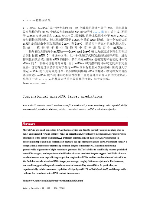
Changes in microRNA expression induced by rabies virus infection in mouse brainsPingsen Zhao a ,b ,Lili Zhao b ,c ,Tao Zhang a ,1,Hualei Wang b ,Chuan Qin a ,Songtao Yang b ,**,Xianzhu Xia a ,b ,*aInstitute of Laboratory Animal Sciences,Chinese Academy of Medical Sciences &Peking Union Medical College,Beijing 100021,PR ChinabKey Laboratory of Jilin Province for Zoonosis Prevention and Control,Institute of Military Veterinary,Academy of Military Medical Sciences,Changchun 130122,PR China cCollege of Animal Science and Veterinary Medicine,Jilin University,Changchun 130062,PR Chinaa r t i c l e i n f oArticle history:Received 30June 2011Received in revised form 29September 2011Accepted 3October 2011Available online xxx Keywords:Rabies virusMicroRNA pro filing Gene pro filing Target prediction Functional analysisa b s t r a c tMicroRNAs (miRNAs)are small RNA (z 22nt)molecules expressed endogenously in cells.They are involved in the regulation of gene expression.Recently,evidence has shown that cellular miRNAs have key regulatory roles in virus e host interactions.The rabies virus (RABV)causes a fatal infection of the central nervous systems (CNS)of warm-blooded animals,yet its pathogenesis remains poorly under-stood.To gain more insight into the pathogenesis of RABV,a miRNA microarray was performed as part of an investigation of changes in host miRNA expression in the brains of mice infected with RABV.The results showed that RABV infection induced modulation of the expression of sixteen miRNA molecules.These data were veri fied by real-time PCR.Functional analysis showed the differentially expressed miRNAs to be involved in many immune-related signaling pathways,such as the RIG-I-like receptor signaling pathway,JAK-STAT signaling pathway,chemokine signaling pathway,T-cell receptor signaling pathway,MAPK signaling pathway,leukocyte transendothelial migration,and natural killer cell mediated cytotoxicity.The predicted expression levels of the target genes of these modulated miRNAs correlated with measurements of gene expression measured by DNA microarray and qRT-PCR.Ó2011Elsevier Ltd.All rights reserved.1.IntroductionThe rabies virus (RABV),a member of the family Rhabdoviridae ,is a highly neurotropic virus that can cause a fatal infection in central nervous systems (CNS)of warm-blooded animals [1,2].Although signi ficant advances have been made in rabies prevention and control,rabies remains a major threat to public health.Every year,it causes 55,000human deaths worldwide [3].The patho-genesis of RABV remains poorly understood.MicroRNAs (miRNA,miR)are endogenous RNA molecules of about 22nt in size.They negatively regulate gene expression by translational repression [4].MiRNA binds to complementary sequences in mRNA,which either prevents translation or accelerates mRNA decay [5].MiRNAs play key roles in cellularprocesses such as development,differentiation,proliferation,and hematopoiesis [6e 11].There is a growing body of evidence showing that cellular miRNAs play key regulatory roles in virus e host inter-actions and that the alterations in cellular miRNA expression that take place during the viral infection may be an important determi-nant of virulence [12,13].In most cases,miRNAs can viral replication.For example,cellular miR-28,miR-125b,miR-150,miR-223,and miR-382inhibit the replication of the human immunode ficiency virus (HIV)in CD4þT-cells [14],and miR-32restricts the replication of primate foamy virus type-1(PFV-1)[15].In contrast,some viruses can use host cellular miRNA to facilitate viral replication.It has been shown that liver-speci fic miR-122binds to sites in the 50-UTR of hepatitis C virus (HCV)RNA and positively regulates the viral life cycle,in part by stimulating HCV translation [16].Recently,microarray analysis has been employed to determine changes in host miRNA expression.This can reveal molecular pathways that govern viral pathogenesis.By using miRNA micro-array pro filing,differentially expressed patterns of cellular miRNAs have been found in the lungs of mice infected with in fluenza [17].Another study showed that miRNAs were signi ficantly modulated in mouse brains upon infection with the Venezuelan equine enceph-alitis virus (VEEV)[18].However,no report on the expression pro file of cellular miRNA in host infected by RABV has yet been published.*Corresponding author.Key Laboratory of Jilin Province for Zoonosis Prevention and Control,Institute of Military Veterinary,Academy of Military Medical Sciences,No.666,Liuying Xilu,Jingyue Economic Development Zone,Changchun 130122,PR China.Tel.:þ8643186985808;fax:þ8643186985828.**Corresponding author.Tel.:þ8643186985515;fax:þ8643186985828.E-mail addresses:zhaopingsen@ (P.Zhao),yst10223@ (S.Yang),xiaxianzhu@ (X.Xia).1Present address:State Key Laboratory of Reproductive Biology,Institute of Zoology,Chinese Academy of Sciences,Beijing 100101,China.Contents lists available at SciVerse ScienceDirectMicrobial Pathogenesisjournal h omepage:ww/locate/micpath0882-4010/$e see front matter Ó2011Elsevier Ltd.All rights reserved.doi:10.1016/j.micpath.2011.10.001Microbial Pathogenesis xxx (2011)1e 8In the current study,we created an expression profile of cellular miRNAs in the CNS of mice infected with RABV and performed target prediction and functional enrichment of miRNAs found to be differentially expressed.Finally,we performed gene microarray analysis and qRT-PCR to verify the expression levels of the pre-dicted targets of the modulated miRNAs in these pathways.Several miRNAs were modulated in RABV-infected mouse brains.Func-tional enrichment revealed that many of the predicted targets of these miRNAs are likely to play key roles in immune response.2.Methods and materials2.1.AnimalsSix-to-eight-week-old female BALB/c mice were obtained from the Changchun Institute of Biological Products,China.Animals had access to food and water ad libitum.All the experiments with live virus challenge were carried out at the bio-safety level2(BSL-2) facilities of the Key Laboratory of Jilin Province for Zoonosis Prevention and Control,Institute of Military Veterinary,Academy of Military Medical Sciences.2.2.Virus challengeRABV strain Evelyn Rokitnicki Abelseth(ERA)was used for this study.Virus stock was prepared as described previously[19]. Briefly,virus samples were grown in mouse neuroblastoma(NA) cells.These were propagated in Dulbecco’s modified Eagle’s medium(DMEM,Gibco)containing10%fetal calf serum(PAA),100 U of penicillin/ml,and100m g of streptomycin/ml at37 C.ERA titer was determined in triplicate on monolayer cultures of NA cells,as described previously in Ref.[20].Eight animals were used for each group.Briefly,mice were anesthetized and105focus-forming units (f.f.u)of RABV in30m l of DMEM were injected intracerebrally(i.c.). Mice mock-infected i.c.with DMEM were included as controls. 2.3.Tissue collection and total RNA isolationMice at7days post-infection(dpi)were anesthetized with ketamine e xylazine(1.98and0.198mg per mouse,respectively) and euthanized.Brains were harvested and stored in RNAlater (Ambion)atÀ80 C for total RNA extraction.Total RNAs were isolated from the entire homogenized brains using the mirVanaÔmiRNA Isolation Kit(Ambion).The integrity of total RNA was analyzed by Agilent2100Bioanalyzer(Agilent Technologies).2.4.MiRNA microarray assaysThree RABV-infected or mock-infected mice at7dpi were randomly selected for miRNA microarray analysis.m Paraflo miRNA microarray assays were outsourced to LC Sciences(Houston,U.S.). The assay was performed on a5-m g total RNA sample,which was size-fractionated using a YM-100Microcon centrifugalfilter (Millipore).The small RNAs(<300nt)isolated were30-extended with a poly-(A)tail using poly-(A)polymerase.An oligonucleotide tag was then ligated to each poly-(A)tail for laterfluorescent dye staining.Hybridization was performed overnight on a m Paraflo microfluidic chip using a micro-circulation pump(Atactic Tech-nologies)[21,22].The microfluidic chips each contained a detection probe consisting of a chemically modified nucleotide coding segment complementary to target microRNA(from miRBase, /sequences/)or other RNA(control or customer defined sequences)and a spacer segment of polyethylene glycol to extend the coding segment away from the substrate.The detection probes were made by in situ synthesis using PGR (photogenerated reagent)chemistry.The hybridization melting temperatures were balanced by chemical modifications of the detection probes.Hybridization used100m l6xSSPE buffer(0.90M NaCl,60mM Na2HPO4,6mM EDTA,pH 6.8)containing25% formamide at34 C.Post-hybridization detection usedfluorescence labeling with tag-specific Cy5dyes.Hybridization images were collected using a laser scanner GenePix4000B(Molecular Devices) and digitized using Array-Pro image analysis software(Media Cybernetics).Raw data were obtained for further analysis.2.5.DNA microarray assaysDNA microarray was carried out using the same archived samples as those used for miRNA microarray.DNA microarray assays were outsourced to Phalanx Biotech Group Inc.(Hsinchu, Taiwan).Fluorescence-labeled cRNA were prepared from5-m g total RNA samples using a Message AMPTM aRNA Kit(Ambion)and Cy5 dye(Amersham Pharmacia).Fluorescent targets were hybridized to a Mouse OneArrayÔWhole Genome DNA Microarray(Phalanx, Taiwan)containing31,802oligonucleotides,including29,922 mouse genome probes and1880experimental control probes.After an overnight hybridization at50 C,non-specific binding targets were washed out in three different washing steps.The slides were dried by centrifugation and scanned using a GenePix4000B (Molecular Devices).The Cy5fluorescent intensity of each spot was analyzed by GenePix4.1software(Molecular Devices).Raw data were obtained for further analysis.2.6.Quantitative real-time PCRThe expression of specific miRNAs was analyzed with quantita-tive real-time PCR using a LNAÔ-based qRT-PCR kit,miRCURY LNAÔUniversal RT microRNA PCR System(Exiqon)according to manu-facturer’s instructions.Total RNA from brain tissues of RABV-infected and mock-infected mice was converted to cDNA using universal reverse transcriptase primers(Exiqon).cDNA samples were diluted1:80in nuclease-free water and then PCR was ampli-fied using SYBR Green master mix and specific LNAÔmiRNA primers (Exiqon)for mmu-miR-200a(target sequence UAACACUGU-CUGGUAACGAUGU),mmu-miR-200b(target sequence UAAUA-CUGCCUGGUAAUGAUGA),mmu-miR-200c(target sequence UAAUACUGCCGGGUAAUGAUGGA),mmu-miR-429(target sequence UAAUACUGUCUGGUAAUGCCGU),mmu-miR-182(target sequence UUUGGCAAUGGUAGAACUCACACCG),mmu-miR-183 (target sequence UAUGGCACUGGUAGAAUUCACU),mmu-miR-203 (target sequence GUGAAAUGUUUAGGACCACUAG),mmu-miR-207 (target sequence GCUUCUCCUGGCUCUCCUCCCUC),mmu-miR-290-3p(target sequence AAAGUGCCGCCUAGUUUUAAGCCC),and mmu-miR-1896(target sequence CUCUCUGAUGGUGGGU-GAGGAG).Samples were run in duplicate on an Mx3005P QPCR system(Agilent Technologies).The data were normalized to a U6 RNA control(Exiqon)and relative expression was calculated using the2ÀDD Ct method[23].For mRNA quantitative real-time PCR,reverse transcription was performed using a Reverse Transcription System(Promega) according to manufacturer’s instructions.The names of the genes and their primers are presented in Table1.Real-time PCR was performed using BrilliantÒII SYBRÒGreen QPCR Master Mix (Agilent Technologies)in an Mx3005P QPCR system(Agilent Technologies)according to the manufacturer’s instructions. Expression of the gene of interest was normalized to glyceraldehyde-3-phosphate dehydrogenase(GAPDH).The expression levels of genes were measured in terms of threshold cycle value(CT)using the2ÀDD Ct method[23].P.Zhao et al./Microbial Pathogenesis xxx(2011)1e8 22.7.Microarray data analysisFirst,raw data both from miRNA and DNA microarrays,with replicates,were averaged.Probes with median detection P value >0.05andflags reported as absent werefiltered out.Expression data were normalized using a variance stabilizing method VSN in Bioconductor()[24].Differential expression of miRNAs was assessed by eBayes(Empirical Bayes Statistics)from LIMMA[25].Differentially expressed miRNAs with P<0.01were selected for further analysis.Microarray data of miRNA and target genes were found to be MIAME compliant and have been submitted to the Gene Expression Omnibus(GEO) database(/geo/;accession number: GSE26269(miRNA data);GSE26270(mRNA data)).2.8.Target prediction and functional enrichment of differentially expressed miRNAsThree continuously updated miRNA target prediction data-bases,TargetScan,MicroCosm Targets,and miRanda,were used to infer the targets of differentially expressed miRNAs.The inter-section of these datasets was taken as reliable.Gene ontology(GO) category and Kyoto Encyclopedia of Genes and Genomes(KEGG) pathway analyses of target genes of differentially expressed miR-NAs were performed using the web-based tool,Database for Annotation,Visualization,and Integrated Discovery[26](DAVID, /).3.Results3.1.MiRNA modulation upon RABV infectionTo determine changes in miRNA expression in mouse brains in response to RABV infection,we evaluated miRNA expression profiles at7dpi.MiRNAs whose relative expression levels showed a fold change(FC)!2and P0.01were considered significantly up-regulated,and those with FCÀ2and P0.01were considered significantly down-regulated.As shown in Fig.1,ten miRNAs, miR-1894-5p,miR-290-3p,miR-1901,miR-207,miR-1896,miR-715, miR-3470b,miR-146b*,miR-203,and miR-770-5p,were found to be significantly up-regulated,and six miRNAs,miR-200a, miR-200b,miR-200c,miR-182,miR-183,and miR-429,were significantly down-regulated upon RABV infection.This indicated that host miRNAs were modulated in the CNS upon rabies virus infection.3.2.Verification of miRNA microarray by qRT-PCRTo validate the differential expression profiles of miRNAs obtained by miRNA microarray,we completed quantitative RT-PCR assessment of ten of the differentially expressed miRNAs shown in Fig.1,miR-200a,miR-200b,miR-200c,miR-429,miR-182,miR-183, miR-1896,miR-203,miR-207,and miR-290-3p.The overall results of qRT-PCR were consistent with those of the microarray analysis. Although differences were observed between these two analyses, most likely due to intrinsic differences between the techniques,the qRT-PCR results showed the same relative regulation of differen-tially expressed miRNAs as the microarray data(Fig.2).3.3.Target prediction and GO analysis of targets of differentially expressed miRNAsFor these sixteen differentially expressed miRNAs,miRanda predicted41,304target genes,TargetScan predicted4,811,and MicroCosm predicted10,135.Of these,6200target geneswereFig.1.MiRNA profile of RABV-infected mouse brain.Two-way hierarchical cluster heat map showing all significantly expressed miRNAs in three independent samples (P<0.01).Each row shows the relative expression level of a single miRNA.Each column shows the expression level of a single sample.Up-regulated miRNAs are shown in red and down-regulated miRNAs are shown in green.(For interpretation of the references to color in thisfigure legend,the reader is referred to the web version of this article.)Table1Primers for selected genes analyzed using quantitative real-time PCR.Gene symbol Gene title Forward primer Reverse primer AmpliconCXCL10Chemokine(C-X-C motif)ligand1050-GCTGCAACTGCATCCATATCGATGA-3050-TAGTAGGGACGCTCGGATAGGAC-30113bpSH2D1A SH2domain protein1A50-GTATCAAGGTTACATCTACACÀ3050-ATCTTTTATGTACTCCAGGTGCTÀ3089bp MAP4K4Mitogen-activated protein kinasekinase kinase kinase450-GGCTGGGTCCATCACAGACCTTGT-3050-TGAGGATTTCCCTGGAGATGTA-3089bpIRF3Interferon regulatory factor350-CTCAAAGATGAGGGGTCCTCAGA-3050-GTGCAGGGTTTAGGAAGTTGTTC-3094bp STAT3Signal transducer and activatorof transcription350-CAAGAGTCCAATGTCCTCTATCA-3050-GCCACGATCCGGGCAATTTCCAT-30101bpTRIM25Tripartite motif-containing2550-GTACAAACTGAGGAATAAACTCACT-3050-TCTCAGCTGTTCCATCTTCCTCTTC-30130bp NFAT5Nuclear factor of activated T-cells550-CAAACGACGAGATTGTGAAGAAT-3050-GAGTTGCCTTTGTTGTCCGTGGT-30108bpIL10Interleukin1050-AGGCAGAGAAGCATGGCCCAG-3050-AGAAATCGATGACAGCGCCTCAG-30100bp IFNAR2Interferon(alpha and beta)receptor250-ATCTGACGAAGGTTAAGAACTGT-3050-TCTGTGCACTATGACGATGGCG-30113bp STAM2Signal transducing adapter molecule(SH3domain and ITAM motif)250-CTATTGAGTTATCGTTGCAAGAGC-3050-CTCGTTGTCCTCAACAGCTTCA-30143bpGAPDH Glyceraldehydes-3-phosphatedehydrogenase50-CTCAACTACATGGTCTACATGTTC-3050-ATTTGATGTTAGTGGGGTCTCGCTC-30142bpP.Zhao et al./Microbial Pathogenesis xxx(2011)1e83predicted by all three systems.These were used for GO analysis.Functional analysis revealed that 177GO terms found to be involved in biological processes,27in molecular function,and 24in cellular components were signi ficantly enriched (P <0.01)(Supplementary Table S1).The ten most common GO categories were cellular process,metabolic process,biological regulation,primary metabolic process,regulation of biological process,cellular metabolic process,regulation of cellular process,macromolecule metabolic process,cellular macromolecule metabolic process,and nitrogen compound metabolic process (Fig.3).3.4.KEGG pathway analysis of targets of differentially expressed miRNAsTo identify the biological pathways that become active in the mouse brain in response to RABV infection,we mapped the targetgenes of differentially expressed miRNAs to canonical signaling pathways in the KEGG.The results showed that 44statistically remarkable categories (P <0.05)were enriched (Supplementary Table S2).As shown in Table 2,the predicted target genes of three up-regulated miRNAs,miR-203,miR-207,and miR-1896,and six down-regulated,miRNAs,miR-200a,miR-200b,miR-200c,miR-429,miR-182and miR-183,were found to be involved in seven immune-related pathways,such as the RIG-I-like receptor signaling pathway,JAK-STAT signaling pathway,chemokine signaling pathway,MAPK signaling pathway,T-cell receptor signaling pathway,natural killer cell mediated cytotoxicity,and leukocyte transendothelial migration.The predicted target genes of some of these up-regulated miRNAs were also found to be involved in immune-related pathways,including the targets of miR-290-3p in the MAPK signaling pathway,chemokine signaling pathway,JAK-STAT signaling pathway,leukocyte transendothelial migration,T-cell receptor signaling pathway,and RIG-I-like receptor signaling pathway;targets of miR-715in the MAPK signaling pathway,chemokine signaling pathway,leukocyte transendothelial migra-tion,T-cell receptor signaling pathway,natural killer cell mediated cytotoxicity,and RIG-I-like receptor signaling pathway;targets of miR-770-5p in the MAPK signaling pathway,chemokine signaling pathway,JAK-STAT signaling pathway,leukocyte transendothelial migration,T-cell receptor signaling pathway,and natural killer cell mediated cytotoxicity;targets of miR-1894-5p in the MAPK signaling pathway,leukocyte transendothelial migration,T-cell receptor signaling,and RIG-I-like receptor signaling pathway;and targets of miR-146b*in the MAPK signaling pathway and chemo-kine signaling pathway;and targets of miR-3470b in the MAPK signaling pathway,chemokine signaling pathway,and natural killer cell mediated cytotoxicity.3.5.DNA microarray assay and qRT-PCR measurement of miRNA targetsMiRNAs predominately function as repressors of target gene expression,therefore,the miRNAs and their targets essentially show mutually antagonistic expression levels.To determine whether any such correlation existed between the deregulated miRNA levels and their corresponding targets,we performed DNA microarray assay and qRT-PCR validation.To investigate genes involved intheFig.2.qRT-PCR veri fication of miRNA microarray.Ten differentially expressed miRNAs were selected from miRNA microarray datasets and examined by qRT-PCR.The fold change from the qRT-PCR was determined using the 2ÀDD Ct method and all miRNA expression values were normalized against the U6endogenous control.Data from qRT-PCR are shown as mean Æstandard deviation (SD)of one representative experi-ment.Similar results were obtained in three independentexperiments.Fig.3.Enriched GO terms in the biological process category among differentially expressed miRNAs.After miRNA microarray assay,signi ficantly enriched GO analysis in the biological process category was performed on differentially expressed genes in the brains of RABV-infected mice was performed using DAVID (P <0.01).Each color represents a different biological process and the number of target genes enriched is shown after the name of the biological process.Only the top ten GO terms are listed here.For other enriched GO terms,please see Supplementary Table 1.(For interpretation of the references to colour in this figure legend,the reader is referred to the web version of this article.)P.Zhao et al./Microbial Pathogenesis xxx (2011)1e 84P.Zhao et al./Microbial Pathogenesis xxx(2011)1e85Table2Predicted targets of modulated miRNAs upon RABV infection involved in immune response pathways.P value miRNAs KEGG pathways Target genes Numbersof targetmmu-miR-200a mmu04010:MAPK signaling pathway ARRB2,GRB2,STK3,PRKACB,MAP2K4,NTRK2,PPP3R1,18 5.46E-05PDGFA,RAPGEF2,MAP3K7,TGFB2,PPP3R2,FAS,DUSP2,TGFBR1,CDC42,DUSP6,TGFBR2100.003982 mmu-miR-200a mmu04062:Chemokine signaling pathway ARRB2,GRB2,TIAM1,PRKACB,STAT5B,CSK,CDC42,PIK3CA,CXCL12,STAT2mmu-miR-200a mmu04630:Jak-STAT signaling pathway GRB2,STAT5A,IFNAR1,IL20RA,STAT5B,SPRY4,IL12B,IFNAR2,130.00711STAT4,CSF3,PIK3CA,IL2,STAT2mmu-miR-200a mmu04670:Leukocyte transendothelial migration CTNNA1,JAM3,CDC42,PIK3CA,CXCL1250.015038 mmu-miR-200a mmu04660:T-cell receptor signaling pathway DLG1,GRB2,PPP3R1,MAP3K7,PPP3R2,CDC42,PIK3CA,IL280.00172790.001221 mmu-miR-200a mmu04650:Natural killer cell mediated cytotoxicity GRB2,KLRC1,IFNAR1,PPP3R1,IFNAR2,PPP3R2,FAS,PIK3CA,CD48mmu-miR-200a mmu04622:RIG-I-like receptor signaling pathway MAP3K7,IL12B20.00249115 5.46E-05 mmu-miR-203mmu04010:MAPK signaling pathway CACNA2D1,MAP3K13,DUSP5,MAP4K3,NLK,AKT2,ATF2,PDGFRA,RAP1A,MAPT,MAP3K1,PPM1B,FGF7,CRK,PRKCAmmu-miR-203mmu04062:Chemokine signaling pathway TIAM1,PXN,XCL1,AKT2,RAP1A,GNG10,CRK,ADCY9,VAV390.003982 mmu-miR-203mmu04630:Jak-STAT signaling pathway IL24,CNTFR,IL22RA2,CCND1,SOCS3,AKT2,IL12B70.00711 mmu-miR-203mmu04670:Leukocyte transendothelial migration PXN,RAP1A,JAM3,VAV3,PRKCA50.015038 mmu-miR-203mmu04660:T-cell receptor signaling pathway AKT2,VAV320.001727 mmu-miR-203mmu04650:Natural killer cell mediated cytotoxicity VAV3,PRKCA20.001221 mmu-miR-203mmu04622:RIG-I-like receptor signaling pathway IRF3,IL12B,MAP3K130.00249114 5.46E-05 mmu-miR-182mmu04010:MAPK signaling pathway MAP3K3,GRB2,HSPA2,NF1,MAPK1,PPP3R1,PDGFA,RASA1,PLA2G12A,MEF2C,RAC1,BDNF,FGF9,MAP2K1110.003982 mmu-miR-182mmu04062:Chemokine signaling pathway GRB2,TIAM1,MAPK1,ELMO1,WASL,RAC1,ROCK1,ADCY6,MAP2K1,SHC4,FOXO3mmu-miR-182mmu04630:Jak-STAT signaling pathway GRB2,SOCS4,SPRY4,VCL30.00711 mmu-miR-182mmu04670:Leukocyte transendothelial migration RAC1,PLCG1,ROCK130.015038 mmu-miR-182mmu04660:T-cell receptor signaling pathway GRB2,MAPK1,PPP3R1,PLCG1,MAP2K150.001727 mmu-miR-182mmu04650:Natural killer cell mediated cytotoxicity GRB2,MAPK1,PPP3R1,RAC1,SH2D1A,PLCG1,MAP2K1,SHC480.001221 mmu-miR-182mmu04622:RIG-I-like receptor signaling pathway IFIH110.002491 mmu-miR-290-3p mmu04010:MAPK signaling pathway RPS6KA5,RAPGEF2,CACNB3,TGFBR24 5.46E-05 mmu-miR-290-3p mmu04062:Chemokine signaling pathway CXCR4,STAT3,20.003982 mmu-miR-290-3p mmu04630:Jak-STAT signaling pathway IL24,IL12RB1,STAT330.00711 mmu-miR-290-3p mmu04670:Leukocyte transendothelial migration CXCR4,CLDN520.015038 mmu-miR-290-3p mmu04660:T-cell receptor signaling pathway PAK7,PDCD120.001727 mmu-miR-290-3p mmu04622:RIG-I-like receptor signaling pathway DDX3X10.002491 mmu-miR-207mmu04010:MAPK signaling pathway HSPA1L,MAP2K4,NTRK2,EGFR,MAP3K55 5.46E-05 mmu-miR-207mmu04062:Chemokine signaling pathway CXCR6,BCAR120.003982 mmu-miR-207mmu04630:Jak-STAT signaling pathway LEP,IL5,IL23A,STAT640.00711 mmu-miR-207mmu04670:Leukocyte transendothelial migration BCAR110.015038 mmu-miR-207mmu04660:T-cell receptor signaling pathway IL5,CD247,LCK30.001727 mmu-miR-207mmu04650:Natural killer cell mediated cytotoxicity CD247,LCK20.001221 mmu-miR-207mmu04622:RIG-I-like receptor signaling pathway TRIM2510.00249119 5.46E-05 mmu-miR-429mmu04010:MAPK signaling pathway MKNK1,MAP4K3,MAPK7,PRKACB,RPS6KA2,NTRK2,AKT2,NTF3,RAPGEF2,RAC1,MAP2K6,PLA2G4A,DUSP1,MAP3K1,RAP1B,PPM1B,MAP4K4,JUN,SRFmmu-miR-429mmu04062:Chemokine signaling pathway HCK,PRKACB,AKT2,ADCY2,RAC1,GNAI3,PTK2B,100.003982RHOA,RAP1B,FOXO3100.00711 mmu-miR-429mmu04630:Jak-STAT signaling pathway EP300,CNTFR,IL13,IFNA11,IL10,AKT2,STAM2,SPRED2,PTPN11,CISHmmu-miR-429mmu04670:Leukocyte transendothelial migration OCLN,RAC1,GNAI3,PTK2B,MSN,PTPN11,PLCG1,100.015038RHOA,CLDN23,RAP1Bmmu-miR-429mmu04660:T-cell receptor signaling pathway FYN,IL10,PAK7,AKT2,CD247,PLCG1,RHOA,JUN80.001727 mmu-miR-429mmu04650:Natural killer cell mediated cytotoxicity FYN,IFNA11,CD247,RAC1,PTK2B,PTPN11,PLCG1,KLRC280.001221 mmu-miR-429mmu04622:RIG-I-like receptor signaling pathway PIN1,IFNA11,DDX3Y,D1PAS1,TBK1,MAP3K1,80.002491RNF125,DDX3X20 5.46E-05 mmu-miR-200b mmu04010:MAPK signaling pathway MKNK1,DUSP4,MAP4K3,MAPK7,PRKACB,RPS6KA2,NTRK2,AKT2,NTF3,RAPGEF2,RAC1,MAP2K6,PLA2G4A,DUSP1,MAP3K1,RAP1B,PPM1B,MAP4K4,JUN,SRF90.003982 mmu-miR-200b mmu04062:Chemokine signaling pathway PRKACB,AKT2,ADCY2,RAC1,GNAI3,PTK2B,RHOA,RAP1B,FOXO3110.00711 mmu-miR-200b mmu04630:Jak-STAT signaling pathway EP300,GHR,CNTFR,SOCS4,IL13,IFNA11,IL10,AKT2,STAM2,SPRED2,CISHmmu-miR-200b mmu04670:Leukocyte transendothelial migration OCLN,RAC1,GNAI3,PTK2B,MSN,CLDN11,PLCG1,90.015038RHOA,RAP1Bmmu-miR-200b mmu04660:T-cell receptor signaling pathway FYN,IL10,PAK7,AKT2,CD247,PLCG1,RHOA,JUN80.001727 mmu-miR-200b mmu04650:Natural killer cell mediated cytotoxicity FYN,IFNA11,CD247,RAC1,PTK2B,PLCG160.001221 mmu-miR-200b mmu04622:RIG-I-like receptor signaling pathway PIN1,IFNA11,DDX3Y,D1PAS1,TBK1,MAP3K1,80.002491RNF125,DDX3X21 5.46E-05 mmu-miR-200c mmu04010:MAPK signaling pathway MKNK1,MAP4K3,MAPK7,PRKACB,RPS6KA2,NTRK2,AKT2,NTF3,RAPGEF2,RAC1,SOS2,MAP2K6,PLA2G4A,DUSP1,MAP3K1,RAP1B,RASGRP1,PPM1B,MAP4K4,JUN,SRF(continued on next page)pathways common to miRNA target prediction and RABV infection,we measured the expression of genes from RIG-I-like receptor signaling pathway (IRF3,TRIM25,IFNAR2,STAM2),JAK-STAT signaling pathway (STAT3),chemokine signaling pathway (CXCL10),T-cell receptor signaling pathway (NFAT5,IL10),natural killer cell mediated cytotoxicity (SH2D1A ),and MAPK signaling pathway (MAP4K4).As presented in Table 3,IRF3,STAT3,TRIM25,and NFAT5,the targets of up-regulated miRNAs miR-203,miR-290-3p,miR-207,and miR-1896,respectively,were found to be down-regulated.IFNAR2,STAM2,IL10,MAP4K4,SH2D1A ,and CXCL10,the targets of down-regulated miRNAs miR-200a,miR-200b,miR-200c,miR-429,miR-182,and miR-183,respectively,were found to be up-Table 2(continued )miRNAs KEGG pathwaysTarget genesNumbers of target P value mmu-miR-200c mmu04062:Chemokine signaling pathway PRKACB,AKT2,ADCY2,RAC1,GNAI3,SOS2,RHOA,RAP1B,FOXO90.003982mmu-miR-200c mmu04630:Jak-STAT signaling pathwayEP300,CNTFR,IL13,IL10,AKT2,STAM2,SOS2,CISH 80.00711mmu-miR-200c mmu04670:Leukocyte transendothelial migration OCLN,RAC1,GNAI3,MSN,PLCG1,RHOA,RAP1B 70.015038mmu-miR-200c mmu04660:T-cell receptor signaling pathway FYN,IL10,PAK7,AKT2,CD247,PLCG1,SOS2,RHOA,RASGRP1,JUN100.001727mmu-miR-200c mmu04650:Natural killer cell mediated cytotoxicity FYN,CD247,RAC1,PLCG1,SOS250.001221mmu-miR-200c mmu04622:RIG-I-like receptor signaling pathway PIN1,DDX3Y,TBK1,MAP3K1,FADD,RNF125,DDX3X 70.002491mmu-miR-715mmu04010:MAPK signaling pathwayFGF22,MAPK112 5.46E-05mmu-miR-715mmu04062:Chemokine signaling pathwayGRK5,SHC420.003982mmu-miR-715mmu04670:Leukocyte transendothelial migration MAPK1110.015038mmu-miR-715mmu04660:T-cell receptor signaling pathwayMAPK1110.001727mmu-miR-715mmu04650:Natural killer cell mediated cytotoxicity SHC410.001221mmu-miR-715mmu04622:RIG-I-like receptor signaling pathway MAPK11120.002491mmu-miR-183mmu04010:MAPK signaling pathwayDUSP10,EGFR,MAP3K4,MAPK8IP1,MAP2K1,PRKCA 6 5.46E-05mmu-miR-183mmu04062:Chemokine signaling pathwayTIAM1,PLCB4,GNAI1,CXCL10,GNB1,GNG5,CX3CL1,MAP2K1,CCL25,SHC4100.003982mmu-miR-183mmu04630:Jak-STAT signaling pathwaySPRY2,SPRY320.00711mmu-miR-183mmu04670:Leukocyte transendothelial migration ITGB1,GNAI1,ACTN3,TXK,PRKCA 50.015038mmu-miR-183mmu04660:T-cell receptor signaling pathwayNCK2,LCK,CD3D,MAP2K140.001727mmu-miR-183mmu04650:Natural killer cell mediated cytotoxicity SH2D1A,LCK,MAP2K1,SHC4,PRKCA 50.001221mmu-miR-183mmu04622:RIG-I-like receptor signaling pathway CXCL10,AZI220.002491mmu-miR-1894-5p mmu04010:MAPK signaling pathwaySTMN1,MAPK142 5.46E-05mmu-miR-1894-5p mmu04670:Leukocyte transendothelial migration THY1,MSN,MAPK1430.015038mmu-miR-1894-5p mmu04660:T-cell receptor signaling pathway MAPK1410.001727mmu-miR-1894-5p mmu04010:MAPK signaling pathwayMAP3K121 5.46E-05mmu-miR-1894-5p mmu04622:RIG-I-like receptor signaling pathway MAPK1410.002491mmu-miR-146b *mmu04010:MAPK signaling pathwayRPS6KA4,MAPK8IP32 5.46E-05mmu-miR-146b *mmu04062:Chemokine signaling pathway PLCB310.003982mmu-miR-1896mmu04010:MAPK signaling pathwayATF2,NRAS,RRAS2,ELK14 5.46E-05mmu-miR-1896mmu04062:Chemokine signaling pathway NRAS 10.003982mmu-miR-1896mmu04630:Jak-STAT signaling pathwayPIAS110.00711mmu-miR-1896mmu04670:Leukocyte transendothelial migration 2900073G15RIK 10.015038mmu-miR-1896mmu04660:T-cell receptor signaling pathwayNFAT5,NRAS 20.001727mmu-miR-1896mmu04650:Natural killer cell mediated cytotoxicity NFAT5,NRAS 20.001221mmu-miR-1896mmu04622:RIG-I-like receptor signaling pathway TRAF3,DDX3X 20.002491mmu-miR-3470b mmu04010:MAPK signaling pathwayMAPT,PPM1B 2 5.46E-05mmu-miR-3470b mmu04062:Chemokine signaling pathwaySHC1,10.003982mmu-miR-3470b mmu04650:Natural killer cell mediated cytotoxicity SHC110.001221mmu-miR-770-5p mmu04010:MAPK signaling pathwayRAF1,RAP1B2 5.46E-05mmu-miR-770-5p mmu04062:Chemokine signaling pathway CCR3,ADCY2,GNAI3,RAF1,BCAR1,RAP1B 60.003982mmu-miR-770-5p mmu04630:Jak-STAT signaling pathwayCSF3R,SPRY1,IFNGR230.00711mmu-miR-770-5p mmu04670:Leukocyte transendothelial migration CLDN6,GNAI3,MSN,BCAR1,RAP1B 50.015038mmu-miR-770-5p mmu04660:T-cell receptor signaling pathwayPAK6,RAF120.001727mmu-miR-770-5pmmu04650:Natural killer cell mediated cytotoxicityRAF1,KLRK1,IFNGR230.001221Table 3DNA microarray and qRT-PCR analysis of expression of miRNA targets.MicroRNAs Accession number MiRNA microarray (Fold:[or Y )qRT-PCR(Fold:[or Y )Targets of microRNAs Accession Number DNA microarray (Fold:[or Y )qRT-PCR(Fold:[or Y )mmu-miR-203MIMAT0000264 3.68Â,[ 4.68Æ2.00Â,[IRF3NM_016849 1.04Â,Y 3.44Æ1.98Â,Y mmu-miR-290-3p MIMAT0004572 3.27Â,[ 5.27Æ1.01Â,[STAT3NM_213659 1.11Â,Y 2.76Æ0.24Â,Y mmu-miR-207MIMAT0000240 4.04Â,[ 6.57Æ2.65Â,[TRIM25NM_009546e a8.66Æ4.22Â,Y mmu-miR-1896MIMAT0007873 3.74Â,[ 4.81Æ2.08Â,[NFAT5NM_133957 1.23Â,Y 3.26Æ1.09Â,Y mmu-miR-200a MIMAT000068258.82Â,Y 60.21Æ14.42Â,Y IFNAR2NM_001110498 1.23Â,[ 4.25Æ2.14Â,[mmu-miR-200b MIMAT000031858.98Â,Y 46.98Æ12.17Â,Y STAM2NM_019667 1.37Â,[ 4.07Æ1.56Â,[mmu-miR-200c MIMAT000061745.66Â,Y 36.97Æ10.01Â,Y IL10NM_010548 1.02Â,[ 2.87Æ1.51Â,[mmu-miR-429MIMAT000153727.99Â,Y 27.66Æ9.50Â,Y MAP4K4NM_008696 1.19Â,[ 3.23Æ1.33Â,[mmu-miR-182MIMAT000021128.62Â,Y 29.95Æ9.02Â,Y SH2D1A NM_011364 2.23Â,[ 6.42Æ3.42Â,[mmu-miR-183MIMAT000026133.46Â,Y26.13Æ12.50Â,YCXCL10NM_0212741.1Â,[2.76Æ1.93Â,[Data from qRT-PCR are the mean Æstandard deviation (SD)of one representative experiment.Similar results were obtained in three independent experiments.aNo signal was detected in DNA microarray.P.Zhao et al./Microbial Pathogenesis xxx (2011)1e 86。