CRESS: A Platform of Infrastructure Resource Sharing for Educational Cloud Computing
- 格式:pdf
- 大小:1.08 MB
- 文档页数:10
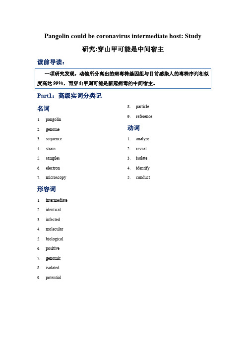
Pangolin could be coronavirus intermediate host: Study研究:穿山甲可能是中间宿主读前导读:Part1:高级实词分类记名词1.pangolin2.genome3.sequence4.strain5.samples6.electron7.microscopy 8.particle9.reference 动词1.analyze2.reveal3.isolate4.identify5.conduct形容词1.intermediate2.identical3.infected4.molecular5.biological6.positive7.genomic8.isolated9.potential其他词类1.jointly2.currentlyPart2:高级词汇拓展1.identical完全相同的adv ___________________2.infected感染的vt ___________________ n ___________________3.biological生物学的n 生物学________________生物学家n ___________________ adv ___________________4.positive积极的adv ___________________5.genomic基因组学的___________________ 基因组基因___________________ 基因的adj__________________6.isolated孤立的隔离vt ___________________7.jointly共同地adj ___________________8.currently目前地adj ___________________9.reference参考v ___________________10.analyze分析n ___________________11.reveal揭示n ___________________12.identify识别adj ___________________ n ___________________ Part3:高级短语荟萃1.conducted by researchers from South China Agricultural University2.related scientific research institutes3.of great significance to prevention and control of4.the source of novel coronavirus5.at a press conference6.molecular biological testing7.positive rate8.electron microscopy9.particle structure10.genomic sequence similarities11.isolated virus strain a12.currently infected human strain13.provide references for further scientific researchPart4:原汁文章思维能力提升训练1.What might be a potential intermediate host of the novel coronavirus according tothe latest research? And why?2.What is the significance of the latest research result?Part5:报刊复杂句式剖析。

Natural disasters are events that occur naturally and cause significant damage to life, property,and the environment.They can be sudden and unexpected,or they may develop gradually over time.Here is a detailed essay on the topic of natural disasters,discussing their types,causes,impacts,and how we can prepare for and mitigate them.IntroductionNatural disasters have been a part of our planets history,and they continue to shape our world in various ways.They are a stark reminder of the immense power of nature and the vulnerability of human societies.The essay will explore the different types of natural disasters,their causes,the devastating effects they have on communities,and the steps that can be taken to reduce their impact.Types of Natural Disasters1.Earthquakes:Sudden movements of the Earths crust cause these seismic events,which can lead to widespread destruction,especially in densely populated areas.2.Tsunamis:Triggered by underwater earthquakes,volcanic eruptions,or landslides, tsunamis are massive waves that can inundate coastal areas,causing significant loss of life and property.3.Hurricanes and Typhoons:These are tropical cyclones that form over warm ocean waters and bring heavy rains,strong winds,and storm surges,leading to flooding and damage.4.Floods:Excessive rainfall,snowmelt,or dam failures can cause floods,which can submerge large areas,disrupt transportation,and lead to waterborne diseases.5.Droughts:Prolonged periods of low precipitation can lead to water scarcity,crop failures,and famine,affecting both humans and wildlife.6.Volcanic Eruptions:The release of molten rock,ash,and gases from the Earths interior can cause widespread devastation,including air travel disruptions and longterm climate effects.ndslides and Mudslides:These occur when soil,rock,and other debris move down a slope,often due to heavy rainfall or earthquakes,causing damage to infrastructure and loss of life.Causes of Natural DisastersNatural disasters are caused by a combination of geological,meteorological,and hydrological processes.The Earths tectonic activity,climate patterns,and water cycles are some of the primary drivers behind these events.Human activities,such as deforestation and urbanization,can exacerbate the conditions that lead to natural disasters.Impacts of Natural DisastersThe impacts of natural disasters are multifaceted,affecting not only human lives but also the economy,infrastructure,and the environment.They can lead to:Loss of life and injuriesDestruction of homes and businessesDisruption of essential services like healthcare and educationEconomic losses due to damage and the cost of recoveryLongterm environmental damage and ecosystem disruptionPreparation and MitigationTo reduce the impact of natural disasters,it is crucial to invest in preparedness and mitigation strategies.Some of these include:1.Early Warning Systems:Developing and implementing systems that can predict and provide early warnings of impending disasters,allowing for timely evacuation and preparation.2.Infrastructure Resilience:Designing and constructing buildings and infrastructure that can withstand the forces of natural disasters,such as earthquakeresistant buildings and floodresistant barriers.nd Use Planning:Implementing land use policies that avoid building in highrisk areas,such as floodplains or earthquakeprone zones.munity Education:Educating communities about the risks of natural disasters and how to respond effectively during and after such events.5.Emergency Response Plans:Establishing robust emergency response plans that include the coordination of resources,evacuation procedures,and postdisaster recovery efforts.ConclusionNatural disasters are an inevitable part of our existence on Earth.While we cannot prevent them,we can take proactive steps to minimize their impact.By understanding the causes,preparing for the inevitable,and investing in mitigation strategies,we can build more resilient communities that are better equipped to face the challenges posed by nature.It is a collective responsibility to ensure that our actions today contribute to a safer and more sustainable future for all.。

TPO27 R-3 原文翻译Predator-Prey Cycles捕食者是怎样影响被捕食者的数量的呢?答案并不是想象中那么简单的。
麋鹿通过穿越冬天的冰层和到达了在苏必略路的罗亚尔岛,在那他们因为和捕食者分隔开来就更自由了。
当狼在晚一点的时候到达那座岛的时候,自然学家都认为,狼对控制麋鹿的数量起到了关键的作用。
但是,严密的研究已经说明,并不是这样的。
狼吃掉的大部分是年老的,或是已经生病的动物,他们本身就不会存活很久了。
总的来说,麋鹿的数量是由食物的可利用性,疾病,和其他的一些条件,而不是因为狼。
当实验性的数量被设置在简单的实验条件下,捕食者经常吃掉了所有的被捕食者,然后因为没有东西吃,自己灭绝了。
但是,如果能给被捕食动物提供如同在野外的安全的区域,被捕食动物的数量会降低到很低的数值,但不会灭绝。
低的被捕食动物数量造成了捕食者的食物不足,使捕食者的数量下降。
当这个情况发生的时候,被捕食动物的数量又会反弹。
这样,在一定的时候,捕食者和被捕食者的数量会持续地在这种循环中变化。
这种数量的循环是小型哺乳动物的特性,有时候这种循环的出现是由捕食者带来的。
生态学家对野兔数量的研究已经显示,北美“雪鞋野兔”一直遵守着大约以十年为一周期的循环。
在一个典型的循环中,其数量会以十倍到三十倍的减少,甚至会出现百倍的改变会出现。
有两种因素会导致这种循环:食物和捕食者。
“雪鞋野兔”比较喜欢的食物是柳木和桦树树枝。
当野兔的密度增加的时候,这些树枝的数量就会减少,迫使野兔去吃一些低质量的,高纤维食物。
随之而来的是低生育率,低成活率,低生长率,所以产生了对应的野兔数量的减少反应。
一旦野兔数量减少了,树枝的数量会需要两到三年去恢复。
雪鞋野兔的主要捕食者是加拿大山猫。
加拿大山猫呈现了平行于野兔的十年鼎盛循环。
当野兔的数量下降的时候,当山猫的食物供给减少,山猫数量也会发生同样的变化。
是什么造成了捕食者和被捕食者的振幅呢?是野兔数量的增长使得植物被过度破坏,随之导致了野兔的减少?还是山猫的增加导致了野兔被过度捕捉?在1992年,Charles Krebs 和其合作者的野外试验提供了答案。

子宫圆韧带的英语名词解释The English Term for the Round Ligament of the UterusIntroductionThe human body is a complex organism, with numerous organs and structures that perform specific functions. One such organ is the uterus, a crucial part of the female reproductive system. The uterus is supported by several ligaments, including the round ligament of the uterus. In this article, we will explore the English term for the round ligament of the uterus and provide a comprehensive explanation of its function and importance.The Round Ligament of the UterusThe round ligament of the uterus, known as Ligamentum teres uteri in Latin, is a band of fibrous tissue that connects the uterus to the labia majora in the female reproductive system. It is present on both sides of the uterus and is approximately four inches in length. Although the round ligament may seem insignificant, it plays a vital role in supporting the uterus during pregnancy and childbirth.Function and ImportanceThe round ligament of the uterus serves multiple functions throughout a woman's life, particularly during pregnancy. Its primary purpose is to provide support to the growing uterus as the fetus develops. As the uterus expands during pregnancy, the round ligament stretches to accommodate the increasing size. This stretching can cause discomfort and mild pain, referred to as round ligament pain, which is a common symptom experienced by many pregnant women.Moreover, the round ligament of the uterus helps to maintain the correct positioning and orientation of the uterus within the pelvic cavity. It prevents excessive movement or displacement of the uterus, providing stability and ensuring proper functioning of thereproductive system. Without the round ligament, the uterus would lack the necessary support, leading to potential complications during pregnancy and childbirth.During childbirth, the round ligament also contributes to the process by helping to guide the fetal head through the birth canal. The ligament aids in the descent of the baby, ensuring a smoother and more efficient delivery. This function highlights the significance of the round ligament in facilitating the natural birth process.In addition to its role in pregnancy and childbirth, the round ligament of the uterus has implications for women's health in general. It has been observed that certain conditions, such as endometriosis, may affect the round ligament and cause pain or discomfort. Understanding the role and function of this ligament can aid in the diagnosis and treatment of such conditions.ConclusionThe round ligament of the uterus, or Ligamentum teres uteri, is a crucial structure within the female reproductive system. Its importance lies in providing support to the uterus throughout pregnancy, maintaining its position within the pelvic cavity, and aiding in the natural birthing process. Although often overlooked, the round ligament plays a significant role in women's health and reproductive well-being. Moreover, understanding this structure assists in the diagnosis and management of conditions that may impact its function. By appreciating the complexity and significance of the round ligament of the uterus, we gain a deeper understanding of the remarkable nature of the female reproductive system.。

The Tyrannosaurus rex,often affectionately referred to as the T.rex,is one of the most iconic dinosaurs that ever roamed the Earth.Its fearsome image has been immortalized in films,books,and scientific studies, captivating the imagination of people across the globe.The process of creating a lifelike representation of this prehistoric beast is a fascinating journey that combines art,science,and a deep understanding of the creatures anatomy and behavior.The first step in the process is research.This involves delving into the extensive body of paleontological literature to gather as much information as possible about the T.rex.Scientists have discovered numerous fossils of this dinosaur,providing a wealth of data about its size,shape,and skeletal structure.By studying these fossils,one can gain a detailed understanding of the T.rexs physical characteristics,which is crucial for creating an accurate representation.Once the research is complete,the next step is to create a blueprint.This involves sketching the T.rexs body,paying close attention to the proportions and the placement of its various features.The blueprint serves as a guide for the rest of the process,ensuring that the final product is as accurate as possible.With the blueprint in hand,the process moves on to sculpting.This is where the artists skill and creativity come into ing materials such as clay or foam,the sculptor carefully shapes the T.rexs body,working from the blueprint to ensure accuracy.This stage requires a keen eye for detail and a deep understanding of the T.rexs anatomy,as even the smallestmistake can affect the overall appearance of the sculpture.After the sculpting is complete,the next step is molding and casting.This involves creating a mold of the sculpture,which can then be used to produce multiple copies of the T.rex.The choice of material for the final product depends on the intended use for example,a museum display might use a durable material like fiberglass,while a toy might be made from a more flexible plastic.Once the T.rex has been cast,the final stage of the process is painting and detailing.This involves carefully painting the sculpture to match the T.rexs natural colors,as well as adding any additional details such as scales, feathers,or skin texture.This stage requires a steady hand and a keen eye for detail,as even the smallest imperfection can detract from the overall realism of the sculpture.Throughout the entire process,its important to keep in mind the T.rexs behavior and environment.For example,the sculpture might be posed in a hunting stance,with its powerful jaws open and its muscular legs tensed for a leap.The surrounding environment,such as a prehistoric forest or a barren plain,can also add to the overall effect,helping to bring the T.rex to life.In conclusion,creating a lifelike representation of a T.rex is a complex and rewarding process that requires a combination of scientific knowledge, artistic skill,and a deep understanding of the dinosaurs natural environment.The result is a stunning piece of art that captures the essenceof this fearsome predator,allowing us to glimpse into the world of the dinosaurs and appreciate the incredible creatures that once roamed the Earth.。

胆囊癌临床诊疗的新进展中华外科杂志普外空间 2022-08-10 10:00 发表于北京作者:杨自逸,刘诗蕾,蔡晨,吴自友,熊逸晨,李茂岚,吴向嵩,全志伟,龚伟文章来源:中华外科杂志, 2022, 60(8)摘要胆囊癌的恶性程度极高,尚缺乏早期诊断方法和有效治疗手段,亟需高质量研究突破诊疗瓶颈。
本文回顾了2021年国内外发表的胆囊癌研究相关文献,对临床诊疗领域的重要进展进行综述,详细介绍了胆囊癌最新流行病学数据及危险因素、新兴的外周血实验室检查和影像学诊断方法、病理学类型新分类、外科治疗的热点与争议及系统性综合治疗动态。
这些研究结果有助于探索更有效的胆囊癌诊治方法,为改善胆囊癌患者的预后带来希望。
胆囊癌是胆道系统常见的恶性肿瘤,具有症状隐匿、发展迅速、早期转移、预后极差的特点。
我国是胆囊癌的高发地区之一,近年来发病率和病死率呈缓慢上升趋势。
目前仍缺乏特异度和灵敏度均较好的胆囊癌早期诊断手段,临床发现的胆囊癌多为中晚期。
尽管医学科技不断发展,早期诊断和根治性手术切除仍是可能治愈胆囊癌的手段,行之有效的系统性治疗方法依然在不断探索中。
本文展示了2021年胆囊癌临床诊疗领域的研究进展,以探索更好的胆囊癌诊疗方法。
一、流行病学特征(一)发病率与死亡率2020年全球癌症统计数据显示,全球胆囊癌新发115 949例(男性41 062例,女性74 887例),死亡84 695例(男性30 265例,女性54 430例)[1],均居消化系统肿瘤第6位。
胆囊癌全球发病率存在明显的地域差异,全球年龄标准化发病率平均为2.3/10万人,以东亚、南美最高,西欧、北美则发病率较低[2];且近年来男性和年轻群体的胆囊癌发病率呈升高趋势。
我国国家癌症中心数据显示,国内胆囊癌发病率为3.95/10万人(男性3.70/10万人,女性4.21/10万人),死亡率为2.95/10万人(男性1.9/10万人,女性2.1/10万人)[3]。
In recent years,the issue of water resource wastage has become a global concern. The following report delves into the various aspects of this critical problem,highlighting its causes,consequences,and potential solutions.Introduction to the IssueWater is a vital resource for life on Earth,yet it is being wasted at an alarming rate.From agricultural runoff to industrial discharges and household leaks,the misuse of water resources is rampant.This report aims to shed light on the gravity of the situation and the urgent need for collective action.Causes of Water Wastage1.Inefficient Irrigation Systems:Traditional irrigation methods,such as flood irrigation, often lead to significant water loss due to evaporation and runoff.ck of Infrastructure:In many regions,the absence of proper water management systems results in leakage and wastage.3.Industrial Overuse:Industries often use large volumes of water for cooling and processing,with inadequate recycling or treatment measures in place.4.Domestic Misuse:Everyday activities like overwatering gardens,long showers,and running taps unnecessarily contribute to water wastage.5.Poor Water Pricing Policies:In some areas,low water prices do not incentivize conservation,leading to overuse.Consequences of Water Wastage1.Environmental Impact:Wastage can lead to the depletion of aquifers,drying up of rivers,and the loss of biodiversity.2.Economic Loss:The economic cost of water wastage is substantial,affecting agriculture,industry,and municipal services.3.Social Implications:Water scarcity can lead to conflicts over resources,affecting community stability and wellbeing.4.Health Risks:Contaminated water from improper disposal can lead to waterborne diseases,impacting public health.Potential Solutions1.Promoting WaterEfficient Technologies:Encouraging the use of drip irrigation and smart watering systems can significantly reduce agricultural water use.2.Upgrading Infrastructure:Investing in modern water supply and distribution systems can minimize leakage and improve efficiency.3.Regulatory Measures:Implementing stricter regulations on industrial water use and enforcing penalties for noncompliance can curb wastage.4.Public Awareness Campaigns:Educating the public about the importance of waterconservation and promoting watersaving practices at home.5.Economic Incentives:Introducing tiered pricing for water usage can encourage users to reduce consumption.ConclusionThe wastage of water resources is a pressing issue that demands immediate attention.By understanding the causes and consequences,and by implementing effective solutions,we can work towards a more sustainable use of water.It is crucial for governments, industries,and individuals to take responsibility and contribute to the preservation of this precious resource.Call to ActionAs we conclude this report,it is essential to call upon all stakeholders to take proactive steps in reducing water wastage.From implementing technological advancements to fostering a culture of conservation,every effort counts towards ensuring a watersecure future for generations to come.。
2833 Commentary2834Journal of Cell Science 116 (14)2835 Fluorescence in situ hybridizationFig. 2.State of the art in FISH. (A) Many transcription sites (10) detected using barcoded probes can determine the gene expression pattern of each cell (Levsky et al., 2002) (B-D). Detection of single mRNA molecules using double or triple colored probes (Femino et al., 1998). (E) Detection of 24 chromosomes using spectral imaging (Macville et al., 1997).2836re-balance multi-color images composed of bands of varying strengths and to correct for spectral signal overlaps (Castleman, 1998).Difficulties inherent in objective analyses of FISH images have impeded but not stopped the development of automated algorithms for interpretation. Detection of DNA targets with large probes and the use of multi-color fluorescent cytometry algorithms (Galbraith et al., 1991) have allowed the production of automated mechanisms for assisting pathologists (Piper et al., 1994). In addition, the use of diagnostic probe sets and dot-counting approaches have yielded independent platforms capable of making simple diagnostic conclusions (Piper et al., 1990). Although methods have been introduced to analyze and optimize these cell classifiers (Castleman, 1985; Castleman and White, 1980; Castleman and White, 1981), manual cytopathology remains the gold standard for reliable tissue analysis, and automated mechanisms that can yield comparable data are still in development. Nevertheless, the benefits of high-throughput analysis of cell preparations, namely objective, computerized interpretation of cell samples on fixed substrates cannot be understated in the future development of diagnostic medicine. Although morphological analyses remain best suited to human operators, the ability to assay many molecular signatures rapidly in cells is only possible through computer-assisted approaches. Automated processing has recently been extended from the detection of specific DNA loci (Lawrence et al., 1988) and sites of transcription (Lawrence et al., 1989) to the determination of functional cell states by multi-gene transcriptional profiling (Levsky et al., 2002). The ability to assess accurately the transcriptional state of individual cells in situ has begun to influence the way we conceptualize single-cell versus tissue-level gene expression as well as study transcription activation, co-expression, and nuclear structure-function (Levsky and Singer, 2003).Advancing technologyWhereas the initial development of FISH involved expansion of the types of probe and number of detectable targets, the outlook for future development of fluorescence techniques will include expansion of the subjects of investigation. Clinical application of fluorescence imaging will require further advances in mechanization that allow the probes to be delivered, imaged, and analyzed automatically, thereby reducing operator-to-operator variability. Specimen thickness has been a limiting factor in the types of sample that can be analyzed with fluorescence microscopy. Until recently microstructure analysis based on methods such as confocal microscopy and optical coherence tomography was limited to specimens of approximately 1-2 mm thickness. A recent technological improvement known as optical projection tomography has allowed image-reconstruction of specimens up to 15 mm thick, setting the stage for more broad application to biological and clinical specimens (Sharpe et al., 2002). FISH techniques for detecting RNAs have been introduced to living cells (reviewed by Boulon et al., 2002), using either fluorophores that can be ‘un-caged’ in vivo (Politz and Singer, 1999) or probes that fluoresce only when hybridized (Tyagi and Kramer, 1996). Both of these innovative approaches circumvent the high background usually found in scenarios with unbound probe present (such as living cells), to allow the investigator to follow the creation and travels of mRNAs. These approaches are more easily applied to different target molecules than non-hybridization based GFP-fusion protein systems that bind a unique nucleic acid motif (Bertrand et al., 1998). One drawback of live-cell in situ hybridization as opposed to GFP-based assays is that FISH requires mechanical disturbance of the cell to introduce probes. These techniques allow deeper study of live gene expression in a minimally disturbed context, but must be interpreted with consideration of the possible artifacts that may result as physiological ramifications of hybridization. The separation drawn between approaches using fluorescent proteins and FISH should not be considered absolute. In fact, the compatibility of FISH and technologies employing fluorescent fusion proteins promises to allow simultaneous monitoring of proteins and nucleic acids of interest.The use of multi-photon approaches will also expand application of fluorescence imaging. In multi-photon microscopy, a laser source fires short bursts of photons that are focused by the microscope to arrive in pairs or triplets such that they summate to excite the fluorophore of interest. Near-infrared excitation light is used, which penetrates biological specimens more deeply and is far less toxic to live samples than visible light. This scheme has already allowed the application of fluorescence imaging to many living systems, including whole animals. Owing to limitations of our ability to introduce synthetic probes into organisms, most current applications of in vivo fluorescence imaging involve naturally occurring fluorescent molecules or bioluminescence (reviewed by Contag and Bachmann, 2002). Native fluorescent signatures that are present in tissues due to normal physiology or pathophysiological processes can encode important clinical information (reviewed by Konig, 2000). Should a form of organism-friendly probe become a reality, the power to discern many specific nucleic acids could be applied to non-invasive diagnostics, providing an informative adjunct to current methods of medical imaging.Diagnostic FISHThe development of in situ technologies has provided us with a wealth of information regarding the locations and expression patterns of genes in single cells. More complete gene expression profiles of single cells will provide a new level of insight into the correlation of gene expression patterns with particular cellular phenotypes. This will be particularly important in studies of development and disease progression, where complicated, finely demarcated gene expression programs are in play.Surprises are in store, however. The stochastic nature of gene expression revealed by this kind of approach indicates that perhaps our conception of a precisely regulated gene expression pattern is too constrained. Higher levels of tolerance for diversity in cell expression patterns may require a different model (Levsky and Singer, 2003). Computer-interpreted FISH assays are now sufficiently advanced to provide enormous amounts of data from a single cell, and even more from a tissue section. Measurement of expression from 20 genes by scoring for activity of neither, both, or one of the two alleles as mere binary ‘on or off’ signals, yields 320or greater than three billion bits of information per cell. If expression informationJournal of Cell Science 116 (14)2837 Fluorescence in situ hybridizationconcerning 100 genes were to be assayed, the information density would increase to 3100 bits (on the order of 1047). This exponential increase indicates that high-throughput data processing of gene expression information will have to evolve with the technology. The mere enormity of data may reveal insights not dreamt of in our philosophy.The ability to visualize RNA movement in living cells will provide models for how and where specific sequences are expressed and the steps by which transcripts are processed and exported from the nucleus. Our understanding of infectious disease will benefit from elucidation of how retroviruses direct nuclear import, trafficking and packaging for export into infectious particles. We are just beginning to understand the mechanisms by which specific RNAs are localized to subcellular regions of oocytes and some somatic cells for the purposes of asymmetric translation and how this is used to effect permanent structural changes – for instance, in synaptogenesis. When the tools become available for us to visualize multiple gene expression patterns in living cells, we will finally be able to fulfill the promise of FISH technology by building and testing models of molecular transcriptional dynamics within the true native context of the cell.The traditional route to diagnosis has been through morphological evaluation of biopsy specimens and the correlation of this analysis with clinical outcomes. The morphological basis of diagnosis has its limitations. It is well known that tumors that look alike under the microscope, and that appear phenotypically similar, may have radically different clinical courses in real patients. The new field of molecular pathology attempts to obviate the ambiguities of morphology by studying the origins of disease through characterization of genetic mutations and gene products. These investigations promise to provide more reliable biomarker information, founded on recent bioinformatics advances made possible by expression studies using micoarrays and serial analysis of gene expression (SAGE). Transcriptional alterations associated with malignant transformation and markers that correlate with cancer progression are being identified. The mechanism by which these data can be incorporated in the pathologist’s ‘tool box’ is currently being developed.The recently developed tissue microarray technology is an ideal platform for the introduction of high-throughput molecular profiling of tumor specimens at the single cell level. To construct a tissue microarray, small core biopsies are taken from representative areas of paraffin-embedded tumor tissues and assembled in a single block. Microtome sections are taken from the tissue microarray and placed on glass slides for rapid and efficient molecular analyses. In addition to pathological specimens such as tumor tissues, microarrays generally contain corresponding normal tissue and internal controls. The entire group of samples is analyzed simultaneously in one experiment, providing enormous amounts of correlative information about specific biomarkers, in the context of rigorous procedural controls. The next challenge will be to apply multi-gene FISH technology to these samples to correlate putative genes of prognostic value with specific morphological features initially, and then extend studies to samples where the morphology is not sufficiently informative. Certain genes can then be associated with the pre-cancerous state, for instance. Through such developments, one can foresee how molecular pathology could eventually surpass the limitations of morphological pathology. This would allow more judicious use of minimally invasive biopsy techniques that sacrifice retrieved tissue morphology in favor of comfort of the patient. FISH has already colored the way that we visualize and conceptualize genes, chromosomes, transcription and nucleic acid movements. What remains to be seen is how exhaustive molecular analysis of single cells and tissue samples will impact how we identify, diagnose and alter the course of genetic pathology. Over the long term, it is expected that databases correlating gene expression patterns on the single cell level will accumulate as investigators and industries employ the technology of FISH with their favored biomarkers. Ultimately, FISH will be the preferred approach to anticipate the complicated components of gene expression leading to disease.Our work is supported by the Innovative Molecular Analysis Technologies program at the National Cancer Institute, and research and training grants from the NIH.ReferencesArndt-Jovin, D. J., Robert-Nicoud, M., Kaufman, S. J. and Jovin, T. M. (1985). Fluorescence digital imaging microscopy in cell biology. Science 230, 247-256.Baschong, W., Suetterlin, R. and Laeng, R. H.(2001). Control of autofluorescence of archival formaldehyde-fixed, paraffin-embedded tissue in confocal laser scanning microscopy (CLSM). J. Histochem. Cytochem. 49, 1565-1572.Bauman, J. G., Wiegant, J., Borst, P. and van Duijn, P.(1980). A new method for fluorescence microscopical localization of specific DNA sequences by in situ hybridization of fluorochromelabelled RNA. Exp. Cell Res. 128, 485-490.Bertrand, E., Chartrand, P., Schaefer, M., Shenoy, S. M., Singer, R. H. and Long, R. M.(1998). Localization of ASH1 mRNA particles in living yeast. Mol. Cell2, 437-445.Bingham, E. and Hyvarinen, A.(1997). A fast fixed-point algorithm for independent component analysis. Int. J. Neural Syst.10, 1-8.Boulon, S., Basyuk, E., Blanchard, J. M., Bertrand, E. and Verheggen, C. (2002). Intra-nuclear RNA trafficking: insights from live cell imaging. Biochimie 84, 805-813.Brown, J., Horsley, S. W., Jung, C., Saracoglu, K., Janssen, B., Brough, M., Daschner, M., Beedgen, B., Kerkhoffs, G., Eils, R. et al. (2000). Identification of a subtle t(16;19)(p13.3;p13.3) in an infant with multiple congenital abnormalities using a 12-colour multiplex FISH telomere assay, M-TEL. Eur. J. Hum. Genet. 8, 903-910.Carrington, W. A., Lynch, R. M., Moore, E. D., Isenberg, G., Fogarty, K.E. and Fay,F. S.(1995). Superresolution three-dimensional images of fluorescence in cells with minimal light exposure. Science268, 1483-1487. Castleman, K. R.(1985). Estimation of sampling errors. Cytometry6, 276-277.Castleman, K. R.(1998). Concepts in imaging and microscopy: color image processing for microscopy. Biol. Bull. 194, 100-107.Castleman, K. R. and White, B. S.(1980). Optimizing cervical cell classifiers. Anal. Quant. Cytol. 2, 117-122.Castleman, K. R. and White, B. S.(1981). The effect of abnormal cell proportion on specimen classifier performance. Cytometry2, 155-158. Contag, C. H. and Bachmann, M. H.(2002). Advances in in vivo bioluminescence imaging of gene expression. Annu. Rev. Biomed. Eng. 4, 235-260.Dirks, R. W., van Gijlswijk, R. P., Tullis, R. H., Smit, A. B., van Minnen, J., van der Ploeg, M. and Raap, A. K.(1990). Simultaneous detection of different mRNA sequences coding for neuropeptide hormones by double in situ hybridization using FITC- and biotin-labeled oligonucleotides. J. Histochem. Cytochem. 38, 467-473.Dirks, R. W., van Gijlswijk, R. P., Vooijs, M. A., Smit, A. B., Bogerd, J., van Minnen, J., Raap, A. K. and van der Ploeg, M.(1991). 3′-end fluorochromized and haptenized oligonucleotides as in situ hybridization probes for multiple, simultaneous RNA detection. Exp. Cell Res. 194, 310-315.2838Fauth, C. and Speicher, M. R.(2001). Classifying by colors: FISH-based genome analysis. Cytogenet. Cell Genet. 93, 1-10.Femino, A. M., Fay, F. S., Fogarty, K. and Singer, R. H.(1998). Visualization of single RNA transcripts in situ. Science280, 585-590. Forozan, F., Karhu, R., Kononen, J., Kallioniemi, A. and Kallioniemi, O. P.(1997). Genome screening by comparative genomic hybridization. Trends Genet. 13, 405-409.Galbraith, W., Wagner, M. C., Chao, J., Abaza, M., Ernst, L. A., Nederlof, M. A., Hartsock, R. J., Taylor, D. L. and Waggoner, A. S.(1991). Imaging cytometry by multiparameter fluorescence. Cytometry12, 579-596. Gall, J. G. and Pardue, M. L.(1969). Formation and detection of RNA-DNA hybrid molecules in cytological preparations. Proc. Natl. Acad. Sci. USA63, 378-383.Han, M., Gao, X., Su, J. Z. and Nie, S.(2001). Quantum-dot-tagged microbeads for multiplexed optical coding of biomolecules. Nat. Biotechnol. 19, 631-635.Hopman, A. H., Wiegant, J., Raap, A. K., Landegent, J. E., van der Ploeg, M. and van Duijn, P.(1986). Bi-color detection of two target DNAs by non-radioactive in situ hybridization. Histochemistry85, 1-4. Hougaard, D. M., Hansen, H. and Larsson, L. I.(1997). Non-radioactive in situ hybridization for mRNA with emphasis on the use of oligodeoxynucleotide probes. Histochem. Cell Biol. 108, 335-344. Kislauskis, E. H., Li, Z., Singer, R. H. and Taneja, K. L.(1993). Isoform-specific 3′-untranslated sequences sort alpha-cardiac and beta-cytoplasmic actin messenger RNAs to different cytoplasmic compartments. J. Cell Biol. 123, 165-172.Konig, K.(2000). Multiphoton microscopy in life sciences. J. Microsc. 200, 83-104.Landegent, J. E., Jansen in de Wal, N., Dirks, R. W., Baao, F. and van der Ploeg, M.(1987). Use of whole cosmid cloned genomic sequences for chromosomal localization by non-radioactive in situ hybridization. Hum. Genet. 77, 366-370.Langer, P. R., Waldrop, A. A. and Ward, D. C.(1981). Enzymatic synthesis of biotin-labeled polynucleotides: novel nucleic acid affinity probes. Proc. Natl. Acad. Sci. USA78, 6633-6637.Lawrence, J. B. and Singer, R. H.(1986). Intracellular localization of messenger RNAs for cytoskeletal proteins. Cell45, 407-415. Lawrence, J. B., Singer, R. H., Villnave, C. A., Stein, J. L. and Stein, G. S.(1988). Intracellular distribution of histone mRNAs in human fibroblasts studied by in situ hybridization. Proc. Natl. Acad. Sci. USA85, 463-467. Lawrence, J. B., Singer, R. H. and Marselle, L. M.(1989). Highly localized tracks of specific transcripts within interphase nuclei visualized by in situ hybridization. Cell57, 493-502.Levsky, J. M. and Singer, R. H. (2003). Gene expression and the myth of the average cell. Trends Cell Biol. 13, 4-6.Levsky, J. M., Shenoy, S. M., Pezo, R. C. and Singer, R. H.(2002). Single-cell gene expression profiling. Science297, 836-840.Levsky, J. M., Braut, S. A. and Singer, R. H. (2003). Single cell gene expression profiling by multiplexed expression fluorescence in situ hybridization: application to the analysis of cultured cells. In Cell Biology: A Laboratory Handbook(in press) (ed. J. E. Celis): Academic Press. Lichter, P., Cremer, T., Borden, J., Manuelidis, L. and Ward, D. C.(1988). Delineation of individual human chromosomes in metaphase and interphase cells by in situ suppression hybridization using recombinant DNA libraries. Hum. Genet. 80, 224-234.Lichter, P., Joos, S., Bentz, M. and Lampel, S.(2000). Comparative genomic hybridization: uses and limitations. Semin. Hematol. 37, 348-357. Macville, M., Veldman, T., Padilla-Nash, H., Wangsa, D., O’Brien, P., Schrock, E. and Ried, T.(1997). Spectral karyotyping, a 24-colour FISH technique for the identification of chromosomal rearrangements. Histochem. Cell Biol. 108, 299-305.Manuelidis, L., Langer-Safer, P. R. and Ward, D. C.(1982). High-resolution mapping of satellite DNA using biotin-labeled DNA probes. J. Cell Biol. 95, 619-625.Nederlof, P. M., Robinson, D., Abuknesha, R., Wiegant, J., Hopman, A.H., Tanke, H. J. and Raap, A. K.(1989). Three-color fluorescence in situ hybridization for the simultaneous detection of multiple nucleic acid sequences. Cytometry10, 20-27.Nederlof, P. M., van der Flier, S., Wiegant, J., Raap, A. K., Tanke, H. J., Ploem, J. S. and van der Ploeg, M.(1990). Multiple fluorescence in situ hybridization. Cytometry11, 126-131.Nederlof, P. M., van der Flier, S., Raap, A. K. and Tanke, H. J.(1992a). Quantification of inter- and intra-nuclear variation of fluorescence in situ hybridization signals. Cytometry13, 831-838.Nederlof, P. M., van der Flier, S., Vrolijk, J., Tanke, H. J. and Raap, A. K.(1992b). Fluorescence ratio measurements of double-labeled probes for multiple in situ hybridization by digital imaging microscopy. Cytometry13, 839-845.Neumann, M. and Gabel, D.(2002). Simple method for reduction of autofluorescence in fluorescence microscopy. J. Histochem. Cytochem. 50, 437-439.Pachmann, K.(1987). In situ hybridization with fluorochrome-labeled cloned DNA for quantitative determination of the homologous mRNA in individual cells. J. Mol. Cell Immunol. 3, 13-19.Palotie, A., Heiskanen, M., Laan, M. and Horelli-Kuitunen, N.(1996). High-resolution fluorescence in situ hybridization: a new approach in genome mapping. Ann. Med. 28, 101-106.Pinkel, D., Straume, T. and Gray, J. W.(1986). Cytogenetic analysis using quantitative, high-sensitivity, fluorescence hybridization. Proc. Natl. Acad. Sci. USA83, 2934-2938.Piper, J., Fantes, J., Gosden, J. and Ji, L. A.(1990). Automatic detection of fragile X chromosomes using an X centromere probe. Cytometry11, 73-79.Piper, J., Poggensee, M., Hill, W., Jensen, R., Ji, L., Poole, I., Stark, M. and Sudar, D.(1994). Automatic fluorescence metaphase finder speeds translocation scoring in FISH painted chromosomes. Cytometry16, 7-16. Politz, J. C. and Singer, R. H.(1999). In situ reverse transcription for detection of hybridization between oligonucleotides and their intracellular targets. Methods18, 281-285.Puvion-Dutilleul, F. and Puvion, E.(1996). Non-isotopic electron microscope in situ hybridization for studying the functional sub-compartmentalization of the cell nucleus. Histochem. Cell Biol. 106, 59-78. Raap, A. K.(1998). Advances in fluorescence in situ hybridization. Mutat. Res. 400, 287-298.Rudkin, G. T. and Stollar, B. D.(1977). High resolution detection of DNA-RNA hybrids in situ by indirect immunofluorescence. Nature265, 472-473. Schrock, E., du Manoir, S., Veldman, T., Schoell, B., Wienberg, J., Ferguson-Smith, M. A., Ning, Y., Ledbetter, D. H., Bar-Am, I., Soenksen, D. et al. (1996). Multicolor spectral karyotyping of human chromosomes. Science273, 494-497.Sharpe, J., Ahlgren, U., Perry, P., Hill, B., Ross, A., Hecksher-Sorensen, J., Baldock, R. and Davidson, D.(2002). Optical projection tomography as a tool for 3D microscopy and gene expression studies. Science296, 541-545.Singer, R. H. and Ward, D. C.(1982). Actin gene expression visualized in chicken muscle tissue culture by using in situ hybridization with a biotinated nucleotide analog. Proc. Natl. Acad. Sci. USA79, 7331-7335. Speicher, M. R., Gwyn Ballard, S. and Ward, D. C.(1996). Karyotyping human chromosomes by combinatorial multi-fluor FISH. Nat. Genet. 12, 368-375.Tanke, H. J., Florijn, R. J., Wiegant, J., Raap, A. K. and Vrolijk, J.(1995). CCD microscopy and image analysis of cells and chromosomes stained by fluorescence in situ hybridization. Histochem. J. 27, 4-14.Tanke, H. J., Wiegant, J., van Gijlswijk, R. P., Bezrookove, V., Pattenier, H., Heetebrij, R. J., Talman, E. G., Raap, A. K. and Vrolijk, J.(1999). New strategy for multi-colour fluorescence in situ hybridisation: COBRA: COmbined Binary RAtio labelling. Eur. J. Hum. Genet. 7, 2-11.Trask, B. J.(2002). Human Genetics and Disease: Human cytogenetics: 46 chromosomes, 46 years and counting. Nat. Rev. Genet. 3, 769-778. Tyagi, S. and Kramer, F. R.(1996). Molecular beacons: probes that fluoresce upon hybridization. Nat. Biotechnol. 14, 303-308.van der Ploeg, M.(2000). Cytochemical nucleic acid research during the twentieth century. Eur. J. Histochem. 44, 7-42.Wiegant, J., Ried, T., Nederlof, P. M., van der Ploeg, M., Tanke, H. J. and Raap, A. K.(1991). In situ hybridization with fluoresceinated DNA. Nucleic Acids. Res. 19, 3237-3241.Journal of Cell Science 116 (14)。
2022考研英语阅读干细胞治疗Stem-cell therapies干细胞治疗Prometheus unbound普罗米修斯自由了Researchers have yet to realise the old dream ofregenerating organs. But they are getting closer亘古以来,器官再生便是人类的夙愿。
如今讨论人员虽然还未真正实现,但胜利的脚步已经越来越近PROMETHEUS, a Titan bound to a rock by Zeus, endured the daily torture of an eaglefeasting on his liver, only to have the organ regrow each night.在希腊神话之中,提坦普罗米修斯被宙斯束缚在一块巨石之上,白天忍受着老鹰啄食他的肝脏,夜晚他的肝脏却又总会再次生长。
Compared with this spectacle, a video on the website of Nature this week seems decidedlydull.与生动的故事情节相比,本周《自然》网站上的一段视频就显得如此枯燥。
It shows a collection of pink dots consolidating into a darker central glob.这段视频向世人展现了很多粉红色的小点向中心聚集,成为一个颜色更深的团状物的过程。
But something titanic is indeed happening.但这正是普罗米修斯自由的曙光。
The pink dots are stem cells, and thevideo shows the development of a liver bud, something which can go on to look and act likea liver.那些粉红色的小点就是干细胞,而视频显示的正是肝芽生长的过程,并且这个肝芽可以进一步发育为,无论是外表还是功能都与肝脏类似的组织。
淀粉肠塌房后有什么感想作文英文回答:The recent collapse of the star known as Amylose Gut has sparked a wide range of emotions among the scientific community. As a researcher in the field of astrophysics, I am deeply saddened by this loss, as it represents the end of a celestial body that has been the subject of countless studies and discoveries.The collapse of Amylose Gut was a sudden and unexpected event. Just days before, the star was observed to be in a stable state, with no signs of impending collapse. However, on the fateful day, a series of violent explosions tore through the star's core, sending shockwaves throughout the galaxy. The once-bright star was extinguished, leaving behind a faint remnant that will eventually fade into oblivion.The loss of Amylose Gut is a significant setback forastrophysics. The star was a unique and valuable object of study, and its demise has left a void in our understandingof stellar evolution. Scientists had hoped to learn more about the processes that drive star formation and death by observing Amylose Gut, but those hopes have now been dashed.Despite the sense of loss, the collapse of Amylose Gut also serves as a reminder of the transient nature of the universe. Stars are born, they live, and they die, and the universe is constantly changing. The collapse of AmyloseGut is a reminder that even the most massive and luminous stars are not immune to the inexorable forces of cosmic evolution.In the wake of this tragedy, the scientific communitywill continue to study the remnants of Amylose Gut and search for answers to the many questions that remain. The star's collapse may have been a setback, but it will also serve as a catalyst for new discoveries and a renewed appreciation for the fragility of the universe.中文回答:淀粉肠塌房事件的发生,让我感到非常震惊和惋惜。