Protein G层析填料说明书
- 格式:pdf
- 大小:408.01 KB
- 文档页数:2

Protein A/G免疫沉淀磁珠Figure 1. General Protocol for ImmunoprecipitationcomplexSDS-PAGE loading buffer Neutralize bufferMagnetic Beads antibodyMagnetic Separator Remove supernatant Pipette Repeat45产品组分产品参数:磁珠粒径100 nm,浓度10 mg/mL,结合量>400 μg human IgG/mL2-8℃保存,保质期2年。
储存方法实验步骤1. 抗原样品制备本操作说明书提供以下三种样品处理方法。
2. 磁珠预处理将磁珠漩涡振荡1 min,使其充分混悬;取25~50 µL磁珠悬液置于1.5 mL EP管中。
加入200 µL结合缓冲液洗涤,进行磁性分离(将离心管置于磁力架上,管底对准①卡口压紧,静置2分钟或待磁珠吸附于管壁),吸弃上清。
抽出②磁条,加入200 µL结合缓冲液重复洗涤一次,插回②磁条,磁性分离并吸弃上清。
加入200 µL结合缓冲液重悬磁珠备用。
血清样品处理:若目标蛋白丰度较高, 建议用结合缓冲液稀释血清样品至目标蛋白终浓度为10~100 µg/mL,置于冰上备用(或置于-20℃长期保存)。
悬浮细胞样品处理:离心收集细胞(4℃, 500 g, 10 min),弃上清后称重,按每毫克细胞50 µL的比例用1×PBS洗涤2次;按每毫克细胞5~10 µL的比例加入结合缓冲液,同时加入蛋白酶抑制剂,混匀后置于冰上处理10 min;离心收集上清液(4℃, 14000 g, 10 min),置于冰上备用(或置于-20℃长期保存)。
贴壁细胞样品处理:移去培养基,按每1.0×10⁵个细胞150 µL的比例用1×PBS洗涤两次;用细胞刮棒刮脱细胞,收集至1.5 mL EP管内,按每1.0×10⁵个细胞20~30 µL的比例加入结合缓冲液,同时加入蛋白酶抑制剂,混匀后置于冰上处理10 min;离心收集上清液(4℃, 14000 g, 10 min),置于冰上备用(或置于-20℃长期保存)。

Protein G Agarose产品编号产品名称包装Agarose 2ml P2009 ProteinG产品简介:¾本Protein G Agarose为进口分装,主要用于免疫沉淀(Immunoprecipitation, IP)或免疫共沉淀(Co-IP),也可以用于抗体的纯化。
¾Protein G Agarose适合于免疫沉淀mouse IgG1,IgG2a, IgG2b, IgG3, rat IgG1, IgG2a, IgG2b, IgG2c, rabbit and goat polyclonal Abs, 以及human IgG1, IgG2, IgG3和IgG4。
¾Protein G共价交联到4% agarose beads上,2ml Protein G Agarose中共含有约2mg重组的Protein G。
2 ml Protein G Agarose共可以结合约11mg human IgG。
¾Protein G Agarose配制在TBS溶液中,含0.05%叠氮钠,2ml中共含有0.5ml Agarose beads。
¾本Protein G Agarose如果用于常规的免疫沉淀,可以免疫沉淀100次。
包装清单:产品编号产品名称包装Agarose 1ml/管,共2管GP2009 Protein—说明书1份保存条件:4℃保存,一年有效。
注意事项:¾请勿冷冻保存本产品。
¾Protein G Agarose使用前一定要充分重悬,即充分颠倒若干次使混合均匀。
¾从蛋白样品收集开始,所有步骤中蛋白样品都必须在4℃或冰上操作。
¾为了您的安全和健康,请穿实验服并戴一次性手套操作。
使用说明:1.免疫沉淀(Immunoprecipitation, IP):A.蛋白样品的准备:A1. 对于10厘米细胞培养皿中的贴壁细胞,吸除细胞培养液,PBS洗涤一次,然后加入500微升至2毫升细胞裂解液裂解细胞。


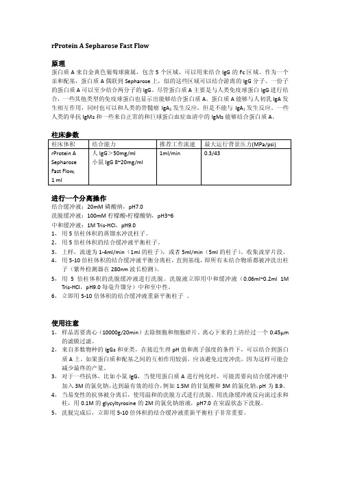
rProtein A Sepharose Fast Flow原理蛋白质A来自金黄色葡萄球菌属,包含5个区域,可以用来结合IgG的Fc区域。
作为一个亲和配基,蛋白质A偶联到Sepharose上,似的这些区域可以结合游离的IgG分子。
一份子的蛋白质A可以至少结合两分子的IgG。
尽管蛋白质A主要是与人类免疫球蛋白IgG进行结合,一些其他类型的免疫球蛋白也显示出能够结合蛋白质A。
蛋白质A能够与人初乳IgA发生相互作用,同时也可以和人类的骨髓瘤IgA2发生反应,但是不能与IgA1发生反应。
一些人类的单抗IgMs和一些来自正常的和巨球蛋白血症血清中的IgMs能够结合蛋白质A。
进行一个分离操作结合缓冲液:20mM磷酸纳,pH7.0洗脱缓冲液:100mM柠檬酸-柠檬酸钠,pH3~6中和缓冲液:1M Tris-HCl,pH9.01,用5倍柱体积的蒸馏水冲洗柱子。
2,用5倍柱体积的结合缓冲液平衡柱子。
3,上样,流速为1-4ml/min(1ml的柱子),或者5ml/min(5ml的柱子)。
收集流穿片段。
4,用5-10倍柱体积的结合缓冲液平衡分离柱,直到基线,即所有未结合物质都被冲洗出柱子(紫外检测器在280nm波长检测)。
5,用5倍柱体积的洗脱缓冲液进行洗脱。
洗脱液立即用中和缓冲液(0.06ml~0.2ml 1M Tris-HCl,pH9.0每毫升馏分)中和至中性。
6,立即用5-10倍体积的结合缓冲液重新平衡柱子。
使用注意1,样品需要离心(10000g/20min)去除细胞和细胞碎片。
离心下来的上清经过一个0.45μm 的滤膜过滤。
2,来自多数物种的IgGs和亚类,在接近生理pH值和离子强度的条件下,可以结合到蛋白质A上。
如果蛋白质和配基之间的互相作用较弱,应该避免过度冲洗,因为这样可能会减少最终的产量。
3,对于一些抗体,比如小鼠IgG,当使用蛋白质A进行纯化时,可能需要向结合缓冲液中加入3M的氯化钠,达到最有效的结合,例如1.5M的甘氨酸和3M的氯化钠,pH为8.9。


Protein A与Protein G一、Protein A和Protein G应用蛋白A、蛋白G是微生物来源的天然蛋白质,可以和哺乳动物的免疫球蛋白结合,基于这一特性,蛋白A、蛋白G的主要应用是在:1 纯化抗体: 将这些蛋白偶联于支持物上,提供了纯化抗体的有效方法。
2 免疫沉淀(immunoprecipitation): 利用蛋白A、蛋白G和免疫球蛋白的Fc段的结合,把Ag-Ab复合物分离出来,从而达到从蛋白复合物中分离出特定蛋白进行下一步的研究分析或者研究蛋白质-蛋白质之间的相互作用。
3 去除或减少非特异性背景:如免疫沉淀技术中在加入特异性抗体前加protein A/G beads到细胞裂解液中,以减低背景。
蛋白A、蛋白G与不同的免疫球蛋白的结合能力并不一样,它们受到后者的来源及亚类的影响。
目前,绝大多数的IgG纯化使用Protein A、Protein G亲和层析,它们是来自于不同微生物的蛋白质,对于IgG具有特异性的亲和作用,而对于其他杂蛋白没有或者只有很弱的结合,因此具有极高的选择性。
通常,仅凭Protein A、Protein G一步亲和层析就可达到90%的纯度。
Protein A和Protein G的多个结构域可与IgG结合二、它俩跟免疫球蛋白结合的情况Protein A :Protein A是一种金黄色葡萄球菌细胞壁蛋白质,与多数哺乳动物的IgG Fc段结合(主要是IgG,包括:人、山羊、绵羊、兔、豚鼠、马、猪、猴、小鼠等),不与狗IgG结合、不结合人IgM、IgD、和IgA。
因而,将Protein A 与琼脂糖凝胶以一定的方式结合,可制备用于抗体纯化的亲和填料。
天然的Protein A,42kDa,含有四个Ig Fc结合位点,其中2个是有活性的,A,无动物来源组分,载量约30mg人IgG/ml。
而用于纯化抗体的,一般是重组的protein A,含有4个Ig Fc区域结合位点。
Protein G:是由G群链球菌株产生的一种细胞壁成分,可结合很多种免疫球蛋白的Fc部分。
Protein A/G MagBeads Cat. No. L00277Technical Manual No. TM0249 Version 08212013 Index1.Product Description2. Instruction For Use3.Troubleshooting4. General Information1.Product Description1.1Intended UseGenScript Protein A/G MagBeads are ideal for small‐scale antibody purification and immunoprecipitation (IP) of proteins, protein complexes or other antigens.1.2PrincipleThe sample containing antibody is added to the Protein A/G MagBeads. The antibody will bind to beads during a short incubation. Then the beads‐bound antibody may be eluted from the beads by using a magnetic separation rack, or used for immunoprecipitation (IP). A cross‐linking procedure may be needed before IP to prevent co‐elution of the primary antibody. Magnetic separation eliminates the changes of micro tubes, minimizes the loss of sample and removes excessive steps of traditional centrifugation method.1.3Description of MaterialMaterial SuppliedGenScript Protein A/G MagBeads are super paramagnetic beads of average 40 μm in diameter, covalently coated with recombinant Protein A/G. The beads are supplied as 25% slurry in phosphate buffered saline (PBS), pH 7.4, containing 20% ethanol. The Protein A/G MagBeads have a binding capacity of more than 10 mg Goat IgG per 1 ml settled beads (e.g. 4 ml 25% slurry).Protein A/G is a genetically engineered protein (MW≈43 kDa) that combines the IgG binding sites of both Protein A and Protein G. 6×His‐tag was attached to its N‐terminal to facilitate the purification. The secreted Protein A/G contains four Fc‐binding domains from Protein A and two from Protein G, making it a more universal tool to bind and purify immunoglobulins.Cat. No. L00277 Size: 2 ml.Additional Material RequiredMixing/Rotation DeviceMagnetic Separation RackTest tubes and pipettesBuffers and solutions (see below)Additional Buffers NeededBinding/Wash Buffer: 20 mM Na2HPO4, 0.15 M NaCl, pH 7.0Elution Buffer: 0.1 M glycine, pH 2‐3Neutralization Buffer: 1 M Tris, pH 8.51×SDS Sample Buffer: 62.5 mM Tris‐HCl (pH 6.8 at 25°C), 2% w/v SDS, 10% glycerol, 50 mM DTT,0.01% w/v bromophenol blue2.Instruction For UseThe protocol uses 100 μl Protein A/G MagBeads, this may be scaled up or down accordingly.2.1Preparation of the MagBeadspletely resuspend the beads by shaking or vortexing the vial.2.Transfer 100 μl beads into a clean tube.3.Place the tube on a magnetic separation rack to collect the beads. Remove and discard the supernatant.4.Add 1 ml Binding/Wash Buffer to the tube and invert the tube several times to mix. Use the magnetic separationrack to collect the beads and discard the supernatant. Repeat this step twice.2.2Separation of Target IgG1.Resuspend the beads in 100 μl Binding/Wash Buffer.2.Add the sample containing target IgG to the tube and gently invert the tube to mix.3.Incubate the tube at room temperature with mixing (on a shaker or rotator) for 30 – 60 minutes.e the magnetic separation rack to collect the beads and discard the supernatant. If necessary, keep thesupernatant for analysis.5.Add 1 ml Binding/Wash Buffer to the tube and mix well, use the magnetic separation rack to collect the beads anddiscard the supernatant. Repeat the wash step three more times.6.Proceed to elution of isolated IgG (Section 2.3).2.3Elution of Isolated IgG1.Add 100 μl Elution Buffer to the tube and mix well. Incubate for five minutes at room temperature with occasionalmixing.e the magnetic separation rack to collect the beads and transfer the supernatant that contains the eluted IgG intoa clean tube.3.Repeat Step 1 and 2 twice.4.Add 10 μl of Neutralization Buffer to each 100 μl eluate to neutralize the pH. If needed, perform a buffer exchangeby dialysis or desalting.2.4ImmunoprecipitationBound IgG will be co‐eluted along with the target when using elution methods. If the presence of IgG does not disturb desired detection system, go directly to section 2.4.2 below. For applications where co‐elution of the IgG is not desired, the primary IgG can be cross‐linked to the Protein A/G MagBeads as described in section 2.4.1 below.2.4.1Cross‐linking IgG to the Beads1.Add 1 ml 0.2 M triethanolamine, pH 8.2 to the Protein A/G MagBeads with immobilised IgG. Wash twice using themagnetic separation rack with 0.2 M triethanolamine, pH 8.2 as the washing buffer.2.Resuspend the beads in 1 ml of 20 mM dimetyl pimelimidate dihydrochloride (DMP) in 0.2 M triethanolamine, pH 8.2(5.4 mg DMP/ml buffer). This cross‐linking solution must be prepared freshly.3.Incubate the beads with rotational mixing for 30 minutes at room temperature. Use the magnetic separation rack tocollect the beads and discard the supernatant.4.Resuspend the beads in 1 ml of 50 mM Tris, pH 7.5 to stop the reaction and incubate for 15 minutes at roomtemperature with rotational mixing.e the magnetic separation rack to collect the beads and discard the supernatant.6.Wash the cross‐linked beads three times with 1 ml PBS, pH7.4.2.4.2Binding Antigen to the IgG Cross‐linked Beads1.Add sample containing target antigen to the beads. For a 100 kD protein, use a volume containing approximate 25 µgtarget antigen/ml beads to assure an excess of antigen. If dilution of antigen is necessary, PBS or 0.1 M phosphate buffer (pH 7‐8) can be used as dilution buffer.2.Incubate with tilting and rotation for one hour at room temperature.3.Place the tube on the magnetic separation rack for 2 minutes to collect the IgG‐coated Beads‐target complex. Forviscous samples, double the time on the rack. Discard the supernatant.4.Wash the beads 3 times using 1 ml PBS.2.4.3Elution of Target ProteinA.Denaturing elution1.Place the tube from section2.4.2 on the magnetic separation rack to collect the beads and discard the supernatant.2.Add 100 µl 1XSDS Sample Buffer to the tube and mix well.3.Heat the tube at 100°C for five minutes.e the magnetic separation rack to collect the beads and transfer the supernatant containing desired sample into anew tube.5.Analyze the sample by SDS‐PAGE followed by Western blot analysis.B.Non‐denaturing elution1.Place the tube from section2.4.2 on the magnetic separation rack to collect the beads and discard the supernatant.2.Add 100 µl Elution Buffer to the tube and mix well. Incubate for five minutes at room temperature with occasionalmixing.e the magnetic separation rack to collect the beads and transfer the supernatant into a new tube.4.Repeat Step 2 and 3 twice.5.Add 10 μl Neutralization Buffer to each 100 μl of eluate to neutralize the pH.3.TroubleshootingReview the information below to troubleshoot your experiments using the GenScript Protein A/G MagBeads. Problem Possible Cause SolutionThe beads are difficult toimmobilize using the magneticseparation rack.Too many beads are used.Decrease the volume of MagBeadssuspension.A considerable amount of samplehas been added, but very fewspecific antibody of interest isdetected.The antibody of interest is at verylow concentration.Use a serum‐free medium for cellsupernatant samples.Affinity‐purify the antibody using itsspecific antigen coupled to anaffinity supporting material.The antibody of interest is purified,but it is degraded (as determined byloss of function in downstreamassay).The antibody is sensitive to low‐pHelution buffer.The downstream application issensitive to the neutralized elutionbuffer.Try another elution reagent, such as3.5 M MgCl2, 10 mM phosphate, pH7.2.Desalt or dialyze the eluted sampleinto a suitable buffer.No antibody is detected in anyeluate.The antibody in the sample cannotbind to Protein A/G.Try GenScript Protein A MagBeadsor Protein G MagBeads.4.General Information4.1Storage and StabilityThis product is stable until the expiration date stated on the COA, when stored unopened at 2–8°C. Do not freeze the product. Keep the MagBeads in liquid suspension during storage and all handling steps. Drying will cause loss of binding capacity and result in reduced performance. Resuspend the beads well before use. Be careful to avoid bacterial/fungal contamination.4.2Technical SupportPlease contact GenScript for further technical information (see contact details). Certificate of Analysis/Compliance is available upon request. The latest revision of the package insert/instructions for use is available on .4.3Warning and LimitationsThis product is for research use only. Not intended for any animal or human therapeutic or diagnostic use unless otherwise stated. This product contains 20 % EtOH as a preservative. Flammable liquid and vapor. Flash point 38°C. R‐10 flammable. Material Safety Data Sheet (MSDS) is available at .4.4Related MagBeads ProductsCat. No. Product NameL00273 Protein A MagBeadsL00274 Protein G MagBeadsL00295 Ni‐Charged MagBeadsL00327 Glutathione MagBeadsL00275 Mouse Anti‐His mAb MagBeadsL00336 Mouse Anti‐GST mAb MagBeadsGenScript USA Inc.860 Centennial Ave.,Piscataway, NJ 08854Toll‐Free: 1‐877‐436‐7274Tel: 1‐732‐885‐9188, Fax: 1‐732‐210‐0262Email: *********************Web: 。
NMab TM Pro Protein A亲和层析介质产品使用说明书文件编号:NM-W-DF-0603版本号:A2NMab TMPro 层析介质使用说明产品简介Protein A 亲和层析是利用Protein A 配基与目标抗体具有专一结合力作用从而达到分离纯化抗体的目的。
Protein A 亲和层析大大简化抗体下游分离纯化工艺,成为抗体分离纯化的标准。
目前市场上的Protein A 亲和层析介质主要分为以多糖(琼脂糖、葡聚糖、纤维素)为基质和以合成高分子(聚丙烯酸酯,丙烯酰胺)为基质两大类。
琼脂糖基质在溶胀状态下具有网状结构,比表面积大,因而亲和载量比较高,但机械强度不高、耐压低。
随着上游发酵技术的进步,抗体表达量越来越高,下游亲和捕获成为抗体生产瓶颈,因此对下游Protein A 亲和层析介质的载量要求越来越高。
为了因应这一需求,纳微科技以琼脂糖为基球,利用特有的微球改性技术以增强其机械强度,并结合自主产权的Protein A图1. NMab TM Pro Protein A 亲和层析介质电镜图配基技术,在NMab TM Protein A 的基础上成功开发出比其具有更高载量的NMab TM Pro Protein A 亲和层析介质。
除了高载量外,NMab TM Pro 延续了其优良的结合特异性、耐碱性以及压力-流速特性,是抗体客户降低单抗纯化成本的最佳选择。
下表1是NMab TM Pro Protein A 层析介质的基本性质参数。
相较市场同类产品有如下独特优势:(a) 载量高:一般单抗项目平均动态载量约60-80 mg/mL ;(b) 耐碱性强:0.5 M NaOH 浸泡下24小时载量只下降20%;(c) 配基脱落低:小于20 ppm ;(d) 宿主蛋白(HCP )残留低:一般在1000 ppm 以内,表现不亚于Prism A ; (e) 回收率高:大于90%;(f) 纯度高:亲和洗脱液纯度99 %以上;(g) 压力流速好:明显强于传统4FF/6FF 系列介质;表1. 纳微科技NMab TM Pro Protein A 亲和层析介质技术参数压力流速曲线测试不同流速下压力变化如下,此压力-流速特性可满足工业生产上对填料流速要求,优于传统4FF/6FF 软胶系列填料。
Protein G Sepharose Fast Flow
原理
蛋白质G ,来自G 链球菌组的细胞表面蛋白,是III Fc-受体型蛋白。
蛋白质G 通过非免疫机制结合抗体的Fc 区域。
蛋白质G 特异性的结合IgG 的Fc 区域,但是对于一些多克隆抗体IgGs 对人源的IgG 抗体能够有更强的结合。
在标准缓冲液条件下,蛋白质G 能够结合所有的人源性的亚类抗体和所有鼠源性的IgG 亚类抗体,包括鼠的IgG ,或者兔的IgGa 和IgGb 。
进行一个分离操作
结合缓冲液:20mM 磷酸纳,pH7.0
洗脱缓冲液:100mM 甘氨酸,pH2.7
中和缓冲液:1M Tris-HCl ,pH9.0
1, 用5倍柱体积的蒸馏水冲洗柱子。
2, 用5倍柱体积的结合缓冲液平衡柱子。
3, 上样,流速为1-4ml/min (1ml 的柱子),或者5ml/min (5ml 的柱子)。
收集流穿片段。
4, 用5-10倍柱体积的结合缓冲液平衡分离柱,直到基线,即所有未结合物质都被冲洗出柱
子(紫外检测器在280nm 波长检测)。
5, 用5倍柱体积的洗脱缓冲液进行洗脱。
洗脱液立即用中和缓冲液(0.06ml~0.2ml 1M
Tris-HCl ,pH9.0每毫升馏分)中和至中性。
6, 立即用5-10倍体积的结合缓冲液重新平衡柱子 。
使用注意
1, 样品需要离心(10000g/20min )去除细胞和细胞碎片。
离心下来的上清经过一个0.45μm
的滤膜过滤。
2, 来自多数物种的IgGs 和亚类,在接近生理pH 值和离子强度的条件下,可以结合到蛋白
质G 上。
对于特别物种IgG 的最佳结合条件,应该查阅近期的文献。
3, 当pH 值为2.7或者低于2.7时,大多数种类的免疫球蛋白能从蛋白质
G 上被洗脱下来。
4, 洗脱完成后,立即用5-10倍体积的结合缓冲液重新平衡柱子非常重要。
在通常使用的水溶性缓冲液中都是稳定的。
储存
用20%的乙醇在中性pH值的条件下冲洗介质和柱子,在4°~8°条件下储存。