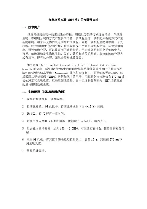
MTT分解比色法测定细胞生存和增殖
- 格式:pdf
- 大小:237.93 KB
- 文档页数:6
MTT reduction - a tetrazolium-based colorimetric assay for cell survival and proliferationContributed byJoan M. ChapdelainePharmakon Research International, Inc.Waverly, PA, 18471INTRODUCTIONIn 1983, a quantitative colorimetric assay for mammalian cell survival and cell prolifera-tion was proposed by Mosmann.1 The assay is dependent on the reduction of the tetrazo-lium salt MTT (3-(4,5-dimethylthazol-2-yl)-2,5-diphenyl tetrazolium bromide) by the mitochondrial dehydrogenase of viable cells to form a blue formazan product. The assay measures cell respiration and the amount of formazan produced is proportional to the number of living cells present in culture. The assay has been shown to be a simple, rapid alternative to counting cells by dye inclusion/exclusion, monitoring the release of 51Cr from lysed cells, or the incorporation of [3H]-thymidine into cellular DNA. The MTT assay has been used with a growing number of cell types including primary cultured cells as well as established cell lines. This colorimetric microplate assay is cost effectivebecause of the number of tests which can be performed at one time without the problem of radio-isotope and contaminated materials disposal.Applications of the MTT assayThe use of the colorimetric assay for cell growth and cell survival offers major advantages in speed, simplicity, cost, and safety over conventional cell counting assays. Figure 1 shows in a block diagram form how the MTT assay can be substituted for the thymidine uptake assay. The MTT assay has fewer steps, uses fewer materials, and does not carry the added burden of radioactive waste disposal.Application Note5The sensitivity of the MTT assay is dependent on the cell type, their average metabolic status, and the technique selected for solubilizing the formazan crystals. For example, the MTT assay was shown to detect as few as 200 EL4.3 cells per well and was linear to 50,000 cells per well.1 Whereas the sensitivity for T-cell blasts, which produce less forma-2The MTT assay, like the dye exclusion assay, must be optimized for seeding conditions and assay duration to obtain satisfactory results. A standard curve for formazan production as a function of the cell number and the growth rate should be determined for each cell type. Once optimized the MTT assay has proven to be adaptable to the needs of the exper-imental design and has been shown to be a practical method in a variety of applications: 1Mosmann1 reported using the MTT assay to measure proliferative lymphokines, mito-gen stimulations, and complement-mediated lysis.2Cell activation levels have been quantified independent of proliferation.33Cell viability assays could be run more rapidly using MTT rather than counting cells using trypan blue exclusion.44This colorimetric assay had been used with anti-proliferative monoclonal antibodies,5 growth factors,2,6 and chemotherapeutic screening.7, 85Tada et.al.6 showed the quantitative assay for interleukin 2 using the MTT assay to be equivalent to the [3H]-thymidine incorporation assay. The mean of the standard errors for the MTT assay was 3.9% as compared to 3.5% observed in the [3H]-thymidine incorporation assay and the results from the two assays were shown to agree.METHODS AND MODIFICATIONS Table 1 is a summary of various MTT colorimetric assay methods. The MTT assay as developed by Mosmann1 uses the same standard 96 well flat bottom tissue culture plate for the initial incubation of the cells and assaying for the cell number. At the end of the ini-tial incubation, a small volume of an MTT solution was added to each well of the culture plate. The plate was incubated to allow the formazan crystals to accumulate. The crystals were solubilized by the addition of an acidic alcohol solution before making an optical density measurement.To obtain complete mixing of the acidic alcohol solution with the media, the liquid in each wells had to be manually agitated with a pipet. The alcohol solubilized the formazan crys-tals and the acid lowered the pH of the medium to minimize interference due to the phenol red indicator which would interfere with the formazan reading. The yellow, acidic form ofthe dye does not interfere. The plates were read at 570 nm with a 630 nm reference to negate the effect of cell debris and precipitated proteins which may be produced by the acidic alcohol addition.Table 1: Summary table of MTT colorimetric assay methods.Denizot and Lang 2 found the tetrazolium dye assay as described by Mosmann to be less sensitive than the [3H]-thymidine uptake assay for their application. They modified the MTT method to improve sensitivity and performance. They used a microplate with a smaller well volume, doubled the MTT concentration, and dissolved it in a serum free media without the phenol red indicator. The unreacted MTT was removed from the centri-fuged plate before solubilizing the formazan crystals with isopropanol. Removing the serum proteins and the phenol red indicator reduced the background optical density and the need for an acidic solvent, respectively. Finally, a different reference wavelength, 690 nm, was selected which increased the signal by about 25%. The authors report that these modifications improved the sensitivity of the MTT assay 2- to 4-fold over the Mosmann procedure.T. Mosmann 1F. Denizot & R. Lang 2 H. Tada et.al. 6JChapdelaine Unpublished M.B. Hansen,et al. 9 J. Carmichael, et al. 7Sample V olume 0.1 mL 0.1 mL 0.1 mL 0.2 mL 0.1 mL 0.2 mL Intermediate Steps None Remove medium None None None ul None MTT V olume 10 µL 10 µL 20 µL 20 µL 25 µL 50 µL Concentratio n 5 mg/mL 5 mg/mL 5 mg/mL 10 mg/mL 5 mg/mL 2 mg/mL MTT Incubation4 Hours3 Hours4.5 Hours4 Hours2 Hours4 HoursAdherent CellsNonadherent Cells Intermediate StepsNoneCentrifuge microplates, remove unreacted MTTNoneRemove 180 µL mediumNoneRemove all mediumCentrifuge microplates, remove 170 µL mediumSolubilize Formazan V olume 150 µL 50 µL 100 µL 180 µL 100 µL 100 µL 100 µL SolventIsopropanol/HCl Isopropanol10% SDS in 0.01 M HCl10% SDS20% SDS in 50% DMF, pH 4.7Mineral oilDMSOIncubation 5 Minutes 1 Minute with shaking Overnight at 37°C Overnight at37°C Overnight at 37°C Overnight at 40°C 5 Minutes Read 570 - 630 nm 560 - 690 nm590 nm570 - 650 nm 570 nm 565 nm540 nmComments:•Serum proteins precipitate with acid/alcohol 1,2,6 •Mechanical mixing required to solubilize formazan 1,6 •Plates read with in 1 hour 1•Use half-area microplates 2 •Use serum free mediumto eliminateproteinprecipitation 2 •No phenol red 2•Use a detergent todissolveformazan 6 •Good mixing without added agitation 6 •Stable color 6 •Less protein precipitation 6•Higher detergent concentration •No acid to precipitate proteins•Phenol red in medium is not a problem since medium is removed•Improved solubilization of formazan crystals 9•More signal relative to acid/alcoholor acid/SDS solubilization 9 •Improved signal relative to alcoholsolubilization 7 •Stable color 7 •Lessreproducible 7 •Signalintensitydepends onresidual medium volume 7•Unstable color7Tada et. al.6 re-examined the MTT assay in detail and modified the assay to make it possi-ble to measure a larger number of samples at one time with less labor and with greateraccuracy. The MTT concentration was doubled and the incubation time was increased bya half an hour. The most significant modification was in how the formazan crystals weredissolved. In place of the acidic alcohol solution, an acidic detergent solution was substi-tuted. This modification resolved two drawbacks with the previously described MTTassay. First, the acidic detergent solution mixed well with the growth medium thus elimi-nating the need to mix each well and, second, the removal of the alcohol reduced the like-lihood of serum protein precipitation. Variations of this method have since beendeveloped. Chapdelaine (unpublished) removes the medium from the well of the tissueculture plate before adding the detergent solution, thus eliminating the need to acidify themedium. Hansen et. al.9 used a buffered detergent and added dimethyl formamide (DMF)to improve the solubilization of the formazan crystals.Carmichael et. al.7,8 modified the MTT assay and developed two protocols: one for adher-ent and the other for nonadherent cell lines. For adherent cells, the media was aspiratedfrom the wells and mineral oil was used to dissolve the formazan crystals because it is rel-atively nontoxic. The plates were read at 570 nm. Plates with nonadherent cells were cen-trifuged after the incubation with the MTT. The medium was aspirated from the wellsleaving about 30 µL behind. The formazan crystals were solubilized with dimethyl sulfox-ide (DMSO) which rapidly solubilized the formazan crystals but produced an unstablecolor. The absorbance plate was read using a 540 nm filter within minutes. CONCLUSION A problem with the MTT assay, the difficulty of solubilizing the formazan crystals with-out precipitation of serum proteins, may be mitigated by using more effective solubilizingagents and alternate procedures. Precipitated protein and cellular debris present in the cul-ture plate wells are a potential source of experimental error because they interfere with theoptical readings. Dual wavelength photometry, reading the plates at a principle and a ref-erence wavelength, is used to cancel out the effect of the debris. If the interfering materialshifts position between readings, the benefit of dual wavelength photometry is lost. TheMAXline microplate readers do not move the microplate during the read thus reducing thechance of debris shifting. The result is better precision and an assurance that the micro-plate reader is not a source of experimental variation.REFERENCES1Mosmann, T. Rapid colorimetric assay for cellular growth and survival: Application toproliferation and cytotoxicity assays. J. Immunol. Methods 65: 55-63 (1983).2Denizot, F. and Lang, R. Rapid colorimetric assay for cell growth and survival. Modi-fication to the tetrazolium dye procedure giving improved sensitivity and reliability.J. Immunol. Methods 89:271-277 (1986).3Gerlier, D. and Thomasset, T. Use of MTT colorimetric assay to measure cell activa-tion. J. Immunol. Methods 94:57-63 (1986).4Penit, C. and Papiernik, M. Regulation of thymocyte proliferation and survival bydeoxynucleosides. Deoxycytidine produced by thymic accessory cells protect thy-mocytes from deoxyguanosine toxicity and stimulates their spontaneous proliferation.Eur. J. Immunol. 16:257-263 (1986).5Vaickus, L. and Levy, R. Antiproliferative monoclonal antibodies: Detection and ini-tial characterization. J. Immunol. 135:1987-1997 (1985).6Tada, H., Shiho, O., Kuroshima, K., Koyama, M., and Tsukamato, K. An improvedcolorimetric assay for interleukin 2. J. Immunol. Methods 93:157-165 (1986).7Carmichael, J., DeGraff, W.G., Gazdar, A.F., Minna, J.D., and Mitchell, J. B. Evalua-tion of a tetrazolium-based semiautomated colorimetric assay. Assessment ofchemosensitivity testing. Cancer Res. 47:936-942 (1987).8Carmichael, J., DeGraff, W.G., Gazdar, A.F., Minna, J.D., and Mitchell, J. B. Evalua-tion of a tetrazolium-based semiautomated colorimetric assay. Assessment of radiosen-sitivity. Cancer Res. 47: 943-946 (1987).9Hansen, M.B., Nielsen, S.E., and Berg, K. Re-examination and further development ofa precise and rapid dye method for measuring cell growth/cell kill. J. Immunol. Meth-ods 119:203-210 (1989).0120-0114A TRADEMARKS: SPECTRAmax, SPECTRAplate, and MAXline are trademarks of Molecular Devices Corporation. SOFTmax is a registered trademark of Molecular Devices Corporation..。