1细胞生物学英文索引
- 格式:doc
- 大小:52.50 KB
- 文档页数:4
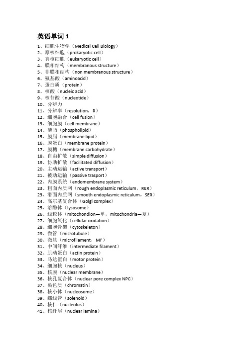
1、细胞生物学(Medical Cell Biology)2、原核细胞(prokaryotic cell)3、真核细胞(eukaryotic cell)4、膜相结构(membranous structure)5、非膜相结构(non membranous structure)6、氨基酸(aminoacid)7、蛋白质(protein)8、核酸(nucleic acid)9、核苷酸(nucleotide)10、分辨力11、分辨率(resolution,R)12、细胞融合(cell fusion)13、细胞膜(cell membrane)14、磷脂(phospholipid)15、膜脂(membrane lipid)16、膜蛋白(membrane protein)17、膜糖(membrane carbohydrate)18、自由扩散(simple diffusion)19、协助扩散(facilitated diffusion)20、主动运输(active transport)21、被动运输(passive trasport)22、内膜系统(endomembrane system)23、粗面内质网(rough endoplasmic reticulum,RER)23、滑面内质网(smooth endoplasmic reticulum,SER)24、高尔基复合体(Golgi complex)25、溶酶体(lysosome)26、线粒体(mitochondion—单,mitochondria—复)27、细胞氧化(cellular oxidation)28、细胞骨架(cytoskeleton)29、微管(microtubule)30、微丝(microfilament,MF)31、中间纤维(intermediate filament)32、肌动蛋白(actin protein)33、马达蛋白(motor protein)34、细胞核(nucleus)35、核膜(nuclear membrane)36、核孔复合体(nuclear pore complex NPC)37、染色质(chromatin)38、核小体(nucleosome)39、螺线管(solenoid)40、核仁(nucleolus)41、核纤层(nuclear lamina)42、核基质(nuclear matrix)43、核骨架(nuclear skeleton)44、遗传密码(genetic code)45、核糖体(ribosome)46、细胞连接(cell junction)46、封闭连接(occluding junction)46、锚定连接(黏着连接,桥粒连接)(anchoring junction)黏着带,黏着斑,桥粒,半桥粒46、通讯连接(communicating junction)47、细胞粘附(cell adhesion)48、整联蛋白(integrin)49、细胞外基质(extracellular matrix,ECM)50、透明质酸(hyaluronic acid,HA)51、胶原(collagen)52、弹性蛋白(elastin)53、信号转导(signal transdution)54、配体(ligand)55、受体(receptor)56、激酶(kinase)57、细胞周期(cell cycle)58、有丝分裂(mitosis)59、减数分裂(meiosis)60、联会(synapsis)61、周期蛋白(cyclin)62、周期蛋白依赖性激酶(cyclin—dependent kinase,Cdk)63、成熟促进因子(maturation promoting factor,MPF)64、细胞分化(cell differentiation)65、细胞决定(cell determination)66、管家基因(housekeeping gene)66、奢侈基因(luxury gene)67、胚胎诱导(embryonic induction)68、细胞衰老(cellular aging,cell senescence)69、细胞凋亡(apoptosis)70、细胞坏死(necrosis)71、干细胞(stem cell)71、全能干细胞(totipotent stem cell)72、多能干细胞(pluripotent stem cell)73、单能干细胞(unipotent stem cell)74、胚胎干细胞(embryonic stem cell)75、干细胞巢(stem cell niche)。

细胞生物学英文注释及名词解释细胞生物学英文注释及名词解释1.Cell biology:细胞生物学以“完整细胞的生命活动”为着眼点,从分子、亚细胞、细胞和细胞社会的不同水平,用动态的和系统的观点来探索和阐述生命这一基本单位的特性。
2.RNA interference:通过促使特定基因的miRNA降解来高效、特异地阻断体内特定基因表达,这种现象称为RNA干扰。
3.Cell membrane:细胞膜是包围在细胞质表面的一层薄膜,又称质膜(plasma membrane)。
4.Lipid rafts:由于鞘脂的脂肪酸尾比较长,因此这一区域比膜的其他部分厚,更有秩序且较少流动,被称为“脂筏”。
5.Endomembrane system:人们把细胞内在结构、功能以及发生上相互密切关联的其他所有膜性结构细胞器统称为内膜系统。
6.Endoplasmic reticulum(ER):在细胞质的内质区分布着一些由小管、小泡相互连接吻合形成的网状结构,称为内质网。
7.Molecular chaperone:热激蛋白虽然能够通过对其各自作用对象的识别、结合来协助它们的折叠组装和转运,但其本身却不参与最终作用产物的形成,也不会改变其自身的基本分子生物学特性,由此被称之为分子伴侣。
8.Signal peptid:指导蛋白多肽链在糙面内质网上进行合成的决定因素,是被合成肽链N 端的一段特殊氨基酸序列,即信号肽。
9.Nuclear import signal:核输入信号是指凡是在细胞质中合成的核蛋白质,其肽链中均含有由7个氨基酸组成的特异性信号序列,负责分拣并指导蛋白质从细胞质通过核孔复合体输入到细胞核内。
又称为核定位信号(nuclear localization signal,NLS)10.Golgi complex (高尔基复合体)11.Lysosome (溶酶体)12.Peroxisome (过氧化物酶体)13.Cytoskeleton:细胞骨架是指真核细胞中与保持细胞形态结构和细胞运动有关的纤维网络,包括微管、微丝、中间丝。

1.I n te rmedia te Filaments (~10 nm)1.Intro1.Main function: enable cells to withstand the mechanical stress that occurs when cells are stretched.2.toughest and most durable of the three types of cytoskeletal filaments.3.Can be found in cytoplasm as well as the nucleus. A mesh of intermediate filaments the nuclear lamina,underlies and strengthens the nuclear envelope in all eucaryotic cells.2.Intermediate filaments are strong and ropelike1.Elongated fibrous protein, each composed of an N-terminal globular head, a C-terminal globular tail, and acentral elongated rod domain, is the subunit of intermediate filaments. The rod domain consists of anextended α-helical region that enables pairs of intermediate filament proteins to form stable dimers bywrapping around each other in a coiled-coil configuration. Two of these coiled-coiled dimers then associateby noncovalent bonding to form a tetramer. The tetramers then bind to one another end to end and side byside, and also by noncovalent bonding, to generate the final ropelike intermediate filament. Eight tetramersare twisted into a ropelike filament.2.the globular head and tail regions, which are exposed on the surface of the filament, allow it to interact withother components of their cytoplasm. The globular domains vary greatly in both size and amino acidsequence from one intermediate filament protein to another.3.Intermediate filaments strengthen cells against mechanical stress1.Present in large numbers: along the length of nerve cell axons, providing essential internal reinforcement tocell extensions; in muscle cells and in epithelial cells.2.In all these cells, intermediate filaments, by stretching and distributing the effect of locally applied forces,keep cells and their membranes from breaking in response to mechanical shear.3.Intermediate filaments can be grouped into four classes: (1)keratin filaments in epithelial cells, hair andnails; (2)vimentin and vimentin-related filaments in connective-tissue cells, muscle cells, and supportingcells of the nervous system (glial cells); (3)neurofilaments in nerve cells; and (4)nuclear lamins, whichstrengthen the nuclear membrane of all animal cells4.Keratin filaments typically span the interiors of epithelial cells from one side of the cell to the other, andfilaments in adjacent epithelial cells are indirectly connected through cell-cell junctions called desmosomes.This cabling of high tensile strength, formed by the filaments through the epithelial sheet, distributes thestress that occurs when the skin is stretched.5.Many intermediate filaments are further stabilized and reinforced by accessory proteins, such as plectin, thatcross-link the filament bundles into strong arrays. In addition to holding together bundles of intermediatefilaments (particularly vimentin), these proteins link intermediate filaments to microtubules, to actinfilaments, and to adhesive structures in the desmosomes.4.The nuclear envelope is supported by a meshwork of intermediate filaments1.Intermediate filaments lining and strengthening the inside surface of the inner nuclear membrane areorganized as a two-dimensional mesh. The intermediate filaments within this tough nuclear lamina areconstructed from a class of intermediate filament proteins called lamins.2.Intermediate filaments of the nuclear lamina disassemble and re-form at each cell division, when the nuclearenvelope breaks down during mitosis and then re-forms in each daughter cell.3.Disassembly and reassembly of the nuclear lamina are controlled by the phosphorylation anddephosphorylation of the lamins by protein kinases. When the lamins are phosphorylated, the consequentconformational change weakens the binding between the tetramers and causes the filament to fall apart.Dephosphorylation at the end of mitosis causes the lamins to reassemble.4.Defects in a particular nuclear lamin are associated with certain types of progeria - rare disorders that causeaffected individuals to appear to age prematurely.2.M icrotubule s1.Intro1.Microtubules are long and stiff hollow tubes of protein that can rapidly disassemble in one location andreassemble in another.2.In a typical animal cell, microtubules grow out from a small structure near the center of the cell calledcentrosome.3.Extending out toward the cell periphery, they create a system of tracks within the cell, along which vesicles,organelles, and other cell components are moved. These and other systems of cytoplasmic microtubules arethe part of the cytoskeleton mainly responsible for anchoring membrane-enclosed organelles within the celland for guiding intracellular transport.4.When a cell enters mitosis, the cytoplasmic microtubules disassemble and then reassemble into an intricatestructure called the mitotic spindle. The mitotic spindle provides the machinery that will segregate thechromosomes equally into the two daughter cells just before a cell divides.5.Microtubules can also form permanent structures called cilia and flagella as a means of propulsion or tosweep fluid over the cell surface. The core of a eucaryotic cilium or flagellum consists of a highly organized and stable bundle of microtubules.2.Microtubules are hollow tubes with structurally distinct ends1.Microtubules are built from subunits-molecules of tubulin-each of which is itself a dimer composed of twovery similar globular proteins called α-tubulin and β-tubulin, bound tightly together by noncovalentbonding.2.The tubulin dimers stack together, again by noncovalent bonding, to form the wall of the hollow cylindricalmicrotubule. This tubelike structure is made of 13 parallel protofilaments, each a linear chain of tubulindimers with α- and β-tubulin alternating along its length. Each protofilament has a structural polarity, with α-tubulin exposed at one end and β-tubulin at the other, and this polarity is the same for all theprotofilaments giving a structural polarity to the microtubule as a whole. β-tubulin end = plus end; α-tubulin end = minus end.3.In vitro, tubulin dimers will add to either end of a growing microtubule, although more rapidly to the plusend than the minus end. This polarity is crucial both of the assembly of microtubules and for their role once they are formed. If they had no polarity, they could not serve their function in defining a direction forintracellular transport, for example.3.The Centrosome is the major microtubule-organizing center in animal cells1.Microtubules are formed by outgrowth from centrosome, which is typically close to the cell nucleus whenthe cell is not in mitosis. It organized the array of microtubules that radiates outward from it through thecytoplasm. Centrosomes contain hundreds of ring-shaped structures formed from another type of tubulin, γ-tubulin ring serves as the starting point, or nucleation site, for the growth of one microtubule. The αβ-tubulin dimers add to the γ-tubulin ring in a specific orientation, with the result that the minus end of each microtubule is embedded in the centrosome and growth occurs only at the plus end.2.The centrosome in most animal cells also contains a pair of centrioles, each made of a cylindrical array ofshort microtubules.3.It is much harder to start a new microtubule from scratch, by first assembling a ring of αβ-tubulin dimers,than to add such dimers to a preexisting microtubule structure. By providing organizing centers containing nucleation sites, and keeping the concentration of free αβ-tubulin dimers low, cells can thus control where microtubules form.4.Growing microtubules show dynamic instability1.Dynamic instability stems from the intrinsic capacity of tubulin molecules to hydrolyze GTP. Each freetubulin dimer contains one tightly bound GTP molecule that is hydrolyzed to GDP (still tightly bound)shortly after the subunit is added to a growing microtubule.2.When polymerization is proceeding rapidly, tubulin molecules add to the end of the microtubule faster thanthe GTP they carry is hydrolyzed. The end of a growing microtubule is therefore composed entirely of GTP-tubulin subunits, forming what is know as a GTP cap. In this situation, the growing microtubule willcontinue to grow. Because of the randomness of chemical processes, however, it will occasionally happen that tubulin at the free end of the microtubule hydrolyzes its GTP before the next tubulin has been added, so that the free ends of protofilaments are now composed of GDP-tubulin subunits. This change tips thebalance in favor of disassembly. Because the rest of the microtubule is composed of GDP-tubulin, oncedepolymerization has started, it will tend to continue, often at a catastrophic rate; the microtubule starts to shrink rapidly and continuously, and may even disappear.5.Microtubules are maintained by a balance of assembly and disassembly1. A microtubule growing out from the centrosome can be prevented from disassembling if its plus end issomehow permanently stabilized by attachment to another molecule or cell structure so as to prevent tubulin depolymerization. If stabilized by attachment to a structure in a more distant region of the cell, themicrotubule will establish a relatively stable link between that structure and the centrosome.2.If a cell in mitosis is exposed to the drug colchicine, which binds tightly to free tubulin and prevents itspolymerization into microtubules, the mitotic spindle rapidly disappears and the cell stalls in the middle of mitosis, unable to partition its chromosomes into two groups.3.The drug taxol has the opposite action at the molecular level. It binds tightly to microtubules and preventsthem from losing subunits. Because new subunits can still be added, the microtubules can grow but notshrink.6.Microtubules organize the interior of the cell1.As cells enter mitosis, microtubules become more dynamic to enable them to disassemble rapidly and thenreassemble into the mitotic spindle. When a cell has differentiated into a specialized cell type and taken on a definite fixed structure, the dynamic instability is often suppressed. Stabilized microtubules serve tomaintain the organization of the cell.2.In the nerve cell, all the microtubules in the axon point in the same direction, with their plus ends toward theaxon terminal.7.Motor proteins drive intracellular transport1.Mitochondria and the smaller membrane-enclosed organelles and vesicles move in small, jerky steps. Thissaltatory movement is more sustained and directional than Brownian movement.2.Both microtubules and actin filaments are involved in saltatory movement. In both cases, the movements aregenerated by motor proteins, which use the energy derived from repeated cycles of ATP hydrolysis to travelsteadily along the actin filament or the microtubule in a single direction. At the same time, these motorproteins also attach to other cell components and thus transport this cargo along the filaments.3.The kinesins move toward the plus end of a microtubule (away from the centrosome), while the dyneinsmove toward the minus end (toward the centrosome). These kinesins and dyneins are both dimers with twoglobular ATP-binding heads and a single tail. The heads interact with microtubules in a stereospecificmanner, so that the motor protein will attach to a microtubule in only one direction. The tail of a motorprotein generally binds stably to some cell component, such as a vesicle or an organelle, and therebydetermines the type of cargo that the motor protein transports.anelles move along microtubules1.As the cell develops and the endoplasmic reticulum grows, kinesins attached to the outside of theendoplasmic reticulum membrane (via receptor proteins) pull it outward along microtubules, stretching itlike a net. Dyneins, similarly attached to the Golgi membranes, pull the Golgi apparatus the other way along microtubules inward toward the cell center.9.Cilia and flagella contain stable microtubules moved dynein1. A single cilium contains a core of stable microtubules, arranged in a bundle, that grow from a basal body inthe cytoplasm; the basal body serves as the organizing center for the cilium.2.Cilia move fluid over the surface of a cell or propel single cells through a fluid.3.The flagella are much like cilia in structure but are usually much longer.4. A cross section through a cilium shows nine doublet microtubules arrange in a ring around a pair of singlemicrotubules.5.The movement of a cilium of a flagellum is produced by the bending of its core as the microtubules slideagainst each other. Ciliary dynein generates the bending motion of the core.3.A c ti n filaments (~7 nm)1.Actin filaments are thin and flexible1.Each filament is a twisted chain of identical globular actin molecules, all of which “point” in the samedirection along the axis of the chain.2.Actin filament has a structural polarity, with a plus end and a minus end.2.Actin and tubulin polymerize by similar mechanisms1.Actin filaments can grow by the addition of actin monomers at either end, but the rate of growth is faster atthe plus end than at the minus end. A naked actin filament is unstable, and it can disassemble from bothends. Each free actin monomer carries ATP which is hydrolyzed to ADP soon after the incorporation of theactin monomer into the filament. Hydrolysis of ATP to ADP in an actin filament reduces the strength ofbinding between monomers and decreases the stability of the polymer. Nucleotide hydrolysis promotesdepolymerization, helping the cell to disassemble filaments after they have formed.2.Toxins such as cytochalasins prevent actin polymerization; toxins such as phalloidin stabilize actinfilaments against depolymerization.3.Many proteins bind to actin and modify its properties1.Cells contain small proteins, such as thymosin and profilin, that bind to actin monomers in the cytosol,preventing them from adding to the ends of actin filaments. These proteins play a crucial role in regulatingactin polymerization.2.When actin filaments are needed, proteins called formins and actin-related proteins (ARPs) both controlactin assembly at the advancing front of a migrating cell.4.An actin-rich cortex underlies the plasma membrane of most eucaryotic cells1.Actin is highly concentrated in cell cortex, a layer just beneath the plasma membrane. In this region, actinfilaments are linked by actin-binding proteins into a meshwork that supports the outer surface of the cell and gives it mechanical strength.5.Cell crawling depends on actin1.Cell crawling includes three essential processes: (1) the cell pushes out protrusions at its “front,” or leadingedge; (2) these protrusions adhere to the surface over which the cell is crawling; and (3) the rest of the celldrags itself forward by traction on these anchorage points.2.In the first step, the leading edge of a crawling fibroblast in culture regularly extends thin, sheetlikelamellipodia, which contain a dense meshwork of actin filaments, oriented so that most of the filamentshave their plus ends close to the plasma membrane. Many cells also extend thin, stiff protrusions calledfilopodia, both at the leading edge and elsewhere on their surface. Both lamellipodia and filopodia areexploratory, motile structures that form and retract with great speed. Both are thought to be generated by the rapid local growth of actin filaments, which assemble close to the plasma membrane and elongate by theaddition of actin monomers at their plus ends. In this way the filaments push out the membrane withouttearing it.3.Actin-related proteins (ARPS) promotes the formation of a web of branched actin filaments in lamellipodia.Formins attach to the growing ends of actin filament and promote the addition of new monomers to formstraight unbranched filaments.4.When the lamellipodia and filopodia touch down on a favorable patch of surface, they stick. Integrinsadhere to molecules in the extracellular matrix that surrounds cells or on the surface of a neighboring cellover which the moving cell is crawling. On the intracellular face of the crawling cell’s membrane, integrinscapture actin filaments, thereby creating a robust anchorage for the system of actin filaments inside thecrawling cell.6.Actin associates with myosin to form contractile structures1.Myosin bind to and hydrolyze ATP, which provides the energy for their movement along actin filamentsfrom the minus end of the filament toward the plus end.2.Myosin-I and myosin-II are most abundant. Myosin-I molecules have only one head domain and a tail.1.Overview of th e ce ll cycl e1.The eucaryotic cell cycle is divided into four phases1.2 most dramatic events: mitosis and cytokinesis. These two processes together constitute the M phase of thecell cycle.2.The period between one M phase and the next is called interphase. It consists of three phases: S phase (thecell replicates its nuclear DNA) and G1 and G2 phase (between S and M). During the gap phases, the cellmonitors the internal and external environments to ensure that conditions are suitable and its preparations are complete before it commits itself to S or M.3.During all of interphase, a cell generally continues to transcribe genes, synthesize proteins, and grow in mass.2.A cell-cycle control system triggers the major processes of the cell cycle1.The cell-cycle control system guarantees that the events of the cell cycle occur in a set sequence and that eachprocess has been complete before the next one begins. The cell-cycle control system achieves this bymolecular brakes that can stop the cycle at various checkpoints.2.3 checkpoints: one checkpoint operates in G1 and allows the cell to confirm that the environment is favorablefor cell proliferation before committing to S phase. If extracellular conditions are unfavorable, cells can delay progress through G1 and may even enter a specialized resting state know as G0. Another checkpoint operates in G2 and ensures that cells do not enter mitosis until damaged DNA has been repaired and DNA replication is complete. A third checkpoint operates during mitosis and ensures that the replicated chromosomes areproperly attached to a cytoskeletal machine, called the mitotic spindle, before the spindle pull thechromosomes apart and distributes them into the two daughter cells.2.Th e ce ll-cycle con tr ol sys tem1.The cell-cycle control system depends on cyclically activated protein kinases called Cdks1.The protein kinases at the core of cell-cycle control systems are present in proliferating cells throughout thecell cycle. The activity of each of these kinases rises and falls in a cyclical fashion.2.Switching the kinases on and off at the appropriate times is partly the responsibility of another set of proteinsin the control system-the cyclins. Cyclins have to bind to the cell-cycle kinases before the kinases canbecome enzymatically active. Kinases of cell-cycle are therefore called cyclin-dependent protein kinases (or Cdks).2.The activity of Cdks is also regulated by phosphorylation and dephosphorylation1.Cyclin concentrations increase gradually, but the activity of associated cyclin-Cdk complexes tends to switchon abruptly at the appropriate time in the cell cycle.2.For a cyclin-Cdk to be maximally active, the Cdk has to be phosphorylated at one site by a specific proteinkinase and dephosphorylated at other sites by a specific protein phosphatase.3.Different cyclin-cdk complexes trigger different steps in the cell cycle1.The cyclin that acts in G2 to trigger entry into M phase is called M cyclin, and the active complex it formswith its Cdk is called M-Cdk. Distinct cyclins, called S cyclins and G1/S cyclins, bind to a distinct Cdkprotein late in G1 to form S-Cdk and G1/S-Cdk, respectively, and trigger S phase. Other cyclins, called G1cyclins, act earlier in G1 and bind to other Cdk proteins to form G1-Cdks, which help drive the cell throughG1 toward S phase.4.The cell-cycle control system also depends on cyclical proteolysis1.The concentration of each type of cyclin rises gradually but falls sharply. This abrupt fall results from thetargeted degradation of the cyclin protein. Specific enzyme complexes add ubiquitin chains to the appropriate cyclin, which is then directed to the proteasome for destruction.5.Proteins that inhibit Cdks can arrest the cell cycle at specific checkpoints1.Some of the molecular brakes rely on Cdk inhibitor proteins that block the assembly or activity of one ormore cyclin-Cdk complexes.2.As a general rule, mammalian cells will multiply only if they are stimulated to do so by extracellular signalscalled mitogens produced by other cells. If deprived of such signals, the cell cycle arrests at a G1 check-point; and, if the cell is deprived for long enough, it will withdraw from the cell cycle and enter the non-proliferating state G0.3.The G1 checkpoint is sometimes called Start.3.S phas e1.S-Cdk initiates DNA replication and helps block re-replication1.DNA replication begins at origins of replication, nucleotide sequences that are scattered along eachchromosome. These sequences recruit specific proteins that control the initiation and completion of DNAreplication. One multiprotein complex, the origin recognition complex (ORC) remains bound to origins ofreplication throughout the cell cycle, where it serves as a sort of landing pad for additional regulatory proteins that bind before the start of S phase.2.One of the regulatory proteins, called Cdc6, is present at low levels during most of the cell cycle, but itsconcentration increases transiently in early G1. When Cdc6 binds to ORCs in G1, it promotes the binding of additional proteins to form a pre-replicative complex. Once the pre-replicative complex has been assembled, the replication origin is ready to “fire”. The activation of S-Cdk in late G1 then “pulls the trigger”, initiating DNA replication.3.S-Cdk also helps prevent re-replication of the DNA. Activated S-Cdk helps phosphorylate Cdc6, causing itand other proteins in the pre-replicative complex to dissociate from the ORC after an origin has fired. Thisdisassembly prevents replication from occurring again at the same origin. In addition to promotingdissociation, phosphorylation of Cdc6 by S-Cdk marks it for degradation, ensuring that DNA replication isnot reinitiated later in the same cell cycle.2.Cohesins help hold the Sister Chromatids of each replicated chromosome together1.The sister chromatids are held together by protein complexes called cohesins. Cohesins form protein ringsthat surround the two sister chromatids, keeping them united.3.DNA damage checkpoints help prevent the replication of damaged DNA1.DNA damage checkpoint in G1 is especially well understood. DNA damage causes an increase in both theconcentration and activity of a protein called p53, which is a transcription regulator that activates thetranscription of a gene encoding a Cdk inhibitor protein called p21. The p21 protein binds to G1/S-Cdk andS-Cdk, preventing them from driving the cell into S phase. The arrest of the cell cycle in G1 gives the celltime to repair the damaged DNA before replicating it. If the DNA damage is too severe to be repaired, p53can induce the cell to kill itself by undergoing apoptosis.2.If p53 is defective, the unrestrained replication of damaged DNA leads to a high rate of mutation and theproduction of cells that tend to become cancerous.3.The activity of cyclin-Cdk complexes is inhibited by phosphorylation at particular sites. For the cell toprogress into mitosis, M-Cdk has to be activated by the removal of these inhibitory phosphates by a specific protein phosphatase. When DNA is damaged, this activating protein phosphatase is itself inhibited, so theinhibitory phosphates are not removed from M-Cdk. As a result, M-Cdk remains inactive and M phase cannot be initiated until DNA replication is complete and any DNA damage is repaired.4.M phas e1.M-Cdk drives entry into M phase and mitosis1.M-Cdk triggers the condensation of the replicated chromosome into compact, rod-like structures, readingthem for segregation, and ti also induces the assembly of the mitotic spindle that will separate the condensed chromosomes and segregate them into the two daughter cells.2.M-Cdk activation begins with the accumulation of M cyclin. Synthesis of M cyclin starts immediately after Sphase. M-Cdk complexes, when they first form, are inactive. The sudden activation of a protein phosphatase (Cdc25) that removes the inhibitory phosphates holding M-Cdk activity in check.3.Once activated, each M-Cdk complex can indirectly activate more M-Cdk, by phosphorylating and activatingmore Cdc25. In addition, activated M-Cdk also inhibits the inhibitory kinase Wee1, further promoting theactivation of M-Cdk. The overall consequence is that, once the activation of M0Cdk begins, there is anexplosive increase in M-Cdk activity that drives the cell abruptly from G2 into M phase.2.Condensins help configure duplicated chromosome for separation1.Protein complexes, called condensins, help carry out this chromosome condensation. The M-Cdk thatinitiates entry into M phase triggers the assembly of condensin complexes onto DNA by phosphorylatingsome of the condensin subunits.2.Both cohesins and condensins form ring structures, and, together, the two types of protein rings help toconfigure the replicated chromosomes for mitosis.3.The cytoskeleton carries out both mitosis and cytokinesis1.The mitotic spindle is composed of microtubules and the various proteins that interact with them, includingmicrotubule-associated motor proteins.2.Contractile ring consists mainly of actin filaments and myosin filaments arranged in a ring around the equatorof the cell. It starts to assemble just beneath the plasma membrane toward the end of mitosis.3.Both structures rapidly disassemble after they have performed their tasks.4.M phase is conventionally divided into six stages1.The first five stages of M phase (prophase, prometaphase, metaphase, anaphase, and telophase) constitutemitosis. Cytokinesis constitutes the sixth stage, and it overlaps in time with the end of mitosis.2.Prophase=replicated chromosomes condense and mitotic spindle begins to assemble outside the nucleus.Prometaphase=the nuclear envelope breaks down, allowing the spindle microtubules to bind to thechromosomes. Metaphase=the mitotic spindle gathers all of the chromosomes to the center (equator) of thespindle. Anaphase=the two sister chromatids in each replicated chromosome synchronously split apart, andthe spindle draws them to opposite poles of the cell. Telophase=a nuclear envelope reassembles around each of the two sets of separated chromosome to form two nuclei. Cytokinesis begins in anaphase and continuesthrough telophase. It is when nucleus and cytoplasm of each of the daughter cells return into interphase,signaling the end of M phase.1.E x tr ace ll ular ma tr ix and connec ti ve ti ssue s1.Intro1.The strength of plant tissue comes from the cell walls, formed like boxes, that enclose, protect, and constrainthe shape of each of its cells.2.The cell wall is a type of extracellular matrix that the plant cell secretes around itself.2.Plant cells have tough external walls1.Osmotic swelling of the cell, limited by the resistance of the cell wall, can keep the chamber distended, and amass of such swollen chambers cemented together forms a semirigid tissue.2.Most newly formed cells in a multicellular plant initially make thin primary cell walls that can slowly expandto accommodate cell growth, the driving force for growth is the swelling pressure, called the turgor pressure, that develops as the result of an osmotic imbalance between the interior of the cell and its surroundings. Once growth stops and the wall no longer needs to expand, a more rigid secondary cell wall is often produced,either by thickening of the primary wall or by deposition of new layers with a different compositionunderneath the old ones.3.Cellulose microfibrils give the plant cell wall its tensile strength1.Plant cells derive their tensile strength from long fibers oriented along the lines of stress. In higher plants, thelong fibers are generally made from the polysaccharide cellulose, the most abundant organic macromolecule on Earth. These cellulose microfibrils are interwoven with other polysaccharides and some structuralproteins, to resists compression as well as tension. In woody tissue, a highly cross-linked network of ligninassociated with cellulose is deposited to make the matrix more rigid and waterproof.2.Because the cellulose microfibrils resist stretching, their orientation governs the direction in which thegrowing cell enlarges.3.Cellulose synthesis is mediated by cellulose synthase complexes. Cellulose is synthesized outside the cell, onthe outer surface of the cell by enzyme complexes embedded in the plasma membrane. These complexestransport sugar monomers across the membrane and incorporate them into a set of growing polymer chains at their points of membrane attachment. Each set of chains forms a cellulose microfibril.4.Just beneath the plasma membrane, microtubules are aligned exactly with the cellulose microfibrils outsidethe cell. These microtubules are thought to serve as tracks to guide the movement of the enzyme complexes.In this way, the cytoskeleton controls the shape of the plant cell and the modeling of plant tissues.4.Animal connective tissues consist largely of extracellular matrix1.4 major types of tissues in animals: connective, epithelial, nervous, and muscular.2.In connective tissues, extracellular matrix is plentiful and carries the mechanical load. In other tissues,extracellular matrix is scanty, and the cells are directly joined to one another and carry the mechanical loadthemselves.3.Animal connective tissues are enormously varied in strength and location. They can be tough and flexible liketendons and ligaments or dermis of the skin, hard and dense like bone, resilient and shock-absorbing likecartilage, or soft and transparent like the jelly that fills the interior of the eye.4.The tensile strength is chiefly provided by collagen.5.Collagen provides tensile strength in animal connective tissues1.Collagen molecule has a long, stiff, triple-stranded helical structure, in which three collagen polypeptidechains are wound around one another in a ropelike superhelix. These molecules in turn assemble into ordered polymers called collagen fibrils. These can pack together into still thicker collagen fibers.2.Connective-tissue cells that manufacture and inhabit the matrix go by various names: in skin, tendon, andmany other connective tissues they are called fibroblasts; int bone they are called osteoblasts. They makeboth the collagen and other organic components of the matrix. Almost all of these molecules are synthesized intracellularly and then secreted in the standard way, by exocytosis.3.Collagen avoids premature assembly before secreting by secreting procollagen, with additional peptides ateach end that obstruct assembly into collagen fibrils. Extracellular enzymes called procollagen proteinases。
Acetylcholine乙酰胆碱ACTH促肾上腺皮质激素Actin filament肌动微丝adenine (A) /腺嘌呤,Adenosine腺苷酸Adenylyl cyclase腺苷酸环化酶adhering junction 粘着连接adhesion plaque 粘着斑Aggregation聚合aleurone grain 糊粉粒allosome, heterochromosome 异染色体Allosteric变构的amino acids /氨基酸amyloplast 造粉体analytical cytology 分析细胞学Anaphase后期annulate lamella, annulate lamellae(复)环孔片层Antenna触须apoplast 质外体Apoptosis凋亡Arginine合成酶arm ratio 臂比Arterial动脉的Asian-Pacific Organization for Cell Biology, APOCB 亚洲及太平洋地区细胞生物学aster 星体astral ray, astral fiber 星射线又称“星体丝”。
astrocenter 星心体astrosphere 星体球Autophosphorylation自磷酸化autosome 常染色体axial filament 轴丝axon 轴突Barr body 巴氏小体basal granule, basal body 基粒basal lamina 基膜又称“基板”。
basement membrane 基底膜belt desmosome 带状桥粒biogenesis 生源论又称“生源说”。
Biotin生物素Bisphosphate二磷酸blepharoplast 生毛体Broadcast spawning/散卵生殖brush border 刷状缘Bud/出芽生殖Catalyze/ 催化cell biology 细胞生物学cell coat 细胞外被cell matrix 细胞基质cell membrane 胞膜cell morphology 细胞形态学cell pathology, cytopathology 细胞病理学cell physiology, cytophysiology 细胞生理学cell sociology 细胞社会学cell theory 细胞学说cell wall 细胞壁cell 细胞cellular immunology 细胞免疫学Cellular respirProkaryotes/原核细胞cellulose/纤维素,chitin/几丁质Central plug中央栓centric fusion 着丝粒融合centric split 着丝粒分裂centriole 中心粒centrodesm 中心体连丝centromere misdivision 着丝粒错分centromere plate 着丝粒板centromere 着丝粒染色体上的一段非编码的DNA,对动粒(或动粒蛋白)有组织和整合作用。
细胞分裂与细胞周期cell division 细胞分裂cell cycle 细胞周期mitosis 有丝分裂meiosis 减数分裂amitosis 无丝分裂prophase 前期prometaphase 前中期spindle 纺锤体kinetochore microtubule 动粒微管polar microtubule 极微管metaphase 中期anaphase 后期telophase 末期cytokinesis 胞质分裂contractile ring 收缩环meiosisⅠ第一次减数分裂meiosisⅡ第二次减数分裂synapsis 联会bivalent 二价体aster 星体tetrad 四分体synaptonemal complex,SC 联会复合体recombination nodule 重组小结chiasma 交叉chiasma terminalization 交叉端化direct division 无丝分裂interphase 分裂间期mitosis,M期分裂期cyclin 细胞周期蛋白cyclin-dependent kinase,Cdk细胞周期蛋白依赖性激酶Cdk inhibitor,CKI Cdk激酶抑制物maturation promoting factor,MPF成熟促进因子checkpoint 检测点growth factor 生长因子chalone 抑素V-oncogene 癌基因proto-oncogene 原癌基因cellular oncogene,C-onc 细胞癌基因anti oncogene 抑癌基因mitotic apparatus 有丝分裂器cytostatic factor,CSF 细胞静止因子细胞的内膜系统ribosome 核糖体endoplasmic reticulum, ER 内质网RER 粗面内质网SER 滑面内质网microsome 微粒体signal hypothesis 信号假说SRP 信号肽识别颗粒SRP-R 信号肽识别颗粒受体Golgi complex 高尔基复合体lysosome 溶酶体residual body 残余小体lipofusion 脂褐质siderosome 含铁小体multivesicular 多泡体myelin figure 髓样结构peroxisome 过氧化物酶体microbody 微体细胞膜与物质的穿膜运输membrane lipid 膜脂phospholipid 磷脂cholesterol 胆固醇glycolipid 糖脂intrinsic protein 内在蛋白integrator protein 整合蛋白transmembrane protein 跨膜蛋白extrinsic protein 外在蛋白peripheral protein 周边蛋白lipid anchored protein 脂锚定蛋白lipid-linked protein 脂连接蛋白cell coat 细胞外被glycocalyx 糖萼liquid-crystal state 液晶态lateral diffusion 侧向扩散 flip-flop 翻转运动 rotation 旋转运动 flexion 弯曲运动 microdomain 微区 lipid rafts 脂筏 passive diffusion 被动扩散 membrane transport protein 膜运输蛋白 carrier protein 载体蛋白 channel protein 通道蛋白 facilitated diffusion 易化扩散 P-class ion pump P-型离子泵 symport 共运输 antiport 对向运输 aquaporin ,AQP 水孔蛋白 vesicular transport 小泡运输 endocytosis 胞吞作用 exocytosis 胞吐作用 microvillus 微绒毛 cilia 纤毛 flagella 鞭毛 ruffle 褶皱 lamellipodium 片状伪足细胞衰老与细胞死亡 cell senescence 细胞衰老 cell death 细胞死亡 Hayflick life span Hayflick 界限 senescence-associated β- galactosidase ,SA-βgal β-半乳糖苷酶 senescence associated gene 衰老相关基因 longevity gene 抗衰老基因 premature senescence 早熟性衰老 necrosis 细胞坏死 apoptosis 细胞凋亡 apoptotic bodies 凋亡小体 programmed cell death, PCD 程序性细胞死亡 phosphatidylserine, PS reactive oxygen species, ROS 活性氧类物质 permeability transition pores, PT pores 线粒体渗透转变孔 anoikis 失巢凋亡 autophagy 细胞自噬 micro-autophagy 微自噬 macroautophagy 巨自噬 chaperone-mediated autophagy,CMA 分子伴侣介导的自噬 cell shrinkage 细胞皱缩 chromatin condensation 染色质凝聚 DNA ladders DNA 梯状条带细胞生物学的研究方法resolution ,R 分辨率 light microscopy 光学显微镜 fixation 固定 embedding 包埋 section 切片 staining 染色 phase contrast microscope 相差显微镜 Fluorescence microscope 荧光显微镜 fluorescence microscope 荧光显微镜transmission electron microscope, TEM透射电子显微镜ultrastructure 超微结构submicroscopic structures 亚显微结构 Scanning electron microscope (SEM ) 扫描电子显微镜 flowcytometer, FCM 流式细胞技术 cell fractionation 细胞分级分离 sedimentation coefficient ,S 沉降系数 Differential centrifugation 差速离心 velocity sedimentation 速度沉降 isodensity centrifugation 等密度离心 column chromatography 柱层析 in vitro cell culture 体外细胞培养 primary culture 原代培养 secondary culture 继代培养 explan 外植体 primary culture 原代培养物 cell line 细胞系cell strain 细胞株cell clone 细胞克隆cell fusion 细胞融合cell hybridization 细胞杂交natural fusion 自然融合artificial induced fusion 人工诱导融合cytochemistry 细胞化学技术enzyme-cytochemistry 酶细胞化学immunocytochemistry 免疫细胞化学细胞的基本概念与分子基础procaryotic cell 原核细胞nucleoid 拟核cell wall 细胞壁mycoplasma 支原体bacteria 细菌peptidoglycan 肽聚糖plasmid 质粒mesosome 中间体eucaryotic cell 真核细胞cell membrane 质膜cytoplasm 细胞质nucleus 细胞核virus 病毒viroid 类病毒prion 朊病毒线粒体outer membrane 外膜inner memebrane 内膜intermembrane space 膜间隙matrix 基质porin 孔蛋白cristae 嵴elemetary particle 基粒intermembrane space 膜间隙Tim 内膜转位子Tom 外膜转semiautonomous organelle半自主性细胞器endosymbiosis theory 内共生起源学说non-endosymbiosis theory非内共生起源学说cellular respiration 细胞呼吸chemiosmotic hypothesis 化学渗透假说ATP synthase ATP合酶mPT 线粒体通透性改变mitochondrial disorders 线粒体疾病细胞骨架和细胞运动cytoskeleton 细胞骨架microfilament 微丝microtubule 微管intermediate filament 中间纤维actin 肌动蛋白globular actin, G-actin 球状肌动蛋白filamentous actin, F- actin纤维状肌动蛋白tonfilament 张力丝myofilament 肌丝neurofilament 神经丝nucleation phase 成核阶段elongation phase 延长阶段steady-state phase 稳定期阶段phalloidin 鬼笔环肽cytochalasin 细胞松弛素microfilament-associated protein微丝结合蛋白myosin 肌球蛋白microvilli 微绒毛stress fiber 应力纤维tubulin 微管蛋白microtubule associated protein, MAP微管结合蛋白protofilament 原纤维assembly 组装disassembly 去组装microtubule organizing center MTOC微管组织中心centrosome 中心体centriole 中心粒dynamic instability 动态不稳定性colchicine 秋水仙素taxol 紫杉酚kinesin 驱动蛋白dynein 动力蛋白intermediate filament,IF 中间纤维tetramer 四聚体cyto-keratin 胞质角蛋白skeleton 骨骼蛋白intermediate filament associated protein,IFAP 中间纤维的结合蛋白cell motility 细胞运动细胞核cell nucleus 细胞核nuclear envelope 核膜inner nuclear membrane 内核膜outer nuclear membrane 外核膜perinuclear space 核周隙nuclear pore complex, NPC 核孔复合体karyophilic protein 亲核蛋白nuclear localization signal, NLS核定位信号exportin 输出蛋白chromatin 染色质chromosome 染色体genome 基因组unique sequence 单一序列middle repetitive sequence中度重复序列highly repetitive sequence高度重复序列replication origin 复制源centromere 着丝粒telomere 端粒histone 组蛋白core histone 核小体组蛋白linker histone 连接组蛋白nonhistone 非组蛋白Euchromatin 常染色质heterochromatin 异染色质barr body 巴氏小体nucleosome 核小体satellite 棒状小体细胞分化Cell Differentiation 细胞分化totipotent nucleus 全能性细胞核cell determination 细胞决定transdetermination 转决定stability 稳定性dedifferentiation 去分化transdifferentiation 转分化cellular reprogramming 细胞重编程induced pluripotent stem cells, iPS细胞诱导多能干细胞differential expression 差异表达housekeeping protein 管家蛋白luxury protein 奢侈蛋白luxury gene 奢侈基因polyploidy 多倍体polyteny 多线体maternal effect gene, MEG 母体效应基因temporal specificity 时间特异性stage specificity 阶段特异性locus control region, LCR 基因座控制区spatial specificity 空间特异性master control gene 细胞分化主导基因combinatory control 组合调控homeobox 同源异形框histone code 组蛋白密码microRNA 微小RNA embryonic induction 诱导或胚胎诱导regeneration 再生。
第一章 绪论cellcell biology cell theory central dogma cytomics cytoplasmelectron microscope ,EM epigenetics gene细胞细胞生物学 细胞学说 中心法则 细胞组学 细胞质 电子显微镜 表观遗传 基因genomicshuman genome project,HGP medical cell biology model animal noncoding RNA proteomics stem cell biology translational medicine基因组学人类基因组计划医学细胞生物学 模式动物 非编码RNA 蛋白质组学 干细胞生物学 转化医学第二章细胞的概念和分子基础anticodon archaea archaebacteria bacteria cell membrane chlamydia codon cytoplasm cytosol deoxyribonucleic acid,DNAenzyme eukarya eukaryotic cell glycolipids glycoproteins message RNA , mRNAmicroRNA, miRNA mycoplasma反密码子 古菌域 古细菌 细菌 细胞膜 衣原体 密码子 细胞质 细胞质溶胶 脱氧核糖核酸 酶 真核域 真核细胞 糖脂 糖蛋白 信使核糖核酸 微小RNA 支原体 nucleoid nucleus peptide plasmid prion prokaryotic cell protoplasm ribonucleic acid ,RNA ribosomeribosome RNA , rRNA ribozyme RNA silencingsmall nuclear RNA, snRNA structural domains transfer RNA, tRNA viroid virus 拟核细胞核 肽 质粒 朊病毒 原核细胞 原生质 核糖核酸 核糖体 核糖体RNA 核酶RNA 沉默 小核RNA 结构域 转运RNA 类病毒 病毒第三章细胞生物学研究方法Abbe limitagaroseatomic force microscope,AFM autoradiographybiochipcell culturecell free systemcell linecell strainChIPAbbe 限度 琼脂糖 原子力显微镜放射性自显影技术 生物芯片 细胞培养 非细胞体系 细胞系 细胞株 染色质免疫沉淀技术 CLIPcolumn chromatography cytochemistry technique differential centrifugation diffraction pattern DNA denaturation dNTPelectrospray ionization mass spectrometry,ESI-MS embedding紫外交联免疫沉淀技术 柱层析细胞化学技术 差速离心 X-射线衍射图 DNA 变性4种脱氧核苷酸 电喷雾电离质谱 包埋emission lightemzyme cytochemistryequilibrium sedimentation excitation lightfixationflow cytometerfluorescence microscope fluorescence resonanceemergy transfer,FRETfreeze-etchfreeze-fracturegene chipgreen fluorescentprotein,GFPhigh performance liquidchromatography,HPLCimmunocytochemistry,ICC immunomagnetic microsphere immuno-precipitation,IPin situ hybridization,ISHisoelectric focusinglaser capturemicrodissection,LCMmatrix-assisted laserdesorptionionization/time-of-flight , MALDI-TOF-MSmetal shadowingNorthern blotnuclear magnetic resonance spectroscopy,NMR over-expression phage display发射光 酶细胞化学技术 平衡沉降激发光 固定 流式细胞仪 荧光显微镜荧光共振能量转移 冰冻蚀刻 冰冻断裂 基因芯片 绿色荧光蛋白 高效液相层析 免疫细胞化学技术 免疫磁珠免疫沉淀 原位杂交技术 等点聚焦 激光俘获显微切割电泳 基质辅助激光解吸/电离飞行时间质谱金属投影法 Northern 印迹杂交 核磁共振光谱过表达 噬菌体展示polymerase chainreaction,PCR primary cultureprobeprotein chipproteomeproteomicsquantitative PCR ,qPCR radioisotoperenaturationresolutionrestriction nucleasescanning electronmicroscope,SEM scanning tunnelingmicroscope,STM SDS polyacrylamid gel electrophoresissecondary culturesectionsingle molecular fluorescence imaging southern blot stainingtandem massspectrometry,MS/MStransfectiontransmission electron microscope,TEM velocity sedimentation yeast two-hybridzation phase contrast microscope聚合酶链式反应原代培养 探针 蛋白质芯片 蛋白质组 蛋白质组学 荧光实时定量PCR 反应放射性同位素 复性 分辨率 限制性内切酶 扫描电子显微镜扫描隧道显微镜SDS-聚丙烯酰胺凝胶电泳 传代培养 切片 单分子荧光成像 Southern 印迹杂交染色 串联质谱 转染透射电子显微镜速度沉降 酵母双杂交 相差显微镜第四章细胞膜与物质的穿膜运输active transport adaptinantiportaquaporin ,AQPbilayerbiomembranecarrier proteincell coatcell membrane主动运输衔接蛋白 反向运输 双分子层 水孔蛋白 生物膜 载体蛋白 细胞外被 细胞膜 channel proteincholesterol clathrin coated pits coated vesicle constitutive secretion endocytosis exocytosis extrinsic protein通道蛋白 网格蛋白 胆固醇 有被小窝 有被小泡 连续性分泌 胞吞作用 胞吐作用 膜外在蛋白facilitated diffusion fluid mosaic model glycolipid intrinsic proteinligand-gated channel lipid raftslipid anchored protein lipid-linked protein lamella structure model membrane lipid membrane proteinmembrane transport protein micellepassive diffusion passive transport 易化扩散 流动镶嵌模型 糖脂膜内在蛋白 配体门控通道 脂筏脂锚定蛋白 脂连接蛋白 片层结构模型 膜脂 膜蛋白 膜运输蛋白 球状分子团 被动扩散 被动运输 peripheral protein phagocytosis phospholipid pinocytosis plasma membrane regulated secretion simple diffusionstress-activated channel symporttransmembrane protein unit membraneunit membrane model vesicular transport voltage-gated channel外周蛋白 吞噬作用 磷脂 胞饮作用 质膜 受调分泌 简单扩散 应力激活通道 同向运输 穿膜蛋白 单位膜 单位膜模型 小泡运输 电压门控通道第五章内膜系统annulate lamellaeautophagic lysosomecalreticulincis Golgi networkcisternaeclathrin-coated vesiclecontinuous secretioncotranslation insertiondiscontinuous secretionendoplasmic reticulumgated transportglucose regulated protein94,GRP94 glycosylationGolgi complexheavy-chain bindingprotein,BiP heterophagic lysosome internal signal peptideinternal start-transfer peptidelysosomemedial Golgi stackmolecular chaperonemyeloid bodyN-linked glycosylationphagolysosome孔环状片层 自噬溶酶体 钙网素 顺面高尔基网 扁平囊泡 网格蛋白有被囊泡 连续分泌 共翻译插入 非连续分泌 内质网 门孔运输 葡萄糖调节蛋白94糖基化 高尔基复合体 重链结合蛋白 异噬溶酶体内信号肽 内开始转移肽 溶酶体 高尔基中间膜囊 分子伴侣 髓样体 N-连接糖基化 异噬溶酶体 phospholipid exchangeproteins,PEP primary lysosome protein disulfide isomerase,PDI retention protein reticulo-plasmin rough endoplasmic reticulum ,RER sarcoplasmic reticulum secondary lysosome signal hypothesis signal patch signal peptide signal recognition particle,SRPSRP-receptor,SRP-R smooth endoplasmic reticulum ,SERtarget-SNAREs,t-SNAREs tertiary lysosome trans Golgi network translocontransmembrane transport unfolded protein response, UPR磷脂交换蛋白初级溶酶体蛋白二硫键异构酶驻留蛋白 网质蛋白 糙面内质网肌质网 次级溶酶体 信号肽假说 信号斑 信号肽信号识别颗粒信号识别颗粒受体 光面内质网靶SNAREs 三级溶酶体 反面高尔基网 转运体 穿膜运输未折叠蛋白反应vacuoles vesicle vesicles 大囊泡囊泡小囊泡vesicle-SNAREs,v- SNAREsvesicular transport囊泡SNAREs囊泡转运第六章线粒体与细胞的能量转换ATP synthase complex biological oxidation cellular oxidation cellular respiration chemiosmotic coupling hypothesiscristaeelementary particle glycolysisinner membrane intercristae space intermembrane space intracristae spacematrixmatrix spacematrix-targeting sequence,MTS ATP合酶复合体生物氧化细胞氧化细胞呼吸化学渗透假说嵴基粒糖酵解内膜嵴间腔膜间腔嵴内空间基质基质腔基质导入序列mitochondrial disordersmitochondrial phaseouter membraneoxidative phosphorylationpostmitochondrial phasepremitochondrial phasereactive oxygen species, ROSsubstrate-levelphosphorylationtranslocation contact sitetranslocon of the innermembrane,Timtranslocon of the outermembrane,Tomtricarboxylic acid cycle,TCAcycle线粒体疾病线粒体期外膜氧化磷酸化线粒体后期线粒体前期活性氧底物水平磷酸化转位接触点内膜转位子外膜转位子三羧酸循环第七章细胞骨架与细胞的运动actincell cortexcochicine contractile ring cytochalasin B cytoskeletondynamic instability model dyneinfilopodiaintegrin intermediate filaments, IF kinesin lamellipodia microfilaments , MF microfilament associated protein,MAP 肌动蛋白细胞皮层秋水仙素收缩环细胞松弛素B细胞骨架非稳态动力学模型动力蛋白丝状伪足整合蛋白中间纤维驱动蛋白片状伪足微丝微丝结合蛋白microtubule organizing center,MTOCmicrotubules, MTmicrotubule-associadedprotein ,MAPmyosinnucleation phasephalloidinpolymerization phasesliding filament modelsteady state phasetaxoltreadmilling modeltubulinvinblastine微管组织中心微管微管结合蛋白肌球蛋白成核期鬼笔环肽聚合期滑动丝模型稳定期紫杉醇踏车模型管蛋白长春新碱第八章细胞核acrocentric chromosome annular subunitbandcentral domaincentral plug centromerechromatidchromatin chromosomecolumn subunit constitutive heterochromatin cytoplasmic ringdense fibrillar component,DFCeuchromatinexportin facultative heterochromatinfibrillar center,FCfish-trapgenomegranular component,GC heterochromatin nucleosomenucleusouter nuclear membrane pairing domain perinuclear space primary constriction replication origin satellitescaffold radial loop structure model 近端着丝粒染色体环状亚单位带型中央结构域中央栓着丝粒染色单体染色质染色体柱状亚单位组成性异染色质胞质环致密纤维组分常染色质输出蛋白兼性异染色质纤维中心捕鱼笼基因组颗粒中心异染色质核小体细胞核外核膜配对结构域核周间隙主溢痕复制源随体染色体骨架-放射环模型histonekaryophilic proteinsecondary constrictionsolenoidkaryotypekinetochorekinetochore domainluminal subunitmetacentric chromosomeminibandmultiple coiling modelnuclear localization signal,NLSnuclear matrixnuclear matrix associatedproteinnuclear matrix proteinnuclear porenuclear pore complex,NPCnuclear ringnucleolar cyclenucleolar matrixnucleolar organizernucleolar organizing regionnucleolusnucleosomal histonespokesubmetacentric chromosomesupersolenoidtelocentric chromosometelomere组蛋白亲核蛋白次溢痕螺线管核型动粒动粒结构域腔内亚单位中着丝粒染色体微带多级螺旋化模型核内定位信号核基质核基质结合蛋白核基质蛋白核孔核孔复合体核质环核仁周期核仁基质核仁组织者核仁组织区核仁核小体组蛋白辐亚中着丝粒染色体超螺线管端着丝粒染色体端粒第九章基因信息的传递与蛋白质合成activating domain adaptive expression alternative splicing aminoacyl site aminoacyl-tRNA antibiotics anticodon antisense strand 转录激活域适应性表达可变剪接A位点氨酰-tRNA抗生素反密码子反义链CAAT boxcentral dogmachaperoninschromatin remoldingcis-acting elementcoding regioncoding strandcodonCAAT盒中心法则伴侣素染色质重塑顺式作用元件编码区编码链密码子commalessconstitutive expressionconstitutive splicingdegeneracydirectionDNA binding domainenhancerexonGC boxgenegene clustergene familygeneral transcription factor genetic codegenomehelix-loop-helix ,HLHhelix-turn-helix ,HTHheterogeneous nuclear RNA ,hnRNAhighly repetitive sequence inducible geneintronleucine zippermiddle repetitive sequence negative regulationnon-coding regiontranslocationtranspeptidaseubiquitin-proteasomeunique sequenceuniversalwobblezinc finger连续性 组成性表达 常规剪接 简并性 方向性 DNA 结合域 增强子 外显子 GC 盒 基因 基因簇 基因家族 通用转录因子遗传密码 基因组 螺旋-环-螺旋 螺旋-转角-螺旋 不均一核RNA高度重复序列可诱导基因 内含子 亮氨酸拉链 中等重复序列负性调控 非编码区 转位 转肽酶 泛素- 蛋白酶体 单一序列 通用性 摆动性 锌指 operonpeptide formation peptidyl-tRNA site polyribosome positive regulation pribnow box Promoter Registerregulatory gene repetitive sequence repressible gene repressor remodelerribosome circulation sense strandShine-Dalgarno sequence silencer small nuclearribonucleoprotein particle ,snRNPspatial specificity spliceosome split gene structural gene sumoylation TATA box template strand temporal specificity terminatortrans-acting factor transcriptiontranscription factor translation操纵子 成肽 P 位点多聚核糖体 正性调控 Pribnow 盒 启动子 进位 调控基因 重复序列 可阻遏基因 阻遏蛋白 染色质重建子 核糖体循环 有意义链 SD 序列 沉默子核糖核蛋白颗粒空间特异性 剪接体 断裂基因 结构基因 sumo 化 TATA 盒 模板链 时间特异性 终止子反式作用因子 转录 转录因子 翻译第十章细胞连接与细胞黏附adhering junctionadhesion beltanchoring junctioncadherincell adhesioncell adhesion molecule ,CAMcell junctionchemical synapse黏着连接 黏着带 锚定连接 钙黏着蛋白 细胞黏附 细胞黏附分子 细胞连接 化学突触 claudincommunicating junction connexon desmosomedesmosome junction electric coupling electronic synapse filamin密封蛋白 连接连接子 桥粒 桥粒连接 电耦联 电突触 细丝蛋白focal adhesiongap junction hemidesmosome heterophilic binding homophilic binding immunoglobin-superfamily, Ig-SF inside outintegrinintracellular anchor protein linker-dependent binding metabolic coupling epithelial-mesenchymal 黏着斑间隙连接半桥粒异亲型结合同亲型结合免疫球蛋白超家族由内向外整联蛋白细胞内锚定蛋白连接分子依赖性结合代谢耦联上皮-间质转型通讯neural cell adhesionmolecule,N-CAMoccludinoccluding junctionoutside inplectinselectinsynapsetalintransmembrane adhesionprotein神经细胞黏附分子闭合蛋白封闭连接由外向内网蛋白选择素突触踝蛋白穿膜黏连蛋白transition,EMT第十一章细胞外基质及其与细胞的相互作用anchorage dependent growth anoikisbasal lamina,basement membrane,BM chondroitin sulfate,CS collagencollagen disease collagenasecore proteindermatan sulfate,DS elastaseelastinextracellular matrix, ECM fibronectin, FN glycosaminoglycan, GAG 锚定依赖性生长失巢凋亡基膜硫酸软骨素胶原胶原病胶原酶核心蛋白硫酸皮肤素弹性蛋白酶弹性蛋白细胞外基质纤连蛋白糖胺聚糖glycosyltransferasesheparan sulfate,HSheparinhyaluronic acid,HAkeratan sulfate,KSlaminin,LNmatrix metalloproteinases,MMPnidogen,entactinperlecanproteoglycan,PGsyndecantriple helixtropoelastin糖基转移酶硫酸乙酰肝素肝素透明质酸硫酸角质素层粘连蛋白基质金属蛋白酶巢蛋白渗滤素蛋白聚糖连接素三股螺旋可溶性弹性蛋白原第十二章细胞的信号转导adenylate cyclase, AC calmodulin,CaM cAMP-dependent protein kinase A,PKA cascadecGMP depedent protein kinase G,PKG cyclic AMP,cAMP cyclic GMP,cGMP diacylglycerol,DAG effector protein 腺苷酸环化酶钙调蛋白cAMP依赖蛋白激酶A级联反应cGMP依赖蛋白激酶G环磷酸腺苷环磷酸鸟苷二酰基甘油效应蛋白first messengerG proteinG protein linked receptorguanylate cyclase,GCinositol trisphosphate,IP3insulinion channel receptorligandprotein tyrosine kinase,PTKreceptorsecond messenger第一信使G蛋白G蛋白偶联受体鸟苷酸环化酶三磷酸肌醇胰岛素离子通道受体配体酪氨酸蛋白激酶受体第二信使serine/threonine kinases ,STKsignal transduction丝氨酸/苏氨酸蛋白激酶 信号转导 signaling networktyrosine-specific protein kinase receptor ,TPKR信号网络酪氨酸蛋白激酶型受体第十三章细胞分裂与细胞周期amitosis anaphase anti oncogene aster bivalentCdk inhibitor ,CKI cell cycle cell divisioncellular oncogene ,C-onc centrosome chalone checkpoint chiasmachiasma terminalization contractile ring cyclincyclin-dependent kinase ,CDKcytostatic factor,CSF direct division growth factor无丝分裂 后期 抑癌基因 星体 二价体Cdk 激酶抑制物 细胞周期 细胞分裂 细胞癌基因 中心体 抑素 检测点 交叉 交叉端化 收缩环 细胞周期蛋白细胞周期蛋白依赖性激酶细胞静止因子 直接分裂 生长因子indirect divisioninterphasekinetochore microtubule maturation promotingfactor ,MPF meiosismetaphasemitosismitosis,Mmitotic apparatuspolar microtubuleprophaseproto-oncogenerecombination nodulespindlesynapsissynaptonemal complex ,SC telophasetetradV-oncogene ,V-onc间接分裂 分裂间期 动粒微管成熟促进因子减数分裂 中期 有丝分裂 分裂期 有丝分裂器 极微管 前期 原癌基因 重组小结 纺锤体 联会 联会复合体 末期 四分体 癌基因 第十五章细胞分化apical ectodermal cap cell determination cell differentiation cellular reprogramming cleavagecombinatory control compensatory regeneration dedifferentiation differential expression ectodermembryonic induction endoderm顶端外胚层帽 细胞决定 细胞分化 细胞重编程 卵裂 组合调控补偿性再生 去分化 差异表达 外胚层 胚胎诱导 内胚层epimorphosis regeneration gastrulation genomic imprinting grafting experiment histone code homeobox homeobox gene homeodomain homeodomain protein homeosis homeotic gene housekeeping gene微变态再生 原肠形成 基因组印记 胚胎移植实验 组蛋白密码 同源异形框 同源异形框基因 同源异形结构域 同源异形域蛋白 同源异形转变 同源异形基因 管家基因induced pluripotent stem cells juxtacrine interaction lateral inhibitionlocus control region,LCR long non-coding RNA,lncRNAluxury geneluxury proteinmaster control gene maternal effect gene,MEG mesoderm metamorphosis microRNA, miRNA morphallaxis regeneration non-coding RNA诱导多能干细胞,iPS细胞近分泌相互作用侧向抑制基因座控制区长链非编码RNA奢侈基因奢侈蛋白细胞分化主导基因母体效应基因中胚层变态微小RNA变形再生非编码RNAorganogenesisparacrine factorpluripotent cellregenerationregeneration blastemasmall interfering RNA, siRNAsomatic recombinationspatial specificitystage specificitytemporal specificitytotipotent celltotipotent nucleustransdeterminationtransdifferentiationunipotency器官发生旁分泌因子多能(干)细胞再生再生胚芽小干扰RNA体细胞重组阶段特异性空间特异性时间特异性全能(干)细胞全能性细胞核转决定转分化单能第十六章细胞衰老与细胞死亡anoikisanti-apoptosis gene apoptosis apoptotic bodies autophagosome autophagycell deathcell senescencecell shrinkage chromatin condensation ClassIIIPI3Kcytochrome C, cyt C death receptor, DR DNA ladders 失巢凋亡抗凋亡基因细胞凋亡凋亡小体自噬泡细胞自噬细胞死亡细胞衰老细胞皱缩染色质凝聚III型磷脂酰肌醇三磷酸激酶细胞色素C死亡受体DNA梯状条带DNA stainabilityendonucleaseHayflick life spaninterleukin-1β convertingenzyme, ICEnecrosisnerve growth factor receptor,NGFRpermeability transition pores,PT poresphosphatidylserine, PSprogrammed cell death, PCDreactive oxygen species, ROStumor necrosis factor receptor,TNFRDNA可染性核酸内切酶Hayflick 界限白细胞介素-1β转换酶细胞坏死神经生长因子受体渗透转变孔磷脂酰丝氨酸程序性细胞死亡活性氧类物质肿瘤坏死因子受体第十七章干细胞与组织的维持与再生adult stem cell asymmetry division cancer stem cell, CSC chimeracorneal stem cell directly induced differentiation 成体干细胞不对称分裂癌干细胞嵌合体角膜干细胞直接诱导分化dedifferentiationembryoid body, EBembryonic carcinoma cell,ECembryonic germ cell,EGembryonic stem cell,ESepidermal stem cell去分化类胚体胚胎癌细胞胚胎生殖细胞胚胎干细胞皮肤干细胞hematopoiesis stem cell ,HSCinner cell mass ,ICMintegral membrane protein induced pluripotent stem cellintegrinintestinal stem cellliver stem celllung stem cellmarrow cavitymesenchymal stem cell, MSCmultipotent stem cellmyogenic stem cellneural stem cells, NSCpancreatic stem cellplasticitypluripotent stem cell造血干细胞 内细胞团 整合膜蛋白诱导多能干细胞,iPS细胞 整联蛋白 小肠干细胞 肝干细胞 肺脏干细胞 骨髓腔 间充质干细胞 专能干细胞 肌肉干细胞 神经干细胞 胰腺干细胞 可塑性 多能干细胞 pluripotenyprogenitor cell renal stem cell self-maintenance somatic stem cellspermatogonial stem cell stage-specific embryonic antigen,SSEA stem cell stem cell niche symmetry division tissue specific stem cell totipotencytotipotent stem cell transdetermination transdifferentiation unipotency多能 前体细胞 肾脏干细胞 自稳定性 成体干细胞 精原干细胞胚胎阶段特异性抗原干细胞 干细胞巢 对称分裂组织特异性干细胞 全 能 全能干细胞 转决定 转分化 专 能。
细胞生物学英语单词细胞的基本结构细胞膜Cell Membrane细胞质cytoplasm细胞核nucleus膜性结构membranous structure生物膜biological membrane单位膜unit membrane双亲媒性分子amphipathic molecules分子团micelle双分子层bilayer脂质体liposome整合蛋白integral proteins边周蛋白Peripheral proteins细胞外被cell coat流动镶嵌模型fluid mosaic model脂筏lipid raft内膜系统endomembrane system内质网endoplasmic reticulum, ER粗面型内质网rough endoplasmic reticulum,RER 滑面型内质网smooth endoplasmic reticulum,SER 微粒体microsome高尔基体Golgi complex, GC溶酶体lysosome过氧化物酶体Peroxisome微体microbody线粒体Mitochondrion基粒elementary particle (ATP synthase)基质导入序列matrix targeting sequence, MTS分子伴侣molecular chaperone核糖体Ribosome多聚核糖体polyribosome细胞骨架Cytoskeleton原纤维protofilaments微管蛋白tubulin微管相关蛋白microtubule-associated protein, MAP 单管singlet二联管doublet三联管triplet纺锤丝--动态微管dynamic microtubule鞭毛--稳定微管stable microtubule空间:MTOC,microtubule organizing center中心外周物质pericentriolar material,PCM微丝microfilament肌动蛋白纤维actin filament中间丝Intermediate filament氨基聚糖Glycosaminoglycan,GAG细胞运输被动运输passive transport单纯扩散Simple diffusion膜通道蛋白channel protein化学门控通道ligand-gated channel电闸门控通道voltage-gated channel帮助扩散Facilitated diffusion主动运输active transport伴随运输co-transport胞吞作用endocytosis胞吐作用exocytosis胞饮作用Pinocytosis吞噬作用Phagocytosis受体介导的胞吞作用receptor-mediated endocytosis 网格蛋白clathrin胞吐作用exocytosis结构性分泌途径constitutive exocytosis pathway调节性分泌途径regulated exocytosis pathway门控转运gated transport信号肽signal peptide信号斑signal patch分子伴侣molecular chaperone信号假说signal hypothesis信号识别颗粒Signal recognition particle信号肽假说signal peptide hypothesis起始转运信号start-transfer signal终止转运信号stop-transfer signal核定位信号nuclear localization signal, NLS动位蛋白dynein驱动蛋白kinesin肌球蛋白myosin6-磷酸甘露糖M6P膜流membrane flow细胞信号传导信号转导signal transduction受体receptor受体酪氨酸激酶tyrosine kinase, trk配体闸门通道ligand-gated ion channelG蛋白偶联受体G-protein-coupled receptorG蛋白guanine nucleotide-binding protein第二信使Second messenger腺苷酸环化酶adenylate cyclase, ACcAMP依赖性蛋白激酶A cAMP dependent protein kinase, PKAcAMP反应元件结合蛋白cAMP responsive element binding protein, CREB 磷脂酶C phospholipase C, PLC4,5二磷酸脂酰肌醇PIP2三磷酸肌醇IP甘油二脂DAG蛋白激酶C Protein Kinase C,PKCCa2+-钙调蛋calmodulin, CaM鸟苷酸环化酶guanylate cyclase, GC一氧化氮合酶nitric oxide synthase,NOScGMP依赖性蛋白激酶G cGMP dependent protein kinase, PKG级联cascade能量细胞呼吸cellular respiration生物氧化biological oxidation细胞氧化cellular oxidation糖酵解glycolysis三羧酸循环tricarboxylic acid cycle,TAC氧化磷酸化oxidative phosphorylation底物水平磷酸化substrate-level phosphorylation电子传递呼吸链electron transport respiratory chainATP合酶ATP synthetase化学渗透假说chemiosmotic coupling hypothesis跨膜的质子动力势能proton motive force结合变构机制binding change mechanism细胞运动动力蛋白motor protein胞质流动cytoplasmic streaming轴突运输axonal transport 动位蛋白dynein驱动蛋白kinesin肌球蛋白myosin。
细胞生物学英文索引A
Ach 乙酰胆碱
AIDS 先天性免疫缺陷综合症
ANP 心房排钠肽
APC 后期促进复合物
AQP 水孔蛋白
ARS 染色体自主复制DNA序列
-ase 酶
ATP 三磷酸腺苷
B
Bip 结合蛋白
C
CaM 钙调蛋白
CAM 景天酸代谢
CBP 辅激活子
CDK 细胞周期蛋白
CEN 着丝粒DNA序列
CGN 顺面高尔基体管网状结构
CI 胆固醇
cis face 顺面
CLIPS 染色单体连接蛋白
CREB cAMP应答元件调控蛋白
D
D(A)G 二酰甘油
DF 原生质小叶断裂面
DFC 致密纤维组分
DNA 脱氧核糖核酸
DP 停泊蛋白
DTT 二硫苏糖醇
E
E.coli 大肠杆菌
EDTA 乙二胺四乙酸(络合剂)
EF 细胞外小叶断裂面
EGF 表皮生长因子
ER 内质网
ES 细胞外表面
F
FAK 黏着斑激酶
FC 纤维中心
FGF 受体酪氨酸激酶
FN 纤连蛋白
FRAP 荧光漂白恢复技术
FRET 荧光共振能量转移
f因子大肠杆菌因子
G
GAP GTP酶活化蛋白
GC 颗粒组分
GDI 鸟苷酸解离抑制剂
GEF 鸟苷酸交换因子
GERL 与高尔基体密切,是内质网的一部分,参与溶酶体的形成GFAP 胶质纤维丝蛋白
GFP 绿色荧光蛋白
Golgi body 高尔基体
GPCRs G蛋白偶联受体
GPI 脂锚定蛋白
GRB2 生长因子受体结合蛋白2
GRK G蛋白偶联受体激酶
Gt 传导素
GV 生发泡
GVBD 生发泡破裂
G蛋白 GTP装饰调整蛋白
H
HIV 人类免疫缺陷病毒
HMG 高组泳动族蛋白
HMM 重酶解肌球蛋白
HRE 激素反应元件
Hsp 热休克蛋白
HU 羟基脲
I
IF 中间丝;起始因子
IFN 干扰素
IFT 鞭毛内运输
IL 白细胞介素
INCENP 内部着丝粒蛋白
IP3 1,4,5-肌醇三磷酸
J
K
KHC 驱动蛋白重链
KLC 驱动蛋白轻链
KRPs 驱动蛋白相关蛋白
L
LBR 核纤层蛋白B受体
LCR 基因座控制区
LDL 低密度脂蛋白
LHC 捕光复合物
LHRH 黄体生成素释放激素
LINE 长散在元件
LMM 轻酶解肌球蛋白
LTP 长时程增强
M
M6P 甘露糖-6-磷酸
MAP 微管结合蛋白
MF 微丝
MPF 有丝分裂促进因子
MPF 卵细胞促成熟因子
MPP 加工肽酶
MT 微管
MTOC 微管组织中心
N
NES 核输出信号
NGF 神经生长因子
NLS 核定位信号
NOR 核仁组织区
NOS NO合酶
NPC 核孔复合体
NSF N-乙基马来酰亚胺敏感因子
Nup 核孔蛋白
O
Operator 操纵基因
Orc 复制起点识别复合体
P
PA 磷脂酸
PC 卵磷脂
PCC 早熟染色体凝集
PDE 环腺苷磷酸二酯酶
PDGF/PDEF 血小板衍生生长因子
PDI 蛋白二硫键异构酶
PE 磷脂酰乙醇胺
PEP 磷酸烯醇式丙酮;加工增强蛋白;磷脂转换蛋白PHA 植物凝集素
pH值酸碱度
PI 磷脂酰肌醇
PIP 磷脂酰肌醇-4-磷酸
PIP2 磷脂酰肌醇-4,5-二磷酸
PKA 蛋白激酶A
PKC 蛋白激酶C
PLC 磷脂酶C
PNBs 前核仁体
Promoter 启动子
PS 原生质表面
PS 磷脂酰丝氨酸
PS 光系统
Q
R
Raf 丝氨酸/苏氨酸蛋白激酶
Ras蛋白大鼠肉瘤蛋白
rER 糙面内质网
RGS G蛋白信号调节子
RNA 核糖核酸
RNP 核糖核蛋白颗粒
RuBP 核酮糖-1,5-二磷酸
S
SDS 十二烷基磺酸钠(去垢剂)
sER 滑面内质网
SINE 短散在元件
SM 鞘磷脂
Smad 胞质基因表达调控蛋白
SNAP 可溶性NSF结合蛋白
SRP 信号识别颗粒
STM 扫描隧道显微镜
SyP 蛋白磷酸酯酶
S值沉降系数
T
TDR 胸腺嘧啶核苷酸
TEL 端粒DNA序列
TGF-β受体受体丝氨酸/苏氨酸蛋白激酶;转化生长因子βTGN 反面高尔基体管网状结构
TPP 焦磷酸硫胺素
trans face 反面
U
UAS 上游激活序列
UV 紫外线
V
W
WGA 麦胚凝集素。