激光扫描共聚焦显微镜图文讲解
- 格式:doc
- 大小:95.22 KB
- 文档页数:31
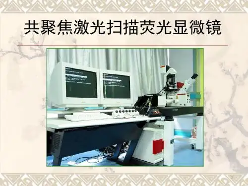

激光扫描共聚焦显微镜技术Laser Scanning Confocal Microscope——基础篇李治国细胞的内在生活显微镜的发展史没有显微镜就不可能有细胞学诞生。
1590年,荷兰眼镜制造商J 和Z.Janssen 父子制作了第一台复式显微镜。
1665年,英国人Robert Hook首次描述了植物细胞(木栓,命名为cella 。
1680年,荷兰人A.van Leeuwenhoek成为皇家学会会员,他一生中制作了200多台显微镜和400多个镜头,用设计较好的显微镜观察了许多动植物的活细胞与原生动物。
Made by A.van Leeuwenhoek (1632-1723.Magnification ranges at 50-275x.显微镜的最重要参数——分辨力显微镜物象是否清楚不仅决定于放大倍数,还与显微镜的分辨力(resolution )有关。
分辨率是指区分开两个质点间的最小距离各种显微镜的分辨能力光学显微镜(light microscopy)0.2μm电子显微镜 (Electro microscopy 0.2nm扫描遂道显微镜 (scanning tunneling microscope 0.2nm以下 1932年,德国人M.Knoll 和E.A.F.Ruska 发明电镜,1940年,美、德制造出分辨力为0.2nm 的商品电镜。
1981年,瑞士人G.Binnig 和H.RoherI 在IBM 苏黎世实验中心(Zurich Research Center)发明了扫描隧道显微镜而与电镜发明者Ruska 同获1986年度的诺贝尔物理学奖。
常用的光学显微镜(light microscopy普通光学显微镜暗视野显微镜相差显微镜偏光显微镜微分干涉显微镜荧光显微镜激光共焦扫描显微镜普通光学显微镜原理普通光学显微镜原理图1. 构成:①照明系统②光学放大系统③机械装置2. 原理:经物镜形成倒立实像,经目镜放大成虚像。

激光扫描共聚焦显微镜吴旭2008.10.14高级显微镜原理正置、倒置显微镜细胞遗传工作站活细胞工作站激光显微分离系统激光共聚焦显微镜概述激光扫描共聚焦显微镜(Laser scanning confocalmicroscope ,LSCM )生物医学领域的主要应用通过一种或者多种荧光探针标记后,可对固定的组织或活体样本进行亚细胞水平结构功能研究高空间分辨率、非介入无损伤连续光学切片、三维图像、实时动态等细胞结构和功能的分析检测……Conventional fluorescence microscope Confocal microscope历史1957年,Marvin Minsky提出了共聚焦显微镜技术的某些基本原理,获得了美国的专利。
1967年,Egger 和Petran 成功地应用共聚焦显微镜产生了一个光学横断面。
1977年,Sheppard 和Wilson 首次描述了光与被照明物体的原子之间的非线性关系和激光扫描器的拉曼光谱学。
1984年,Biorad 为公司推出了世界第一台商品化的共聚焦显微镜,型号为SOM-100,扫描方式为台阶式扫描。
1986年MRC-500型改进为光束扫描,用作生物荧光显微镜的共聚焦系统。
Confocal microscopy comes of ageJG White & WB Amos. Nature 328, 183 -184 (09 July 1987Zeiss 、Leica 、Meridian 、OlympusZeiss LSM510 激光扫描共聚焦显微镜Zeiss LSM510 META 激光扫描共聚焦显微镜Zeiss LSM510 META 激光扫描共聚焦显微镜Nikon A1R 激光扫描共聚焦显微镜Prairie UltimaIV 活体双光子显微镜国家光电实验室(武汉)自制随机定位双光子显微镜Leica TCS SP5 激光共聚焦扫描显微镜基本原理相差、DIC 常用荧光标记共聚焦原理Two ways to obtain contrast in light microscopy. The stained portions of the cell in(A reduce the amplitude of light waves of particular wavelengths passing through them.A colored image of the cell is thereby obtained that is visible in the ordinary way. Light passing through the unstained, living cell (B undergoes very little change in amplitude, and the structural details cannot be seen even if the image is highly magnified. The phase of the light, however, is altered by its passage through the cell, and small phase differences can be made visible by exploiting interference effects using a phase-contrast or a differential-interference-contrast microscope.D. Phase-contrast or adifferential-interference-contrast microscopeFour types of light microscopy. (A The image of a fibroblast in culture obtained by the simple transmission of light through the cell, atechnique known as bright-field microscopy.The other images were obtained by techniques discussed in the text: (B phase-contrast microscopy, (C Nomarski differential-interference-contrast microscopy, and (D dark-field microscopy.常用荧光探针Proteins Nucleic Acids DNA Ions pH Sensitive Indicators Oxidation States Specific Organelles荧光显微镜原理明场:透射荧光:落射落射的优点:物镜的聚光镜作用使视场均匀,发射光强度高。


激光扫描共聚焦显微镜
吴旭
2008.10.14
高级显微镜原理
正置、倒置显微镜
细胞遗传工作站
活细胞工作站
激光显微分离系统激光共聚焦显微镜
概述
激光扫描共聚焦显微镜
(Laser scanning confocalmicroscope ,LSCM )生物医学领域的主要应用
通过一种或者多种荧光探针标记后,可对固定的组织或活体样本进行亚细胞水平结构功能研究
高空间分辨率、非介入无损伤连续光学切片、三维图像、实时动态等细胞结构和功能的分析检测……
Conventional fluorescence microscope Confocal microscope
历史
1957年,Marvin Minsky提出了共聚焦显微镜技术的某些基本原理,获得了美国的专利。
1967年,Egger 和Petran 成功地应用共聚焦显微镜产生了一个光学横断面。
1977年,Sheppard 和Wilson 首次描述了光与被照明物体的原子之间的非线性关系和激光扫描器的拉曼光谱学。
1984年,Biorad 为公司推出了世界第一台商品化的共聚焦显微镜,型号为SOM-100,扫描方式为台阶式扫描。
1986年MRC-500型改进为光束扫描,用作生物荧光显微镜的共聚焦系统。
Confocal microscopy comes of ageJG White & WB Amos. Nature 328, 183 -184 (09 July 1987
Zeiss 、Leica 、Meridian 、Olympus
Zeiss LSM510 激光扫描共聚焦显微镜
Zeiss LSM510 META 激光扫描共聚焦显微镜
Zeiss LSM510 META 激光扫描共聚焦显微镜
Nikon A1R 激光扫描共聚焦显微镜
Prairie UltimaIV 活体双光子显微镜
国家光电实验室(武汉)自制随机定位双光子显微镜
Leica TCS SP5 激光共聚焦扫描显微镜
基本原理相差、DIC 常用荧光标记共聚焦原理
Two ways to obtain contrast in light microscopy. The stained portions of the cell in
(A reduce the amplitude of light waves of particular wavelengths passing through them.
A colored image of the cell is thereby obtained that is visible in the ordinary way. Light passing through the unstained, living cell (
B undergoes very little change in amplitude, and the structural details cannot be seen even if the image is highly magnified. The phase of the light, however, is altered by its passage through the cell, and small phase differences can be made visible by exploiting interference effects
using a phase-contrast or a differential-interference-contrast microscope.
D. Phase-contrast or a
differential-interference-
contrast microscope
Four types of light microscopy. (A The image of a fibroblast in culture obtained by the simple transmission of light through the cell, atechnique known as bright-field microscopy.The other images were obtained by techniques discussed in the text: (B phase-contrast microscopy, (C Nomarski differential-interference-contrast microscopy, and (D dark-field microscopy.
常用荧光探针
Proteins Nucleic Acids DNA Ions pH Sensitive Indicators Oxidation States Specific Organelles
荧光显微镜原理
明场:透射荧光:落射
落射的优点:
物镜的聚光镜作用使视场均匀,发射光强度高。
激发光损失小,荧光
效应高。
共聚焦原理
由于pinhole 的存在,使得部分杂散光(虚线部分)没有被PMT 探测器探测到,从而提高了成像效果。
通过对样品在x-y 方向上的逐点扫描,可以形成二维图像。
如果调解焦平面在z 方向的位置,连续扫描多个不同z 位置的二维图像,则可以形成一个3D 图像,3D
的重建需要软件的支持。
The unique scanning module is thecore of the LSM 510 META.
It contains motorized collimators, scanning mirrors,individually adjustable and positionablepinholes, and highly sensitive detectors including the META detector.
All these components are
arranged to ensure optimum specimen illumination and efficient collection of reflected or emitted light. A highly efficient optical grating provides an innovative way of separating the fluorescence emissions in the META detector.
The grating projects the entire fluorescence spectrum onto the 32 channels of the META detector. Thus, the spectral signature is acquired for each pixel of the scanned image and subsequently can be used for the
digital separation into component dyes.
分光镜组件结构
共聚焦构成
•光学显微镜
•激光光源
•扫描模块(包括共聚焦光路通道和针孔、扫描镜)•光检测器•计算机系统(数据采集、处理、转换应用)•图形输出设备
LCSM 照片,蓝色为细胞核,绿色为微管图像相关概念
RGB
三基色
图像数据位
亮度
对比度
饱和
分辩率
怎样得到样品图像
样品的标记物有合适的激发波长和发射波长荧光下观察选择合适的图象设置激光扫描的通道参数
设置图象的属性
调节每一个通道的亮度、对比度、清晰度、背景亮度
启动
共聚焦和CCD 照片的比较更大的动态范围
高灵敏度
图像背景的消除
灵活的拍照区域选择 ZOOM 功能
伪彩色
环境要求高
多通道图象合成
时间序列
Ca 离子成像
FRAP :Fluorescence Redistribution After Photo bleaching
FRAP 是用来测定活细胞的动力学参数,借助于高强度脉冲激光来照射细胞某一区域,造成该区域荧光分子的光粹灭,该区域周围的非粹灭荧光分子会以一定的速率向受照射区域扩散,这个扩散速率可通过低强度激光扫描探测, 因而可得到活细胞的动力学参数。
LSCM 可以控制光粹灭作用,实时监测分子扩散率和恢复速率,反映细胞结构和活动机制。
广泛用于研究细胞骨架构成,核膜结构跨膜大分子迁移率,细胞间通讯等领域。
荧光漂白恢复(FRAP )
实用文档
人卵细胞的局部光漂白
FRAP 研究ER-GFP 的核转位文案大全。