肺鳞癌中Furin和E-cadherin的表达研究
- 格式:pdf
- 大小:969.00 KB
- 文档页数:3

RhoB和E-cadherin在非小细胞肺癌组织中的表达及临床意义谭祎媚;张逊;徐美林;王菁;徐医军【摘要】Objective To investigate the expressions of RhoB and E-cadherin in non-small-cell lung cancer (NSCLC), and their clinical significances thereof. Methods Immunohistochemical staining was applied to detect expres-sions of RhoB and E-cadherin in 116 samples of NSCLC (NSCLC group) and 116 samples of normal lung tissues (control group). Correlations of expressions of RhoB and E-cadherin to clinical pathological parameters and prognosis were analyzed in two groups. Results The expression intensities of RhoB and E-cadherin were significantly lower in NSCLC group than those in control group (57.76%vs 87.07%,54.31%vs 85.34%,P<0.01). There were significant differences in the expres-sion of RhoB between different pathological types, differentiation and lymph node metastasis in NSCLC group. There were significant differences in the expression of E-cadherin between different TNM stages, differentiation and lymph node metas-tasis in NSCLC group. The expression of RhoB was positively correlated with the expression of E-cadherin ( r=0.503,P<0.01). The 3-year survival rates were significantly higher in patients with high expression of RhoB (83.93%) than those in pa-tients with low expression of RhoB (40.00%, Log-rank χ2=18.992,P<0.01). The 3-year survival rates were significantly higher in patients with high expression of E-cadherin (85.11%) than those in patients with low expression of E-cadherin (44.93%, Log-rankχ2=16.680,P<0.01). Further multivariate analysis suggest ed that both lower expressions of RhoB and E-cadherin and lymph node metastasis were prognostic indicators for NSCLC (P<0.001). Conclusion The expressions of RhoB and E-cadherin showed a good correlation in NSCLC. Detecting the expression of RhoB combined with E-cadherin may give a clue on clinicopathological features and prognosis in patients with NSCLC.%目的:探讨非小细胞肺癌(NSCLC)中RhoB和E-钙黏蛋白(E-cadherin)的表达及其临床意义。

非小细胞肺癌组织中Snail、E-cadherin及N-cadherin的表达及临床意义俞婷婷;刘莉;王若峥【摘要】目的探讨指转录因子Snail、上皮细胞钙粘蛋白(E-cadherin)及神经钙黏素(N-cadherin)在非小细胞肺癌组织中的表达及其之间的相关性.方法采用免疫组化法检测63例非小细胞肺癌(NSCLC)手术切除标本及30例癌旁组织中Snail 、E-cadherin、N-cadherin蛋白的表达,并分析相互间关系及其临床意义.结果与癌旁组织比较,NSCLC组织中E-cad-herin表达显著减少,N-cadhern和Snail表达明显增加(P<0.01).NSCLC组织中,Snail的表达与N-cadhenn呈明显的正相关(P <0.05,r=0.33),与E-cadherin呈显著负相关(P<0.05,r=-0.41),N-cadherinl的表达与E-cadheri也呈明显负相关(P<0.05,r=-0.56).Snail表达阳性率与TNM分期和有无转移相关(P<0.05);E-cadherin表达阳性率与TNM分期和有无转移相关(P <0.05);N-cadherin表达阳性率与组织学分型、TNM分期和有无转移相关(P<0.05).结论 NSCLC癌组织中Snail、E-cadherin和N-cadherin表达水平与肿瘤的浸润转移密切相关,可作为临床诊断和治疗靶点的参考指标.【期刊名称】《实用临床医药杂志》【年(卷),期】2016(020)017【总页数】4页(P29-31,56)【关键词】非小细胞肺癌;锌指转录因子;上皮细胞钙粘蛋白;神经钙黏素;免疫【作者】俞婷婷;刘莉;王若峥【作者单位】新疆医科大学附属肿瘤医院肺内科,新疆乌鲁木齐,830011;新疆医科大学附属肿瘤医院肺内科,新疆乌鲁木齐,830011;新疆医科大学附属肿瘤医院头颈放射治疗科,新疆乌鲁木齐,830011【正文语种】中文【中图分类】R734.2在中国,原发性肺癌是常见的恶性肿瘤,早期诊断困难和转移率高导致其在发病率和死亡率居恶性肿瘤的首位,其中非小细胞肺癌(NSCLC)约占85%[1]。
中山园地・中山园地・E2cadherin及相关指标的临床病理学意义美国G BI公司Lily chen 北京中山生物技术有限公司研发部:戚 闻(摘译) 恶性肿瘤具有从原发灶向其他部位转移的特点,而转移过程中的许多环节都与肿瘤细胞的黏附特性发生改变有关。
在原发病灶肿瘤细胞脱离相邻细胞后经血或淋巴转移,这预示着肿瘤细胞与黏附相关的分子发生了变化,这些分子包括整合素(intergrin)、选择素(selectin)、C D44和钙黏附蛋白(cadherin)等。
上皮性钙黏附蛋白(E2cadherin)是一种分子量为120kd 的穿膜糖蛋白,其胞浆部分直接与某种catenin或另一种catenin相连,两者所构成的复合体再通过某种catenin与肌动蛋白细胞骨架相连。
最近的许多研究表明,E2cadherin所介导的细胞黏附性下降或丧失是许多上皮性肿瘤发生浸润和转移的原因之一。
芝加哥大学的Dr.Ruth及其研究小组的实验表明,在无淋巴结转移的乳腺癌患者中,E2cadherin蛋白的低水平表达(无论是做为独立的指标还是与其他相关的生物学指标联合应用),均提示病人预后不良。
他们共检测了1972~1987年间的乳腺癌标本168例,这些病例均无淋巴结转移,术前术后均未接受化疗和激素治疗,并随访长达14年。
免疫组化结果证实,当病人E2cadherin为高或中等水平表达时,14年生存率达80%;而E2cadherin低水平表达,则14年生存率仅有56%;反之nm232H1高表达和微血管计数低的病例14年生存率可达90%。
他们认为:E2cadherin表达下降或丧失与肿瘤转移密切相关,低水平表达是预后差的重要指标。
综合考虑E2cadherin、nm232H1和微血管计数所得出的关于预后的结论往往比肿瘤的大小、分级、年龄、ER表达水平等因素还要重要。
如果能综合考虑以上指标,可有90%以上的病人不实施化疗。
Chow等人应用免疫组化方法检测了85例舌癌、9例伴有淋巴结转移和7例原位复发性肿瘤标本的E2cadherin和3种catenin。

E—cadherin与肿瘤侵袭、转移相关性研究进展恶性肿瘤的侵袭、转移特性使其成为癌症患者死亡的首要原因,不仅给治疗带来了一定的难度,更让患者承受了身体及心理的巨大痛苦。
探究恶性肿瘤侵袭、转移的机制,并针对其相关分子标志物寻找抑制靶点,是临床医务工作者及基础医学研究者面临的巨大挑战。
恶性肿瘤的侵袭、转移机制是一个多方面因素相互作用的复杂过程,包括了肿瘤细胞自身生物学行为、肿瘤细胞生存的微环境、及各种细胞因子、信号通路等因素。
上皮-间质转化是近年来肿瘤侵袭、转移领域的研究热点,而上皮型钙黏蛋白是上皮-间质转化过程中一种主要的蛋白标志物,更好的理解上皮型钙黏蛋白与上皮-间质转化的关系及它们在癌症进展中所涉及的机制,能够为晚期转移性肿瘤治疗及预防的相关研究提供理论及实验依据。
因此本文就上皮-间质转化与肿瘤侵袭、转移的研究作一综述。
Abstract:The invasion and metastasis of malignant tumor make it the leading cause of death in cancer patients.It not only brings some difficulties to the treatment,but also endures great physical and psychological suffering.To explore the mechanism of invasion and metastasis of malignant tumor and find the target of inhibition against its related molecular markers is a great challenge for clinical medical workers and basic medical researchers.Malignant tumor invasion and metastasis mechanism is a complex process of interaction of many factors,including the biological behavior of tumor cells themselves,the microenvironment of tumor cells survival,and various cytokines,signaling pathways and other factors.Epithelial-mesenchymal transition (EMT)is a hotspot in the field of tumor invasion and metastasis in recent years.E-cadherin is a major protein marker in EMT,and better Understanding the relationship between E-cadherin and EMT and their mechanisms involved in cancer progression can provide theoretical and experimental evidence for the research on the treatment and prevention of advanced metastatic tumors.Therefore,this article reviews E-cadherin and tumor invasion and metastasis.Key words:E-cadherin;Epithelial-mesenchymaltransition;Tumor;Invasion;Metastasis侵袭、转移是恶性肿瘤致患者死亡的首要原因。
Ezrin和E-Cadherin在肺癌中的表达及意义的开题报告引言:肺癌是世界范围内最常见的恶性肿瘤之一,对人类健康和社会经济都造成了严重影响。
早期肺癌的诊断和治疗是降低肺癌病死率的关键所在。
近年来,越来越多的研究表明,肺癌的发生和发展与多种分子的表达异常密切相关,这些分子的表达状态与肿瘤的恶性程度以及预后密切相关。
本研究旨在探讨 Ezrin 和 E-Cadherin 在肺癌中的表达及其意义。
一、 Ezrin 的概述Ezrin 是一种跨膜蛋白,属于 ERM 家族(Ezrin、Radixin 和 Moesin)。
它在细胞质骨架(cytoskeleton)重要作用,参与细胞形态的维持、细胞膜与质膜的互动和细胞信号转导等过程。
Ezrin 在肿瘤发生和发展过程中发挥了重要作用,被认为是一种肿瘤相关蛋白(tumor-associated protein)。
二、Ezrin 在肺癌中的表达及其意义研究表明,肺癌组织中 Ezrin 的表达水平明显升高,Ezrin 的表达水平与肿瘤的分化程度、淋巴结转移、预后等指标均有关联。
Ezrin 可能通过影响细胞增殖、迁移和侵袭等过程来促进肺癌的发生和发展。
此外,Ezrin 的表达还与肺癌患者的预后密切相关,高表达的 Ezrin 可能预示着较差的预后。
三、E-Cadherin 的概述E-Cadherin 是一种细胞间粘附分子,广泛存在于生物体内各种组织和细胞上,其功能是维持细胞间的稳定粘附联系。
当 E-Cadherin 表达异常时,细胞间的粘附能力减弱,会促进肿瘤细胞的浸润和迁移,从而促进肿瘤的发生发展。
四、E-Cadherin 在肺癌中的表达及其意义肺癌组织中 E-Cadherin 的表达水平常常降低,可能是肺癌侵袭和迁移的一个重要因素。
某些研究表明,E-Cadherin 表达水平与肺癌的淋巴结转移和预后密切相关。
因此,通过研究 E-Cadherin 的表达状态,可以更好地了解肺癌的发生和发展过程。
E—cadherin在肺腺癌中的差异性表达目的探讨E-cadherin mRNA及其编码蛋白在肺腺癌中的表达是否存在差异性。
方法选取肺腺癌20例,应用RT-PCR法和WESTERN-BLOT法分别检测肺腺癌区和正常区E-cadherin mRNA及其编码蛋白表达情况。
结果肺腺癌中E-cadherin mRNA水平与蛋白水平存在明显不一致的情况,即尽管40%的肺腺癌病例的E-cadherin在mRNA水平有表达,但在蛋白水平却难以检出。
结论在肺腺癌细胞内很可能存在某种转录后调节(沉默)机制,如RNA选择性剪接、microRNA。
因此,该文为深入研究E-cadherin的表达调节机制及其肿瘤生物学意义提供颇有价值的线索。
标签:E-cadherin;肺腺癌;差异性上皮钙粘素(E-cadherin)是一类介导上皮细胞相互黏附的钙依赖性跨膜糖蛋白。
国内外大量研究证实,恶性肿瘤局部浸润和远处转移与细胞间粘附力的降低有关,其中E-cadherin具有重要作用[1-3]。
但是目前对E-cadherin的研究大多数仍局限于蛋白或转录的单一层面,关于该基因转录产物和蛋白表达之间的关系尚罕有报导。
2010年2月—2012年9月,该科研小组对20例肺腺癌进行了相关研究,力图阐明E-cadherin mRNA及其编码蛋白在肺腺癌中是否存在差异性的问题,现报道如下。
1 资料与方法1.1 一般资料20例肺腺癌病理资料来自辽河油田总医院病理科。
其中女性8例,男性12例;年龄38~76岁,平均65岁。
对同一患者的新鲜肺腺癌组织和正常肺组织标本分别提取mRNA和蛋白质,然后分别应用RT-PCR法和WESTERN-BLOT法检测E-cadherin mRNA及其编码蛋白表达情况。
1.2 RT-PCR反转录反应条件为1.3 WESTERN-BLOT按《分子克隆》实验操作指南进行,一抗为鼠抗人E-cadherin单克隆抗体、兔抗人β-actin多克隆抗体;二抗为辣根过氧化物酶标记的羊抗兔/鼠IgG,应用ECL法标记结合信号,通过荧光自显影感光在Kodak XAY胶片上。
²临床论著²肺鳞癌中Furin和E-cadherin的表达研究牛灵1陶琳2董雅璐3【摘要】目的检测肺鳞癌中弗林蛋白酶(Furin)和上皮型黏附素(E-cadherin)的表达,分析二者在不同临床特征中的表达差别,关注其临床意义。
方法选择65例肺鳞癌作为观察组,选择50例正常肺组织作为对照组,应用免疫组化方法检测Furin和E-cadherin的表达,统计学分析应用卡方检验或相关性分析。
结果观察组中Furin表达的阳性率明显高于对照组(53.85% vs. 4.00%,P<0.000 1),观察组中E-cadherin表达的阳性率明显低于对照组(16.92% vs. 98.00%,P<0.000 1)。
观察组中Furin和E-cadherin的表达均与肿瘤的最大径、分化程度和脉管浸润密切相关(P<0.05)。
察组中Furin和E-cadherin的表达无相关性(P>0.05)。
结论肺鳞癌中Furin高表达、E-cadherin低表达是促进肿瘤发生和进展的重要因素,Furin和E-cadherin无明显的协同作用。
【关键词】肺;费林蛋白酶;免疫组织化学;上皮型黏附素;鳞癌Study on expressions of Furin and E-cadherin in lung squamous cell carcinoma Niu Ling1, Tao Lin2,Dong Yalu3. 1Department of Respiratory, Xinjiang People’s Hospital, Urumqi 830002, China; 2The SecondMedical Department of Midong Branch, Xinjiang People’s Hospital, Urumqi 830000, China; 3Departmentof Oncology, Lanzhou Military Region Urumqi General Hospital, Urumqi 830000, ChinaCorresponding author: Niu Ling, Email: 363141843@【Abstract】Objective To detect the expressions of Furin and E-cadherin in lung squamous cellcarcinoma, analyze the clinical significance. Methods The observation group included 65 cases with lungsquamous cell carcinoma, the control group included 50 cases with normal lung tissue. Expressions ofFurin and E-cadherin were detected by IHC. Chi-square test and correlation analysis were used in study.Results The expression of Furin was higher in the observation group than in the control group (53.85%vs. 4.00%, P<0.000 1). The expression of E-cadherin was lower in the observation group than in the controlgroup (16.92% vs. 98.00%, P<0.000 1). Expressions of Furin and E-cadherin were associated with diameter,differentiation and vascular invasion (P<0.05). The expressions of Furin and E-cadherin were not correlated inobservation group (P>0.05). Conclusion The higher expression of Furin, lower of E-cadherin maypromote the occurrence and development of lung squamous cell carcinoma. The expressions of Furin andE-cadherin are not obvious synergistic effect in observation group.【Key words】Lung; Furin; Immunohistochemistry; E-cadherin; Squamous cell carcinoma肺癌中以鳞癌最常见,其发病时进展快,预后不理想[1]。
近年研究显示基因和蛋白调节失常对肿瘤的发展具有明显促进作用。
弗林蛋白酶(Furin)作为蛋白前体加工酶家族中相关蛋白,不仅与肿瘤的生长相关,还对调节肿瘤的侵袭有重要作用[2-3]。
上皮型黏附素(E-cadherin)是细胞间重要的黏附蛋白,在正常情况下高表达,对维持细胞间的连接有DOI:10.3877/cma.j.issn.1674-0785.2015.23.008作者单位:830002 乌鲁木齐,新疆维吾尔自治区人民医院呼吸二科1;830000 新疆维吾尔自治区人民医院米东分院内二科2;830000 兰州军区乌鲁木齐总医院肿瘤内科3通讯作者:牛灵,Email: 363141843@ 重要作用,当肿瘤性病变形成及进展时,E-cadherin 的表达下降,使细胞间的连接松动,细胞易于离开原发部位出现播散[4-5]。
本研究应用免疫组化方法检测肺鳞癌中Furin和E-cadherin的表达,分析其临床意义,旨在为临床判定生物学行为提供帮助。
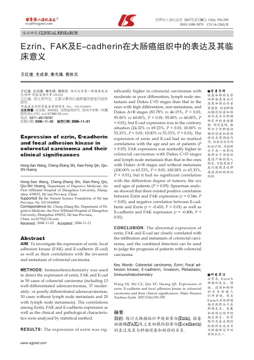
资料和方法一、一般资料选择我院于2012年1月至2014年6月行肺鳞癌根治的患者作为观察组,术后病理均诊断为鳞癌,共65例,其中男35例,女30例,年龄45~85岁,平均(58.1±6.5)岁,其中高分化20例,中表1 两组中Furin和E-cadherin表达阳性率的比较[例,(%)]组别例数Furin E-cadherin+ -χ2值P值+ -χ2值P值观察组65 35(53.85) 30(46.15)32.177 2 <0.000 1 11(16.92) 54(83.08)74.449 5 <0.000 1对照组50 2(4.00) 48(96.00) 49(98.00) 1(2.00)表2 Furin和E-cadherin在观察组不同临床特征中的表达[例,(%)]临床特征例数Furin E-cadherin+ -χ2值P值+ -χ2值P值肿瘤直径6.610 1 0.010 1 10.942 8 0.000 9 ≤4 cm 20 6(30.00) 14(70.00) 8(40.00) 12(60.00)>4 cm 45 29(64.44) 16(35.56) 3(6.67) 42(93.33)分化程度18.251 6 <0.000 1 5.740 6 0.016 6 高+中38 12(31.58) 26(68.42) 10(26.32) 28(73.68)低27 23(85.19) 4(14.81) 1(3.70) 26(96.30)脉管浸润9.960 4 0.001 6 5.469 0 0.019 4 无46 19(41.30) 27(58.70) 11(23.91) 35(76.09)有19 16(84.21) 3(15.79) 0(0) 19(100)分化18例,低分化27例。
选择50例正常肺组织作为对照组,其中男25例,女25例,年龄47~80岁,平均(57.6±5.6)岁。
两组在年龄、性别等资料的比较无明显差别。
二、Furin和E-cadherin表达的检测Furin和E-cadherin的检测应用免疫组化SP法,DAB染色,严格按说明书操作,并由主管技师完成,做好质量控制。
三、Furin和E-cadherin结果判断标准Furin以细胞质中出现棕黄色为阳性,E-cadherin 以细胞膜出现棕黄色为阳性,以表达中至强等强度为阳性(弱阳性不计数),计数10个上皮细胞集中的区域(³400倍),计算阳性率,取平均值,以<25%为阴性,≥25%为阳性。
四、统计学分析SAS 6.12进行分析,应用卡方检验或线性相关性分析,以P<0.05为差别有统计学意义。
结果一、两组中Furin和E-cadherin阳性率的比较两组中Furin和E-cadherin表达的阳性率差别有意义。
即观察组Furin的表达明显高于对照组,E-cadherin的表达明显低于对照组(表1,图1~4)。
二、Furin和E-cadherin在观察组不同临床特征中的表达观察组中Furin和E-cadherin的表达均与肿瘤的最大径、分化程度和脉管浸润密切相关,见表2。
三、观察组中Furin和E-cadherin的相关性分析对观察组中Furin和E-cadherin的表达进行线性相关分析,结果显示Furin和E-cadherin未见明显相关性(P>0.05)。
讨论肺鳞癌近年的发病率有增高趋势,吸烟是重要的发生因素。
临床最常见的类型是中央型,即发生于主支气管、叶支气管或段支气管内,临床表现为气道梗阻症状,部分患者由于咳痰受阻,表现为阻塞性肺炎和黏液分泌受阻,肿瘤较大时,常出现坏死[6-7]。
肿瘤发生和进展过程中蛋白表达明显失调,肺鳞癌可能是一种低黏附性的癌,易于出现迁移和转移。
E-cadherin是细胞间重要的黏附蛋白,也是细胞间紧密连接的重要物质,不仅对具有维持细胞间的结构有重要作用,还对细胞间信息传递起重要作用[8]。
正常情况下E-cadherin高表达,而在肿瘤性病变和肿瘤进展时E-cadherin表达下降,此时肿瘤化的细胞易于离开原发灶,形成明显的浸润和播散[9-10]。
Furin在发挥功能时可以参与多种细胞间的作用,如蛋白质的合成、受体的形成及活化、血浆蛋白前体的激活等[11]。
Furin在细胞内主要定位于核周高尔基体区域,也在细胞质的其他区域中散在分布,对参与底物分子的活化起重要作用[12]。
本实验结果显示两组中Furin和E-cadherin的阳性率差别有统计学意义,提示Furin高表达、E-cadherin低表达是肿瘤形成的因素。