医学影像学专业英语消化系统 戚云杰 (1)
- 格式:ppt
- 大小:11.55 MB
- 文档页数:13
No significant differences were found between the area under the receiver operating characteristic curve of the digital and that of the conventional radiography method for almost all investigated criteria .The only exception (例外;异议)was mediastinal abnormalities, for which the digital method provided better results than the conventionvl method (P<0.05)Invasive breast cancer detection by mammography may be improved through attention to correct positioning.For CT of the brain, there are there standard reference lines; the orbitomeatal line, the Reid baseline, and the supraorbitomeatal line. The orbitomeatal line runs through the external canthus and the center of the external auditory meatus. This is the most widely used line, because acquisition of fewer sections is necessary to cover the entire brain. The Reid baseline runs though the inferior orbital wall and the superior border of the external auditory meatus. This line is paraller to the optic nerve and provides the best demonstration of the orditai contents. The supraorbitomeatal line runs through the superior orbital wall and the center of the external auditory meatus. This line approximately parallels the skull base. We developed reference lines for use at MR imaging that are analogous to the three standard reference lines used at CT, on the basis of anatomic landmarks that are visible on midsagittal MR images. These lines can be used to prescribe subsequent oblique axial sequences.During the past decade , many investigators have addressed the feasibility of picture archiving and communication system (PACS) as an alternative to film-based radiology. To successfully implement the PACS, a number of factors such as film resolution, interpretation conditions, and a cost-effective viewer system must be taken into consideration. The most important clinical criterion for using the PACS technology is the ability to achieve acceptable accuracy when interpreting radiologic images at a soft-copy viewing workstation. Most previous reports have focused on a comparison of observer performance with soft-copy and hard-copy imajes, and such studies have been performed using chest radiography. However, observer performance in projection radiography of another anatomic section must also be compared to achieve that the PACS can completely replace film-based radiology.CT in an invaluable tool in the diagnosis of the chest diseases with high-resolution CT (HRCT) providion exquisite visualization of the pulmonary interstitium. However, CT examinations do not come without a cose.. A report from the 2002 National Conference on Dose Reduction in CT indicates that CT now represents the largest single source of medical radiation exposure. Although CT constitutes only 15% of the total number of examinations in some university departments, it can account for 70% of the dose delivered.。
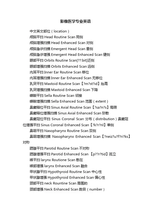
影像医学专业英语中文英文部位(location)颅脑平扫Head Routine Scan同侧颅脑增强扫描Head Enhanced Scan对侧颅脑急诊扫描Emergent Head Scan患侧颅脑急诊增强Emergent Head Enhanced Scan健侧眼部平扫Orbits Routine Scan[??:bit]近侧眼部增强扫描Orbits Enhanced Scan远侧内耳平扫Inner Ear Routine Scan移位内耳增强扫描Inner Ear Enhanced Scan无移位乳突平扫Mastoid Routine Scan【?m?st?id】抬高乳突增强扫描Mastoid Enhanced Scan下降蝶鞍平扫Sella Routine Scan邻接蝶鞍增强扫描Sella Enhanced Scan范围(extent)鼻窦轴位平扫Sinus Axial Routine Scan【?sa?n?s】局限鼻窦轴位增强扫描Sinus Axial Enhanced Scan弥散鼻窦冠位平扫Sinus Coronal Scan分布(distribution)鼻窦冠位增强平扫Sinus Coronal Enhanced Scan【?k?r?nl】单侧鼻咽平扫Nasopharynx Routine Scan双侧鼻咽增强扫描Nasopharynx Enhanced Scan【?neiz?u?f?ri?ks】对称腮腺平扫Parotid Routine Scan不对称腮腺增强平扫Parotid Enhanced Scan 【p??r?tid】孤立喉平扫larynx Routione Scan散在喉部增强larynx Enhanced Scan融合甲状腺平扫Hypothyroid Routine Scan中心性甲状腺增强Hypothyroid Enhanced Scan偏心性颈部平扫neck Rountine Scan周围的颈部增强Neck Enhanced Scan数目(number)肺栓塞扫描lung Embolism Scan单发胸腺平扫Thymus Rountine Scan多发胸腺增强Thymus Enhanced Scan增多胸骨平扫Sternum Rountine Scan减少胸骨增强Sternun Enhanced Scan大小(size)胸部扫描Chest Routine Scan大胸部薄层扫描High Resolution Chest Scan小胸部增强扫描Chest Enhanced Scan稳定胸部穿刺Chest Puncture Scan扩大轴位胸部穿刺Axial Chest Puncture Scan扩张上腹部平扫upper abdomen routine scan 膨胀中腹部平扫mid- abdomen routine scan 缩小上腹部增强扫描upper abdomen enhanced scan体积缩小中腹部增强扫描mid- abdomen enhanced scan 狭窄腹部穿刺Abdomen Puncture Scan闭塞轴位腹部穿刺Axial Abdomen Puncture Scan形状(shape)颈椎胸椎腰椎平扫C/T/L-Spine Routine Scan点状盆腔平扫/增强扫描Pelis Routine /Enhanced Scan斑点状骶髂关节扫描SI Joint Scan粟粒状肩关节扫描Shoulder Joint Scan结节状上肢软组织平扫/增强Upper Extremities Soft Tissue Scan团块状肘关节平扫Elow Joint Routine Scan圆形腕关节平扫Wrist Routine Scan卵圆形手部平扫Hand Routine Scan椭圆形髋关节平扫Hip Joint Routine Scan长方形膝关节平扫Keen Joint Routine Scan分叶形踝关节平扫Ankle Joint Routine Scan片状下肢软组织平扫增强Lower Extremities Soft Tissue Scan/Enhance条索状足部平扫foot routine scan线状血管造影和三维成像网状头部血管造影Head CT Angiography弧线形颈部血管造影Neck CT Angiography星状心脏冠脉造影Coronal Artery Angiography纠集心脏冠脉钙化积分Cardiac Calcium Scoring Scan舟状胸部血管造影Chest CT Angiography哑铃状腹部血管造影AbdomenCT Angiography不规则形上肢血管造影Upper Extremities CT Angiography细致下肢血管造影Lower Extremities CT Angiography粗糙五官三维成像3D Facial Scan变形胃部三维成像3D Stomach CT Scan增粗结肠三维成像3D Colon CT Scan增厚颈椎胸椎腰椎三维成像3D C/T/L -Spine变细肩关节三维成像3D Should Joint变平肘关节三维成像3D Elbow Joint边缘腕关节三维成像3D Wirst Joint轮廓髋关节三维成像3D Hip Joint光滑膝关节三维成像3D KneeJoint锐利踝关节三维成像3D Ankle Joint清晰模糊毛刺状分叶状密度,回声,信号密度透亮阴影不透光致密低密度混杂密度信号低信号高信号性质囊性空洞壁外壁渗出浸润实变增值实性结节状肿块纤维化钙化ipsilateralcontralateralaffected sideintact sideproximal sidedistal sidedeviation;shift;displacement nondisplaced elevation;descent;fallabutting;nextto;secondaryto localized,regional diffuseunilateralbilateralsymmetricasymmetricsolitaryscatteredconfluencecentraleccentricperipheralsolitarymultipledecreaselargesmallstabilityenlargementdilatationdistentionshrinkloss of volumestenosis,narrowingocclusion;obliteration,emphraxis punctual mottingmiliarynodularmasscircular,round,roudedovalellipseoblonglobulatedpathystripelinarreticularcurvilinearstellatecrowding,convergingboat-shaped;navicular;scaphoiddumb-bellfinecoarsedeformitythickenthickenthinningflattenedborder,margin,rim,edgeoutline,contoursmoothsharp,well-defined,well-circumscribed clear ,distinct hazy,indistinct,ill-defined,obscured spiculated multilobulateddensitylucency,transparentshadowhaziness,opacificationdensityhypodense,low densityhyperdense,high densitymixed densityhypointensityhyperintensitycystcavitationwallouter surface of the wall exudationinfiltration consolidationhyperplsiasolid nodule mass fibrosis calcification。
用英语介绍医学影像专业自我介绍A Journey into Medical Imaging: My Professional Self-Introduction.Hello, I am delighted to have the opportunity to introduce myself and share my journey into the fascinating world of medical imaging. My passion for this field has grown over the years, and I am excited to be part of a discipline that plays a crucial role in modern healthcare.My interest in medical imaging started when I was still in high school. I was fascinated by the idea of using technology to peer into the human body and gain insights into its workings. This curiosity led me to pursue a degree in Biomedical Engineering, where I had the opportunity to delve deeper into the principles of imaging technology and its applications in medicine.During my undergraduate studies, I developed a strong foundation in physics, computer science, and biomedicalengineering principles. This interdisciplinary approach provided me with a unique perspective on medical imaging, enabling me to understand not only the technical aspects but also the clinical implications of the images produced.I also had the chance to work on several research projects that explored the use of advanced imaging techniques in diagnosing and treating various medical conditions.After graduating, I decided to specialize in medical imaging, and I pursued a graduate degree in the field. This decision was further influenced by the growing importance of imaging in modern medicine and the ever-evolving technological advancements in this area. My graduate studies focused on advanced imaging modalities such as magnetic resonance imaging (MRI) and computed tomography (CT). I gained extensive knowledge in image acquisition, processing, and interpretation, as well as the clinical applications of these techniques.As part of my graduate program, I also had the opportunity to complete a clinical rotation at a major hospital. This experience was extremely valuable as itallowed me to see how medical imaging is used in real-world clinical settings. I observed radiologists and other healthcare professionals as they interpreted images, made diagnoses, and developed treatment plans. This hands-on experience gave me a deeper understanding of the role of medical imaging in patient care and the responsibilities of a medical imaging professional.Since completing my graduate degree, I have beenworking as a medical imaging specialist in a leading healthcare facility. In my current role, I am responsiblefor performing a range of imaging procedures, including MRI, CT, and ultrasound. I also collaborate with otherhealthcare professionals to ensure accurate image interpretation and effective patient care. The most rewarding part of my job is being able to contribute to the diagnosis and treatment of patients, often playing acrucial role in their recovery and well-being.As a medical imaging specialist, I am constantlylearning and adapting to new technologies and imaging modalities. The field of medical imaging is constantlyevolving, and it is essential to stay up-to-date with the latest advancements and best practices. I am committed to further developing my skills and knowledge by participating in continuing education programs and professional development opportunities.In conclusion, medical imaging has been a fulfilling and rewarding journey for me. I am passionate about this field and excited about the opportunities it presents. I am committed to providing high-quality imaging services and contributing to the delivery of excellent patient care. I am looking forward to continuing my professional growth and making a positive impact in the field of medical imaging.。

(1)To prospectively evaluate the effect of heart rate, heart rate variability, and calcification dual-source computed tomography (CT) image quality and to prospectively assess diagnostic accuracy of dual-source CT for coronary artery stenosis. by using invasive coronary angiography as the reference standard.前瞻性评价心率、心率变异性及钙化双源计算机断层扫描成像质量的影响及对冠状动脉狭窄的双源性冠状动脉狭窄诊断的准确性评价。
以侵入性冠状动脉造影为参照标准。
(2)Chest radiography plays an essential role in the diagnosis of thoracic disease and is the most frequently performed radiologic examination in the United States. Since the discovery of X rays more than a century ago, advances in technology have yieled numerous improvements in thoracic imaging. Evolutionary progress in film-based imaging has led to the development of excellent screen-film systems specifically designed for chest radiography.胸部X线摄影中起着至关重要的作用在胸部疾病的诊断,是最常用的影像学检查在美国。
团队的力量 Strength of our teamStrength of our team!!湘雅医院2008级五年制临床医学、麻醉医学及口腔七年制18组同学合作完成本文的翻译Double-Contrast Upper Gastrointestinal Radiography: A Pattern Approach for Diseases of the StomachAbstractThe double-contrast upper gastrointestinal series is a valuable diagnostic test for evaluating structural and functional abnormalities of the stomach. This article will review the normal radiographic anatomy of the stomach. The principles of analyzing double-contrast images will be discussed. A pattern approach for the diagnosis of gastric abnormalities will also be presented, focusing on abnormal mucosal patterns, depressed lesions, protruded lesions, thickened folds, and gastric narrowing.This article presents a pattern approach for the diagnosis of diseases of the stomach at double-contrast upper gastrointestinal radiography. After describing the normal appearance of the stomach on double-contrast barium studies and the principles ofdouble-contrast image interpretation, we will consider abnormal surface patterns of the mucosa, depressed lesions (erosions and ulcers), protruded lesions (polyps, submucosal masses, and other tumors), thickened folds, and gastric narrowing.上消化道双重对比造影:上消化道双重对比造影:一种用于一种用于胃部疾病诊断的成像方法摘要上消化道双重对比造影系列是用于评估胃部结构性和功能性病变的一种极有价值的诊断方法。

医学影像学英语Medical ImagingMedical imaging plays a crucial role in the diagnosis and treatment of various medical conditions. It is a branch of medicine that utilizes different imaging techniques to create visual representations of the interior of the body. These images help healthcare professionals to identify and diagnose diseases, plan appropriate treatment, and monitor the effectiveness of interventions. In this article, we will explore the importance of medical imaging in modern healthcare and discuss some of the commonly used imaging modalities.X-ray imaging is one of the oldest and most commonly used imaging techniques in medicine. It involves exposing the body to a small dose of ionizing radiation to create images of the internal structures such as bones and soft tissues. X-rays are frequently used to diagnose fractures, pneumonia, and certain types of cancer. Despite its widespread use, x-ray imaging has limitations in visualizing certain structures and tissues, such as the brain and blood vessels.Computed tomography (CT) is another valuable imaging modality that utilizes x-rays to produce detailed cross-sectional images of the body. CT scans are especially useful for detecting tumors, internal bleeding, and injuries to internal organs. The high-resolution images generated by CT scans provide healthcare providers with valuable information for surgical planning and treatment monitoring. However, the use of ionizing radiation in CT scans raises concerns about potential health risks, especially with repeated exposures.Magnetic resonance imaging (MRI) is a non-invasive imaging technique that uses a strong magnetic field and radio waves to create detailed images of the body's organs and tissues. MRI is particularly useful for visualizing soft tissues, such as the brain, spinal cord, and muscles. It is an essential tool in the diagnosis of neurological disorders, musculoskeletal injuries, and certain types of cancer. Unlike x-ray and CT imaging, MRI does not involve ionizing radiation, making it a safer option for imaging pregnant women and children.Ultrasound imaging, also known as sonography, uses high-frequency sound waves to create real-time images of the body's internal organs. Ultrasound is commonly used to monitor fetal development during pregnancy, diagnose abdominal conditions, and guide minimally invasive procedures. It is a safe and cost-effective imaging modality that allows healthcare providers to visualize the anatomy and function of organs in real-time. However, ultrasound has limitations in imaging structures that are located deep within the body or obscured by gas or bone.Nuclear medicine is a specialized branch of medical imaging that involves the use of radioactive substances to diagnose and treat diseases. This imaging modality includes techniques such as positron emission tomography (PET) and single-photon emission computed tomography (SPECT). Nuclear medicine is essential for evaluating cancer, heart disease, and neurological disorders by detecting changes in cellular function and metabolism. While nuclear medicine imaging provides valuable information, it requires careful handling of radioactive materials and exposure risks for both patients and healthcare providers.In conclusion, medical imaging plays a critical role in modern healthcare by enabling the non-invasive visualization of the body's internal structures. Each imaging modality has its unique advantages and limitations, allowing healthcare providers to choose the most appropriate technique based on the patient's clinical condition. As technology continues to advance, medical imaging will continue to evolve, providing more accurate diagnostic information and improving patient outcomes.。
(1)To prospectively evaluate the effect of heart rate, heart rate variability, and calcification dual-source computed tomography (CT) image quality and to prospectively assess diagnostic accuracy of dual-source CT for coronary artery stenosis. by using invasive coronary angiography as the reference standard.前瞻性评价心率、心率变异性及钙化双源计算机断层扫描成像质量的影响及对冠状动脉狭窄的双源性冠状动脉狭窄诊断的准确性评价。
以侵入性冠状动脉造影为参照标准。
(2)Chest radiography plays an essential role in the diagnosis of thoracic disease and is the most frequently performed radiologic examination in the United States. Since the discovery of X rays more than a century ago, advances in technology have yieled numerous improvements in thoracic imaging. Evolutionary progress in film-based imaging has led to the development of excellent screen-film systems specifically designed for chest radiography.胸部X线摄影中起着至关重要的作用在胸部疾病的诊断,是最常用的影像学检查在美国。
医学影像学英语Medical Imaging in EnglishMedical imaging is a crucial aspect of modern healthcare, providing valuable insights into the internal structures of the human body. In this article, we will explore the important role that medical imaging plays in the diagnosis and treatment of various medical conditions, as well as key terminologies and concepts related to the field.Types of Medical ImagingThere are several different modalities of medical imaging, each utilizing different technologies to produce images of the body. Some of the most common types of medical imaging include:1. X-ray: X-rays use electromagnetic radiation to create images of the bones and other dense structures within the body. They are commonly used to diagnose fractures, infections, and other conditions.2. Computed Tomography (CT): CT scans use a combination of X-rays and computer technology to create detailed cross-sectional images of the body. They are particularly useful for detecting internal injuries and tumors.3. Magnetic Resonance Imaging (MRI): MRI scans use a magnetic field and radio waves to generate detailed images of the body's soft tissues, such as muscles and organs. They are commonly used to diagnose conditions affecting the brain, spine, and joints.4. Ultrasound: Ultrasounds use high-frequency sound waves to create real-time images of the body's internal structures. They are often used tomonitor fetal development during pregnancy and to diagnose conditions affecting the abdomen and heart.Key Terminologies in Medical ImagingIn order to better understand medical imaging reports and discussions, it is important to be familiar with key terminologies commonly used in the field. Some essential terms include:1. Radiologist: A physician specially trained to interpret medical imaging studies and diagnose medical conditions based on the images.2. Contrast Agent: A substance that is injected into the body to enhance the visibility of certain structures on medical imaging studies.3. Radiopaque: A term used to describe substances that block X-rays and appear white on imaging studies, such as bone.4. Radiolucent: A term used to describe substances that do not block X-rays and appear dark on imaging studies, such as air.5. Tumor: An abnormal growth of tissue that may be benign (non-cancerous) or malignant (cancerous).Importance of Medical Imaging in HealthcareMedical imaging plays a crucial role in the diagnosis and treatment of a wide range of medical conditions. By providing detailed images of the body's internal structures, medical imaging allows healthcare providers to accurately diagnose and monitor conditions such as:- Fractures and other bone injuries- Tumors and other abnormalities in the body- Heart disease and other cardiovascular conditions- Brain injuries and neurological disorders- Abdominal conditions such as appendicitis and gallstonesIn addition to diagnosis, medical imaging is also used to guide surgical procedures, monitor the progress of treatments, and screen for early signs of disease. As technology continues to advance, medical imaging techniques are becoming increasingly sophisticated, offering more precise and detailed images than ever before.ConclusionIn conclusion, medical imaging is an essential tool in modern healthcare, providing valuable information that helps healthcare providers diagnose and treat a wide range of medical conditions. By understanding key terminologies and concepts related to medical imaging, patients and healthcare professionals can better communicate and collaborate to achieve optimal health outcomes. As technology continues to advance, the field of medical imaging will undoubtedly play an increasingly important role in the future of healthcare.。