Lab-7
- 格式:ppt
- 大小:1.88 MB
- 文档页数:32

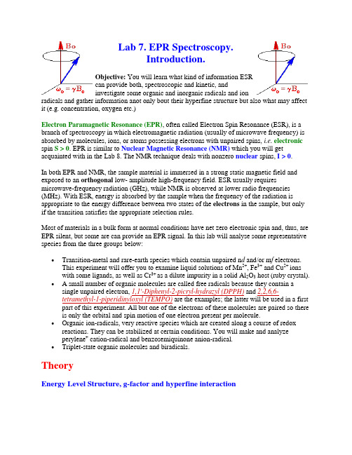
Lab 7. EPR Spectroscopy.Introduction.Objective: You will learn what kind of information ESRcan provide both, spectroscopic and kinetic, andinvestigate some organic and inorganic radicals and ionradicals and gather information anot only bout their hyperfine structure but also what may affect it (e.g. concentration, oxygen etc.)Electron Paramagnetic Resonance (EPR), often called Electron Spin Resonance (ESR), is a branch of spectroscopy in which electromagnetic radiation (usually of microwave frequency) is absorbed by molecules, ions, or atoms possessing electrons with unpaired spins, i.e.electronic spin S > 0. EPR is similar to Nuclear Magnetic Resonance (NMR) which you will get acquainted with in the Lab 8. The NMR technique deals with nonzero nuclear spins, I > 0.In both EPR and NMR, the sample material is immersed in a strong static magnetic field and exposed to an orthogonal low- amplitude high-frequency field. ESR usually requires microwave-frequency radiation (GHz), while NMR is observed at lower radio frequencies (MHz). With ESR, energy is absorbed by the sample when the frequency of the radiation is appropriate to the energy difference between two states of the electrons in the sample, but only if the transition satisfies the appropriate selection rules.Most of materials in a bulk form at normal conditions have net zero electronic spin and, thus, are EPR silent, but some are can provide an EPR signal. In this lab will analyse some representative species from the three groups below:∙Transition-metal and rare-earth species which contain unpaired n d and/or m f electrons.This experiment will offer you to examine liquid solutions of Mn2+, Fe3+ and Cu2+ ionswith some ligands, as well as Cr3+ as a dilute impurity in a solid Al2O3 host (ruby crystal).∙ A small number of organic molecules are called free radicals because they contain a single unpaired electron, 1,1'-Diphenyl-2-picryl-hydrazyl (DPPH) and 2,2,6,6-tetramethyl-1-piperidinyloxyl (TEMPO) are the examples; the latter will be used in a first part of this experiment. All but one of the electrons of these molecules are paired so there is only the orbital and spin motion of one electron present per molecule.∙Organic ion-radicals, very reactive species which are created along a course of redox reactions. They can be stabilized at certain conditions. You will make and analyzeperylene+ cation-radical and benzosemiquinone anion-radical.∙Triplet-state organic molecules and biradicals.TheoryEnergy Level Structure, g-factor and hyperfine interactionIn EPR, because of the interaction of the unpairedelectron spin moment (given by two projections, m s= ± 1/2, for a free electron) with the magnetic field,the so-called Zeeman effect, different projections ofthe spin gain different energies, as shown below andon the figure to the right:E m s = g μB B o m s (1)Here B o is the field strength of the external magnetic field. The SI units for magnetic field is tesla,T, but, historically in EPR, gauss (1 G = 0.0001 T) is still used. Other terms in Eq.(1): m s - is a spin projection on the field (m s = ± 1/2 for a free electron), μB is the Bohr magneton: μB = |eh/ 4πm e |= 9.2740 x 10⁻²⁴ J/T (2)with e and m e beeing electron charge and mass, respectively, and h-Planck's constant. Parameter g for free electron, g e , has the value close to two: g e = 2.0023193. If the electron has nonzero orbiatl angular moment, L , the g -value (sometimes called factor Landé) becomes:g = 1 + S(S + 1) - L(L + 1) + J(J + 1) (3)2J(J + 1) The overall magnetic momentun, μeff , can be expessed via overall angular momentum, J , and the g-value:μeff = g μB [J (J + 1)]1/2 (4)For most of organic radicals and radical ions, unpaired electrons have L close to zero and the total electron angular momentum quantum number J is pretty much the spin quantum number, S . As result, their g-values are close to 2. Situation becomes much more complicated with transition metals. Not only they have large L 's and S 's, but these values depend on the surrounding electric fields of ligands, making everythings messier but also more interesting. In this case, Eq.(1) should be written as E mJ = g J μB B o m J , stating that the Zeeman splitting appears in accordance with the total angular momentum projection, m J . If L = 0 then J = S, and Eq.(1) will define the energies of all the possible projection of m s from -S to S - 1, S (2S + 1 of such).If the molecule contains nuclei with magnetic moments, such as protons, their interaction with external field and the electronic magnetic moment will change stationary energies of Eq.(1). The nuclear angular momentum quantum number I determines the nuclear magnetic moment the same way as for the electron:μ = g N μN [I (I + 1)]1/2 (5)with μN now being the nuclear Bohr magneton:μN = eh/ 4πm p = 5.051 x 10⁻²⁷ J/T (6)much smaller value because of the ~2000 times more heavier proton m p . The nuclear g factor, g N , is obtained from a knowledge of the structure of the nucleus. Some of them are given here . Interaction with external field splits the nuclear sublevels due to Zeeman interaction the same way as for electron spin:E m I = g N μN B o m I (7)where m I is one of the 2I + 1 projections of the nuclear spin. Electron-nuclear interaction will depend on the projections of both, electron and nuclear spins:E electron-nuclear = A m I m s (8)where coefficient A , a so-called hyperfine coupling constant/interaction (hfi), depends not only on the g-values for the electron and the nucleus but also on the distance between them and their orientation with respect to the external field (dipole-dipole interaction). In solutions, the anisotropic part of this interaction averages out because of the fast molecular rotation. The remaining isotropic part is given by the Fermi contact interaction in the form:A = (8π/3)gN μN g e μB ρ(0) (9)where ρ(0) = |ψ(0)|2 is the unpaired electron density at the nucleus.For pure 1s electron on hydrogen atom, A equals h x 1420 MHz.Some other hyperfine constants can be found here . As a matter ofconvinience, hyperfine constants are usually given in Gauss, a =A /g e μB . For example, hydrogen it corresponds to a = 508 G. Summing up all energies, we arrive at modified Eq. (1):E = g eμB m s(B o + ∑ a i m Ii) - g NμN B o m I(10)You should note two important things:∙the signs in front of g e and g N are different because of the opposite charges of the electron and the proton, causing α spin of the electron to be higher in energy than β, but βN spin of the proton to be higher in energy than αN∙the magnetic field on the electron differs from the B o by a integer times a i and the sign of this cange is opposite for α and β electron spinsHydrogen atom example is given on the right. In EPR, allowed transitions correspond to the change of only m s:∆m s = ± 1, ∆m I = 0 (11)i.e. electronic spin "flips" but the nuclear one remains unchanged. Thus, in the figure on the right for hydrogen atom, only two transitions can be observed for a single resonant frequency hν. (This is a typical mode of EPR signal detection: microwave frequency is kept constant and magnetic field is varied). These transitions are separated by the hyperfine constant a (508 G for hydrogen atom). If there are more than one hydrogens, each of them will contribute to the changein magnetic field on the electron. All combinationsof the nuclear spin projections should be includedbut (EPR allowed) transitions only betweensublevels with the same configurstions of nuclearspins will be realized. For the case of four equivalentprotons, a simple ladder propagation scheme (Figureon the left) illustrates how to construct the EPRspectrum. Each new step corresponds to one moreproton added. In the first step, two lines appearcorresponding to the two different orientations of m I= -1/2 and m I = + 1/2. In the second step, each linesplits into two, corresponding to the two differentorientations of m I = -1/2 and m I = + 1/2, of thesecond proton.Since the two protons are equivalent,the splitting is the same on each step, resulting inthree lines of 1:2:1 intesity ratio. For the four protons,there will be 5 lines with the 1:4:6:4:1 intensity ratio.Lines with the lengths of these ratio separated byappropriate constants are called to represent a stickdiagram for the radical.If the spin of the nucleus is greater than 1/2, as in thecase of 14N ( I = 1), it splits each of the electroniclevels, α and β, into the 2I + 1 sublevels resulting in2I + 1 obeserved lines of equal intensity. In mostcases, one observes a combination of different nuclei with different constants which sometimes is not as easy to interpret. Example of a radical with twodifferent groups of hfi constants and different nuclei isshown on the right for a radical with one nitrogen and twoequivalent hydrogens (e.g. NH2).The magnitude of the hyperfine constants in a radical orradical ion reflects the extent of s character in its orbital(only s-orbital has nonzero density at the nucleus).Sometimes it is quite tricky how s -orbital gets populated.For example, we think of aromatic ion radicals as beingpurely π-systems, as you will see for yourselves, there aresubstantial hfi constants on there hydrogens. McConnell6showed that a aprt of the unpaired π-electron density istransferred through the C-H sigma bonding electrons to theH nucleus via exchange interactions. He provided a simpleformular which links the a H on a hydrogen with theunpaired electron spin density on the nearest carbon atomρπ:a H (G) = - 22.5ρπ(12)The signal intensity in EPR depends on how much absorption of the microwave power takes place. This value will be the larger the greater is the difference between populations of the α and β electron spin states. Following Boltzmann statistics, the latter can be estimated :(nα- nβ)/nβ = e-∆E/kT - 1 ~ gμB B o/k B T (13)a small value for regular X-band EPR spectrometer (9.5 GHz), which can be substanially increased by lowering the temperatute.The Lineshape andKineticsFigure on the rightshows how EPR signalshape of a stable radical,TEMPO (see itsstructure on the left),varies withconcentration(intensities were scaledto the same hight). It illustrates that thelinewidths of the resonant transitions are notinfinitely small and can be altered by theenvironment. At low concentrations, each line has a width because of the heterogeneous broadening, i.e. unresolved structure of the small hyperfine constants and the homogeneous broadening, Γo:Γo = 1/γe T2(14)which is caused by a limited lifetime T2 (also called a spin-spin relaxation time) of a radical at a particular spin state (uncertainty principle related). The constant γe is called the magnetogyric (or gyromagnetic) ratio and relates to Bohr's magneton, μB,γe = 4πμB/h = e/m e c = 1.77 x 10⁷ s-1 G-1(15)The full width half maximum, Γ, is 31/2/2 = 0.866 times greater than the measured distance between maximum and minimum of the experimentaly detective derivative of absorption, i.e.Γ= 0.866 ∆. This lifetime can be affected by the dipole-dipole (and exchange) interaction with other radicals. If another unpared electron is at a distance r, the magnitude of its field at the spin can take any value between ± 2μB/r3, depending on orientation. Due to the fast rotation of both radicals, the interaction averages this field to zero except for a very short period of time during collision after which the radical can end up at a random configuration of its nuclear spins. Result of such randomization is equivalent to the jumping from one spectral position to another. For nitroxyl radical, such as TEMPO, with three equivalent spectral lines (S = 1), only 2/3 of such collisions result in a shift. Beacuse of that,the width will increase only at 2/3 of the rate of collision:Slow Exchange:Γ = Γo + 2k[R]/3γe(16)where k is the collision rate constant and [R] is the concentration of radicals. With further increase of the radical concentration, the continuing broadening will be accompanied by a measureable decrease in separation between the peaks, ∆B:Intermediate Exchange:(aN 2 - ∆B2 )1/2 = 2k[R]/3γe? (17)where a N is the hyperfine constant on nitrogen (separation between lines). After they coalesce, the overall width of now a single-line spectrum, starts decreasing:Rapid Exchange:Γ = γe(aN2)/k[R] (18)The radicals can exchange spins not on themselves but on any other radical as well. Oxygen has nonzero spin and can also cause the line broadening by the same mechanism. So if your solvent is nonviscous and you want to detect linewidths below 3 G, you may want to get rid of O2.Experimental apparatusThe ESP-300 ESR spectrometer (Bruker)consists of an electromagnet with powersupplies to generate and modulate a uniformmagnetic field of several thousand Gauss, aswell as the components that generate anddetect microwave power. The operationmanual is awailable on the web, courtesy ofthe University of Illinois at Urbana-Champaign. I am putting our version as well.A static magnetic field is provided by anelectromagnet with a current- regulatedpower supply. A homogeneous field isrequired for best results. A Hall probe, drivenfrom a stable constant-current power system,with a digital multimeter (DMM) reading theHall voltage, is used to measure the value ofthe magnetic field between the poles of themagnet.The microwave system consists of a microwave power supply (on a table above the magnet) which uses a clystron. The ouput of the microwave power (mw) supply is connected via rectangular waveguide (10 GHz) and through a circulator to a high-Q resonant cavity. The samples to be investigated are mounted in the middle of the cavity, where magnetic component of the microwave power has a maximum and is oriented perpendicular to the static field. A microwave diode, which detects the mw resides inside the same box as the power supply. The higher the quality of the resonant cavity the greater the microwave field can be obtained on the sample. Very polar solvents like water absorb efficiently on microwave frequencies themselves (remember microwave ovens?). Because of that, water samples have to be made in narrow cappilaries and carefully located in the center of the cavity. Most of our measurements will be done in the 0.1 - 1.0 mW range.There are variety of schemes for detectingresonant EPR transitions. Historically, mostpopular became a detection of absorption of themicrowave power. To minimize the noise fromthe mw diode in steady state measurements, amagnetic field modulation scheme with phasesensitive detection is usually employed. As aresult, the detected signal appears as a firstderivative (see figure on the right). In the Brukerspectrometer there is choice between 12.5 kHzand 100 kHz modulation frequency. Theamplitude of field modulation (MA) can bevaried up to 40 G (4mT). Yo have to be carefulin choosing appropriate MA. If MA is to big, thewidth of detected signal derivative would be wider than the real width of absorption, but if it is too small, the signal intensity would be less. For most of your case it is good first approximation to set MA between 0.5 and 1.0 G.References1.Pake G. E., Paramagnetic Resonance, (W. A. Benjamin, 1962).2.Wertz J.E., Bolton J.R. Electron spin resonance. - New York, McGraw-Hill BookCompany, 1972.3. C. P. Slichter, Principles of Magnetic Resonance (CLAS), 3rd ed., (Springer-Verlag,1992 and Harper & Row, 1963), p. 65.4.Van Vleck J. H., Phys. Rev., 74, 1168, (1948).5.Feynman, Leighton, and Sands, Lectures on Physics, (Addison-Wesley PublishingCompany, 1965), Vol. II, Chap. 23 & 24.6.McConnell, H.M. J. Chem. Phys. 24, 764 (1956)Last updated .。

Lab工作职责1. 简介实验室(Lab)是一个专门用于科学研究和实验的场所。
在实验室中,科学家、研究人员和学生进行各种实验、观察和测试,以推进科学知识的发展和应用。
Lab工作职责是指在实验室中从事的具体工作内容和职责。
2. 实验设备维护在实验室中,有各种各样的实验设备,如显微镜、离心机、pH计等。
Lab的工作人员负责对这些设备进行维护和保养,确保其正常运行。
具体职责包括:•定期检查设备的工作状态,发现问题及时修理或更换损坏部件;•清洁设备,保持其清洁卫生;•校准设备,确保测量结果准确可靠;•制定设备使用规范,并培训用户正确使用设备。
3. 实验材料采购与管理实验室中需要大量的实验材料,如试剂、培养基、玻璃器皿等。
Lab的工作人员负责采购这些材料,并进行管理。
具体职责包括:•根据实验需求,制定采购计划;•寻找供应商,比较价格和质量,选择合适的材料;•跟踪订单进展,确保材料按时到达实验室;•对收到的材料进行检查和验收,确保其符合要求;•做好库存管理,避免材料短缺或过期。
4. 实验数据记录与分析在实验过程中产生大量的数据,Lab的工作人员负责记录和分析这些数据。
具体职责包括:•设计和建立数据记录系统,确保数据的准确性和完整性;•及时记录实验过程中的关键信息和结果;•对实验数据进行整理、归档和备份;•进行统计分析,并根据需要生成报告或图表。
5. 实验安全管理实验室中存在一定的安全风险,因此Lab的工作人员需要负责实验室的安全管理工作。
具体职责包括:•制定并执行实验室安全规章制度,确保人员遵守安全操作规范;•提供必要的安全培训,并组织演习、讲座等活动提高人员的安全意识;•定期检查实验设备、仪器和实验室环境,发现安全隐患并及时处理;•处理实验事故,提供急救措施和紧急处理指导。
6. 科研支持与合作Lab的工作人员为科研人员提供支持,并与其他实验室合作开展科研项目。
具体职责包括:•协助科研人员进行实验设计和方案制定;•提供技术支持,解决实验过程中的问题;•参与科研项目的讨论和交流,并提出建议;•与其他实验室建立联系,开展合作项目。

人音版六年级上册第7课《七色光彩》教学设计一. 教材分析《七色光彩》是人音版六年级上册第7课的一首歌曲,歌曲旋律优美,歌词富有想象力,通过七个颜色的描绘,展现了丰富多彩的世界。
歌曲为C大调,2/4拍,共四乐句,结构简洁明了。
这首歌曲旨在培养学生的音乐审美能力,提高他们对音乐的感知和表现力。
二. 学情分析六年级的学生已经具备了一定的音乐基础,对旋律、节奏、音色等方面有了一定的认识。
他们在学习过程中善于观察、思考,勇于表达自己的观点。
然而,部分学生对音乐理论知识的掌握仍有待提高,因此在教学过程中需要注重理论与实践相结合。
三. 教学目标1.让学生能够熟练地演唱《七色光彩》这首歌曲,体会音乐的美感。
2.培养学生对色彩的感知和表现能力,提高他们的音乐审美水平。
3.引导学生掌握简单的音乐理论知识,如音高、节奏、音色等。
4.培养学生的团队协作精神,提高他们的综合素质。
四. 教学重难点1.音高:歌曲中有一些音高较高的音符,学生需要正确把握。
2.节奏:歌曲中的节奏变化较多,学生需要熟练掌握。
3.音色:歌曲中采用了多种音色,学生需要了解并能够正确表现。
4.音乐理论知识:学生需要掌握简单的音乐理论知识,如音高、节奏、音色等。
五. 教学方法1.情境教学法:通过创设情境,让学生在情境中感受音乐的美感。
2.互动教学法:引导学生相互交流、合作,提高他们的团队协作能力。
3.启发式教学法:引导学生主动思考、探索,培养他们的创新精神。
4.视听结合法:利用多媒体设备,让学生在视听中感受音乐的魅力。
六. 教学准备1.教学课件:制作课件,展示歌曲旋律、歌词、图片等。
2.教学乐器:准备钢琴、吉他等乐器,为学生提供音乐伴奏。
3.教学素材:准备与歌曲相关的色彩图片、音乐理论知识资料等。
4.录音机:用于播放歌曲,让学生跟唱。
七. 教学过程1.导入(5分钟)利用课件展示歌曲旋律、歌词、图片等,引导学生初步感受《七色光彩》的魅力。
同时,提问学生对色彩的认知,激发他们的学习兴趣。


品种:黎平杂边禾野生型lab7测定时间:2014年3月记录人:
备注:地点:三亚南繁基地
品种:9311 lab1测定时间:2014年3月记录人:
备注:地点:三亚南繁基地
品种:黎平杂边禾半矮化lp srs-1lab11测定时间:2014年3月记录人:
备注:地点:三亚南繁基地
品种:lpsrs-1×9311 F0代lab15测定时间:2014年3月记录人:
备注:需要采集叶片提取大量的DNA。
地点:三亚南繁基地
品种:Bgsd-2×银桂粘lab52测定时间:2014年3月记录人:
备注:白色颖壳、黑色颖壳、矮化、高杆各采集30株叶片提取DNA。
地点:三亚南繁基地
品种:Bgsd-2lab9测定时间:2014年3月记录人:
备注:地点:三亚南繁基地
品种:osgr lab63测定时间:2014年3月记录人:
备注:地点:三亚南繁基地
品种:osgr×银桂粘 F2lab64 测定时间:2014年3月记录人:
备注:地点:三亚南繁基地
品种:银桂粘lab64 测定时间:2014年3月记录人:
备注:地点:三亚南繁基地
水稻农艺性状测定记录表
品种名称:测定时间:年月日记录人:。
官网:https:///安装参考网址:https:///downloads/环境Ubuntu 14.04如果是虚拟机的话要改成桥接模式让局域网内其他计算机访问网络设置1.宿主机网络配置:连接特定的 DNS 后缀: pnp.gw描述: Intel(R) WiFi Link 1000 BGN物理地址: 74-E5-0B-99-52-22已启用 DHCP: 是IPv4 地址: 172.31.0.229IPv4 子网掩码: 255.255.248.0IPv4 默认网关: 172.31.0.1IPv4 DHCP 服务器: 172.31.0.1IPv4 DNS 服务器: 223.5.5.5, 223.6.6.6根据宿主机手动配置虚拟机的网络2.虚拟机网络配置:IPv4 地址: 172.31.0.199IPv4 子网掩码: 255.255.248.0IPv4 默认网关: 172.31.0.1IPv4 DNS 服务器: 223.5.5.5, 223.6.6.6检查下虚拟机能否上网。
如果不能上网的会影响后面GitLab发送邮件。
检查下能否被局域网的其他计算机访问安装下载1.Download the package and install everything默认管理员账号密码:Username: rootPassword: 5iveL!feFor troubleshooting and configuration optionshttps:///gitlab-org/omnibus-gitlab/blob/master/README.md 配置1.配置postfixsudodpkg-reconfigure postfix选择internetdomain:hostname:smtpserver:destination:localhost备份下原来的配置sudocp /etc/postfix/main.cf/etc/postfix/main.cf_bak修改配置填写smtp服务器登录信息:添加smtp登录信息:sudopostmap /etc/postfix/sasl_passwdsudo postfix reloadsudo postfix stopsudo postfix start2.配置GitLabexternal_url,虚拟机的ip地址external_url "http:// 172.31.0.199"GitLab的邮件地址:最好是smtp邮件发送服务器的邮箱地址gitlab_rails['gitlab_email_from'] = 'yuanzhencai1990@'重启GitLabsudo gitlab-ctl reconfigure查看GitLab状态查看GitLab日志sudogitlab-ctl tail至此,GItLab配置结束使用访问使用虚拟机ip地址访问:172.31.0.199,默认管理员账号登录。