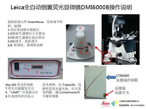
LeicaDMILM倒置金相显微镜-金相显微镜-USFEN
- 格式:pdf
- 大小:570.21 KB
- 文档页数:9
卡尔·蔡司:论金相显微镜倒置显微镜金相显微镜倒置的最初使用是通过传统金相显微镜倒置显微镜来了解微观世界,然而传统金相显微镜倒置显微镜的存在分辨率的极限。
继而不断发展出适合研究分析的高分辨率金相显微镜倒置显微镜,目前市场上的金相显微镜倒置显微镜不仅具有分辨率高的优势,同时满足ICCS无限远复消色差校正及反差增强型光学系统;更高衬度、更高分辨率的EC物镜;ADF高级暗场;圆偏光观测技术;高端FEM设计可以隔热、无振动观测。
蔡司金相显微镜倒置显微镜为您提供全系统、全方位的金相显微镜倒置显微镜实验室应用、金相显微镜倒置显微镜检测解决方案。
蔡司金相显微镜倒置显微镜拥有各领域客户群,金相显微镜倒置显微镜客户覆盖全国各地区,我们拥有专业的服务团队,为您提供金相显微镜倒置显微镜咨询、金相显微镜倒置显微镜报价、金相显微镜倒置显微镜技术服务、金相显微镜倒置显微镜免费试样拍摄等多项高端订制服务!。
leica dmi8倒置荧光显微镜工作原理
Leica DMI8倒置荧光显微镜工作原理基于倒置的光路径设计,通过逆向放大观察样品。
具体工作原理如下:
1. 光源:Leica DMI8倒置荧光显微镜通常采用激光或者弧光
灯作为光源。
光线通过光源进入显微镜系统。
2. 激发光源与滤光片:激发光源产生的光通过滤光片进行过滤。
滤光片选择能够激发样品荧光的波长,而过滤其他多余的光线。
3. 物镜:经过滤光片后的光线进入物镜。
物镜有不同的倍数,可以放大样品。
光在物镜中被聚焦,形成一个局部的高强度光点。
4. 分光镜:经过物镜后的光线进入分光镜。
分光镜表面涂有特殊的薄膜,将激发光和荧光信号分开。
激发光线被反射到目镜或相机,荧光信号则透过分光镜射向样品。
5. 样品:样品通常是放置在一个液体培养皿或者玻璃底片上。
荧光染料标记的生物标本或者细胞,会在激发光的作用下发出荧光信号。
6. 荧光信号:样品中的荧光染料吸收激发光子,高能激发光子的能量会导致荧光染料的电子跃迁到一个高能态,当电子回到基态时,会释放出激光波长较长的荧光信号。
7. 探测荧光信号:荧光信号通过目镜或者相机进行观察和记录。
荧光信号的特点可以提供有关样品的信息,如位置、结构和代谢活性等。
通过上述步骤,Leica DMI8倒置荧光显微镜能够通过荧光信
号对样品进行观察和分析。
徕卡光学显微镜操作方法徕卡光学显微镜是一种高质量的显微镜,使用起来需要一些操作方法和技巧。
下面,我将为您详细介绍徕卡光学显微镜的操作方法。
第一步:调节光源1. 打开显微镜底部的电源开关,通过转动亮度调控旋钮来调节光源的亮度。
2. 使用多种光源如白光、蓝光、绿光等,可以根据所观察的材料和需要来选择合适的光源,并通过切换按钮进行调节。
第二步:准备样本1. 取出待观察的样本,并使用切片机或切片刀等工具将样本切割成适当大小的薄片。
2. 将样本置于载物台上,通过定位螺丝将样本固定在载物台上,确保样本位置合适。
第三步:调焦1. 打开眼镜盒,并将目镜与眼镜盒上的刻度对齐。
2. 通过调节观察管的升降装置,将目镜与样本尽可能靠近,但不接触样本,然后通过调节调焦轮转动机构,使样本清晰可见。
3. 要注意调节焦距时,先用粗焦轮将样本基本对焦,再通过微调焦轮进行精细调节。
第四步:调节放大倍数1. 徕卡光学显微镜通常使用10倍至100倍的倍率,通过旋转目镜上的物镜切换器或者直接换上不同倍数的物镜,可以改变显微镜的放大倍数。
2. 每次切换倍率后,都需要重新调节焦距以保持样本的清晰可见。
第五步:调节光源方向和强度1. 通过显微镜底部的光源调节旋钮,可以调节光源的方向和强度,以便更好地观察样本。
2. 正常情况下,光源应该从样本底部照射并通过样本透射到目镜中。
第六步:观察和记录1. 调节好焦距和放大倍数后,即可通过目镜观察样本。
2. 可以使用调焦轮进行微调,以获取更清晰的图像。
还可以通过调节光源以改变样本的亮度和对比度,以便更好地观察细节。
3. 可以使用显微镜上的调节旋钮来对样本进行聚焦,旋转显微镜的聚焦盘或调节螺丝,观察到不同焦点的图像。
4. 还可以使用显微镜上的目镜游标盘来测量或记录样本的尺寸。
第七步:注意事项1. 在使用显微镜之前,要确保工作区域干净整洁,并保护好显微镜以防止刮擦或污染。
2. 使用显微镜时,要轻柔地操作,避免碰撞和震动。
倒置金相显微镜操作规程一、介绍倒置金相显微镜是一种用于观察金相组织和微观结构的特殊显微镜。
本文将详细介绍倒置金相显微镜的操作规程,帮助读者正确地使用该仪器,准确地观察样品,并获取准确的分析结果。
二、操作步骤2.1 准备工作在使用倒置金相显微镜之前,需要进行一系列的准备工作,包括: 1. 清洁显微镜外壳,保持仪器的整洁。
2. 准备好所需要的显微镜玻璃片、草图纸、铅笔等实验工具。
3. 根据实验需要,选择适当的目镜和物镜。
2.2 样品处理1.将需要观察的样品制备成适当的尺寸和形状,并确保表面平整和清洁。
2.对于金相显微镜观察,通常需要对样品进行打磨和腐蚀处理,以显现出金属组织的结构和特征。
2.3 装置样品1.打开显微镜盖板,将准备好的样品放置在镜台上。
2.利用显微镜的调焦装置,将样品调至合适的焦距。
2.4 观察样品1.调节目镜的放大倍数,选择适当的放大倍数以便观察样品。
2.利用显微镜的聚焦装置,调节合适的焦点,使得样品的细节清晰可见。
3.使用显微镜的可调光源,调整光源的亮度和角度,以便更好地观察样品。
2.5 记录和分析1.使用草图纸和铅笔,将观察到的样品结构绘制下来,标注重要的特征。
2.根据所观察到的样品结构,进行分析和解释,并与相关的理论知识进行比较。
三、维护和保养3.1 每次使用后1.关闭显微镜的电源,将镜头调至最低位置,盖好显微镜盖板。
2.清理镜头和物镜,使用干净的纱布轻轻擦拭,避免使用硬物或化学溶液接触镜头表面。
3.2 定期维护1.保持显微镜的清洁和整洁,定期清理外壳和镜头。
2.定期校准显微镜的聚焦装置,确保焦距的准确性。
3.定期检查显微镜的光源,确保亮度和角度的稳定性。
4.如发现异常情况或故障,应及时联系维修人员进行维修和保养。
四、安全注意事项1.使用显微镜时,应注意保护眼睛和身体安全,避免直视强光源和对镜头施加过大的压力。
2.使用化学腐蚀剂时,应佩戴防护手套和护目镜,避免溅溶液伤及皮肤和眼睛。
Leica荧光倒置显微镜使用指南1、按下接线板电源,打开计算机屏幕电源和主机箱电源,双击桌面上Leica Application Suite图标进入程序。
2、打开显微镜光源,如需使用荧光灯,打开荧光灯光源,在记录本上记录下荧光灯使用时间。
3、显微镜物镜放大倍数有5倍、10倍、20倍、40倍,旋转物镜下转盘改变放大倍数。
4、明场使用方法:将透射光装置的聚光镜转盘旋转至BF档。
选择合适的物镜,调节焦距,用明视光源强度旋转及透射光装置上的光圈调节光源强度。
将滤色块放在第4或5档。
5、相差使用方法:将透射光装置的聚光镜转盘转至PH1档(物镜为10×、20×)或PH2档(物镜为40×时)。
用明视光源强度旋转及透射光装置上的光圈调节光源强度。
将滤色块放在第4或5档。
(5×物镜无相差功能)6、荧光使用方法:放下明场遮拦,拉开荧光遮拦棒,旋转滤色块转盘,选择合适的激发波长。
用荧光光源视野调节旋钮和荧光光源光圈调节旋钮调节荧光的范围和强度。
7、图像处理:在“摄取”菜单栏Micl卡片下根据所选的物镜倍数选择放大倍数。
拉开摄像头操纵杆,单击鼠标左键选择一块白色区域,单击鼠标右键,进行白平衡。
可以通过改变摄取菜单栏摄像头卡片下曝光,饱和,伽马和增益改变现实图像的效果。
明场或者相差模式,建议将增益放在零。
单击拍摄,新建自己的文件夹,保存图片。
8、荧光拍摄完毕,推进荧光遮拦棒,看一下显微镜使用登记表。
(1)如果下一个使用登记时间小于或等于1个小时,不需要关闭荧光灯光源,记录下荧光灯光源使用时间。
将明场电源旋至最低。
将物镜旋至空挡,关闭应用程序。
填写完整仪器使用记录。
(2)如果下一个使用登记时间大于1个小时,使用完毕荧光及时关闭荧光灯光源,注意需长于半个小时,记录下荧光灯光源使用时间。
将明场电源旋至最低,关闭明场电源。
将物镜旋至空挡,关闭应用程序,关闭电脑。
填写完整仪器使用记录。
倒置荧光显微镜使用注意事项1、使用显微镜前,请和仪器负责人联系。
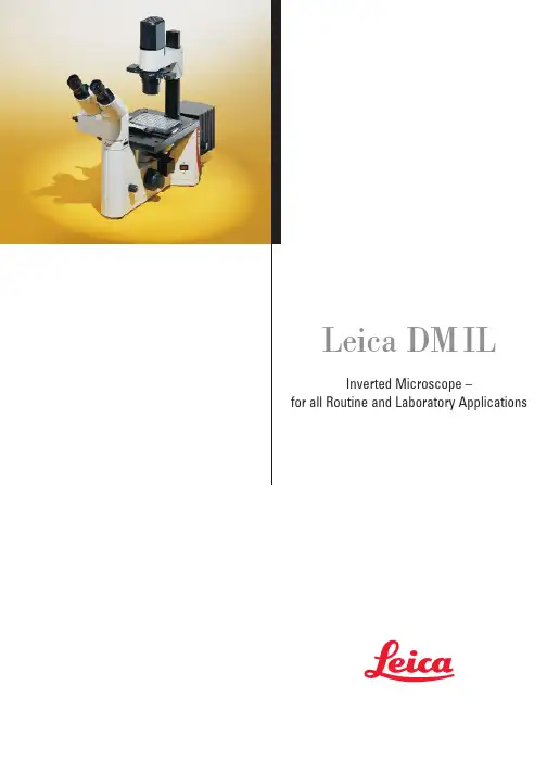
Leica DM ILInverted Microscope –for all Routine and Laboratory Applications2Leica Design by Ernest Igl /Christophe ApothélozLeica DM IL – compact inverted microscope for laboratory routine The new inverted Leica microscope blends ergonomy, a compactdesign and effective contrasting methods into a system for virtuallyunlimited life science applications.The integration of Leica HCS* optics extends the range of objectivesfor inverted microscopy.For the first time, high-quality relief contrast can be producedwithout special objectives with our new Integrated ModulationContrast (IMC) technique.Optimised phase contrast and brilliant incident light fluorescencemake the Leica DM IL the number one choice in contrastingmicroscopes.3With its unbeatable modularity, ergonomy and free view of thespecimen, teamed with newly developed and optimised contrastingtechniques, the Leica DM IL offers you a top level introduction toinverted microscopy.The adaption of the Leica DM IL to infinity optics allows theintegration of Leica HCS* components for superb image resolution,brilliant contrast and precise colour rendering. The DM IL is theinverted equivalent of our successful upright DM microscopes ofthe L and R class.The Leica DMIL is a microscope for all applications in microbiologyor the cell culture laboratory. A universal inverted microscope forroutine use: stable and space-saving, flexible and upgradablewith optics from Leica’s research microscopes.* Harmonic Component System Microinjection of oocytes in mouseRNA microinjection of frog oocytes(Xenopus)4The stand The stand of the Leica DM IL microscope is stable, aluminium-cast and excellently designed. There are two versions for biological applications:The DM IL for brightfield, phase and modulation contrast and the DM IL with additional incident light fluorescence unit.Appreciated by users for many years, the stable T-shaped micro-scope base offers plenty of valuable space round the microscope and ensures comfortable, fatigue-free microscopy. The micro-scope’s footprint is optimised to provide the necessary room for experiments, and all controls are ergonomically located. Many different components can be adapted to suit individual require-ments. The high stability, low centre of gravity and four vibration damping feet eliminate vibrations even in extreme conditions. The excellent stability of the DM IL also makes it the ideal solution for photography with long exposure times. In addition, finite element calculations and thorough practical testing in a wide variety of applications guarantee focusing which is not only ultra precise but also stable over long periods of time.The Leica DMIL contrasting microscopeThe System Discussion unitCardiac muscle cells5Due to its modularity, the Leica DM IL is particularly suitable forliving cell microscopy.Its modern, practical design, the integration of state-of-the-art,top quality optics and the excellent standard of the adaptedcontrasting techniques prove useful in research tasks as well asroutine applications.You will be convinced by the DM IL’s first-class technology andmany innovative and practical ideas.We at Leica believe microscopy should be a pleasurable experienceand designed the DM IL to be associated with enjoyment at thelaboratory workplace.Trinocular tube with DC 100Ergonomic phototubeTrinocular tube with MPS DM IL with illumination column the other way round6Nosepiece focusingSamples are focused with the quadruple objective nosepiece.The reliability, stability and precision of the focusing is not influenced by the microscope stage and the samples on it or by accessory components such as the object guide and manipulators.Illumination system The compact brightfield illumination unit is attached to a column and can be comfortably adjusted. Setting the correct height for the condenser used or the specimen on the stage is facilitated by markings along the column. The pre-centred, extremely powerful 6V 35W halogen lamp provides optimal illumination even of critical specimens. The transmitted light illumination concept is rounded off by the integration of the contrast slide (for phase and modulation contrast), the module for light filters with 32 mm diameter and the aperture diaphragm. The microscope stage with illumination arm can be turned round by 180°and is freely accessable for sample positioning from three sides.Light filters The filter module on the illumination column accommodates 32 mm diameter filters in a spoon-shaped holder. We are constantly adding to our wide range of filters, enabling you to selectively optimise the illumination for observation and image documentation.Built-in 6V 35W power supply The 6V 35W power supply, which provides the full lamp power including on/off switch, status indicator and brightness adjustment,is fully integrated in the microscope stand. Apart from the ergonomic advantages, this saves space on the microscope desk and allows the microscope to be easily picked up as a unit and moved elsewhere.The Technology7CondensersThe Leica DM IL offers you a choice of two condensers.The S 90/0.23 condenser with a free working distance of 90 mm and a numerical aperture of 0.23 is designed for brightfield and phase contrast and is particularly suitable for specimens in bulky laboratory vessels.The S 55/0.35 condenser with a free working distance of 55 mm and a numerical aperture of 0.35 for bulky containers is designed for brightfield, phase and integrated modulation contrast (IMC)and is particularly suitable for higher magnifications or thick specimens.Without a condenser, the maximum free working distance is 200 mm.Stage and accessoriesThe DM IL offers a wide variety of stages with a whole array of accessories and different inserts for your specimen vessels:The standard stage is a fixed stage plate of 252 x 212 mm. The stage can be widened by 70 mm on both sides by adapter plates. The interchangeable stage inserts (20-50 mm) allow smaller petri dishes to be used as well without losing the focus when the objective nosepiece is rotated.Object guides can be attached to both the left and right of the stage and have a minimum adjustment range of 83 x 127 mm. The control of the coaxial drive is in an ergonomically low position so that you can rest your hands on the desk while scanning specimens.The object guides accommodate special and multi-purpose frames for all types of culture vessels.A heating stage up to max. 45°C, a 3-plate mechanical stage and scanning stages round off the range of stages for the DM IL.Micromanipulation ...... and scanning stage8DM LB tubeEyepieces and objectivesThe optics are the heart of every microscope and decisive for the quality of information. We have set new standards here by intro-ducing our HC optics.The Leica DM IL is designed for brightfield, phase contrast, Inte-grated Modulation Contrast (IMC) and incident light fluorescence.All infinity-corrected high performance objectives in the Leica range with 25 mm screw thread are compatible with the DM IL microscope.Even earlier-type Leica objectives can be adapted for use on the DM IL. We offer a wide range of special objectives for inverted microscopy applications with long free working distances (L objectives) and/or with correction mounts (Corr objectives) to compensate for different vessel thicknesses. The latest Leica optics brochure features our whole range of objectives.Depending on the tube configuration, there is a wide choice of eyepieces with magnifications 10x, 12.5x, 15x, 16x or 25x, suitable for fields of view up to 20 mm. Besides special high-point eyepieces for eyeglass wearers, we also supply eyepieces with adjustable eyelens (M eyepieces), into which different types of graticule can be inserted.The DM IL range also comprises many different observation and photo tubes. The tubes are interchangeable and can be individually rotated by 360°in the tube mount and then fixed in position. All tubes are fitted with an infinity tube lens 1x. The following tubes are used on the DM IL:•Binocular tube ILB, with 45°viewing angle, for eyepieces with 23.2 mm outer diameter •Trinocular (photo)tube ILT, with 45°viewing angle, for eyepieces with 23.2 mm outer diameter, with vertical photo/TV exit with switchable light path for either 100% visual or 100% photo/TV.The position of the photo/TV exit 88 mm to the side of the tube has the special advantage that it allows an unobstructed view of the specimen.Other tubes from the Leica DM L range can be used via an IL/L adapter:•Binocular tube HC LB 45°viewing angle •Binocular tube HC LVB 0-35°ergotube •Trinocular (photo)tube HC L1T 45°viewing angle •Trinocular (photo)tube HC L3T 45°viewing angle •Trinocular (photo)tube HC LV1T 0-35°ergotubeThe Optics351+364 mn UVAr 488mn 568mn 647mn Ar/Kr 4905205575766486703504004505005506006507009Developments in diagnostics and med./biol. research (e.g. fluores-cence applications) and the increasing use of video technology and electronic image processing have to be paralleled by intelligent technical adaptations of the microscope system.The new HCS optics concept introduced with the Leica DM R microscope meets this requirement. It is the result of an integral system consideration, harnessing all technological potential.The abbreviation HCS stands for Harmonic Component System.Its special features are:•well-balanced optical and mechanical fitting dimensions•harmonious balance of all optical system components, i.e. the parameters contributing to the microscope’s performance (objectives, tube lenses, tubes, eyepieces, TV cameras/adapters,etc.) have been harmonised throughout the entire optic system.This has created scope for even greater optical opportunities.The HCS system is the answer to your application requirements not only today, but in future, too.Rat testicles, IMC10The Leica DM IL is the microscope for all requirements in the cell culture lab. A universal inverted microscope for routine application: stable and space-saving, flexible and upgradable with optics from Leica research microscopes.BrightfieldThe whole range of objectives from 4x-100x magnification can be used for brightfield applications. Samples in almost any kind of vessel can be examined with or without a condenser, while a 6V 35W halogen lamp ensures optimal illumination.Phase contrastIn vivo/ in vitro microscopy specimens are mostly living cultures or microorganisms and are examined under sterile conditions. Contrast of the transparent tissue can only be enhanced by optical methods.Phase contrast is a useful technique for high-contrast imaging of unstained specimens. The phase contrast technique used by Leica on the DM IL has been optimised for inverted microscopy applications and produces equally excellent contrast in watery solutions and of dry preparations in petri dishes.Spinal cord, catContrastingLymphocyte toxicity testdouble staining, strongly positiveLymphocyte toxicity testdouble staining, weakly positive11Human brain Femtotip microinjection needle (Photo: Eppendorf)FibroblastsIMC (Integrated Modulation Contrast)The innovative technique of Integrated Modulation Contrast (IMC)now introduced by Leica in the DM IL is based on Hoffman’sprinciple and produces this contrast without the need for specialobjectives–ordinary brightfield or phase contrast objectives canbe used. The Leica IMC provides a high-contrast, 3D image oftransparent objects similar to that of interference contrast. Plasticculture vessels do not impair the quality of the image as thetechnique is polarisation-neutral.The diaphragm slide on the side of the illumination and theswitchable modulator in the intermediate image of the pupilproduce the type of contrast named after Hoffman without modi-fying the objectives. High contrast, high resolution, a halo-freerelief image of either stained or unstained specimens make Leica’sIMC a new standard in the class of inverted routine microscopy.FluorescenceThe fluorescence model of the DM IL reflects the growing signifi-cance of fluorescence for in vivo/in vitro microscopy. The maincomponents of this configuration are an incident light axis integrat-ed in the microscope stand, incorporating a fluorescence slide forthree filter cubes. A wide range of light sources with multi-lens,chromatically corrected collectors brighten up even the weakestfluorescence. The fluorescence filter cubes comprise an optimallymatched combination of excitation, reflection, band-pass andbarrier filters. We are constantly updating our range of filtercubes to keep pace with the latest challenges in biology andmedicine.Transmitted light techniques can be used simultaneously or inalternation in order to clearly allocate fluorescent and non-fluorescent structures.The Leica DM IL offers you a powerful system for immunology,cytopathology, virology – in fact wherever fluorescence techniquesare used in combination with inverted microscopy.Leica Microsystems – the brand for outstanding products Microscopes Compound Stereo Surgical Laser Scanning Photomicrography Video Microscopy Measuring Microscopes Advanced Systems Image Analysis Spectral Photometry Automated Inspection Stations Measurement Systems Electron Beam Lithography Laboratory equipment Tissue Processors Embedding Systems Routine- & Immunostaining Coverslippers Refractometers Microtomes Sliding, Rotary & Disc Cryostats Ultramicrotomes EM Sample Preparation Leica Microsystems’ Mission is to be the world’s first-choice provider of innovative solutions to our customers’ needs for vision, measurement, lithography and analysis of microstructures.Leica, the leading brand for microscopes and scientific instruments, has developed from five brand names, all with a long tradition: Wild, Leitz, Reichert, Jung and Cambridge Instruments. Leica symbolizes not only tradition, but also innovation.Leica Microsystems – an international company with a strong network of customer servicesAustralia:North Ryde/NSW Tel. +61 2 9879 9700Fax +61 2 9817 8358Austria:Vienna Tel. +43 1 495 44 160Fax +43 1 495 44 1630Canada:Willowdale/Ontario Tel. +1 416 497 2860Fax +1 416 497 8516Denmark:Herlev Tel. +45 4454 0101Fax +45 4454 0111Finland:Espoo Tel. +358 9 6153 555Fax +358 9 5022 398France:Rueil-Malmaison Tel. +33 1 473 285 85Fax +33 1 473 285 86CedexGermany:Bensheim Tel. +49 6251 136 0Fax +49 6251 136 155Italy:Milan Tel. +39 0257 486.1Fax +39 0257 40 3273Japan:Tokyo Tel. +81 3 5435 9600Fax +81 3 5435 9618Korea:Seoul Tel. +82 2 514 65 43Fax +82 2 514 65 48Netherlands:Rijswijk Tel. +31 70 4132 100Fax +31 70 4132 109Portugal:Lisbon Tel. +351 1 388 9112Fax +351 1 385 4668Republic of China:Hong Kong Tel. +852 2564 6699Fax +852 2503 4826Singapore:Tel. +65 779 7823Fax +65 773 0628Spain:Barcelona Tel. +34 93 494 95 30Fax +34 93 494 95 32Sweden:Sollentuna Tel. +46 8 625 45 45Fax +46 8 625 45 10Switzerland:Glattbrugg Tel. +41 1 809 34 34Fax +41 1 809 34 44United Kingdom:Milton Keynes Tel. +44 1908 246 246Fax +44 1908 609 992USA:Deerfield/Illinois Tel. +1 847 405 0123Fax +1 847 405 0030and representatives of Leicain more than 100 countries.Leica Microsystems Wetzlar GmbH Ernst-Leitz-Straße D-35578 Wetzlar (Germany)Tel. +49 (0)6441-290Fax +49 (0)6441-292599 ©L e i c a M i c r o s y s t e m s W e t z l a r G m b H •E r n s t -L e i t z -S t r a ße •35578 W e t z l a r •T e l. (06441) 29-0 •F a x (06441) 29-2599 G e d r u c k t a u f c h l o r f r e i g e b l e i c h t e m P a p i e r .B e s t e l l -N u m m e r n d e r A u s g a b e n i n : D e u t s c h 913 761•E n g l i s c h 913 762 •F r a n z ös i s c h 913 763 •I t a l i e n i s c h 913 764 •S a c h -N r . 501-068 G e d r u c k t i n D e u t s c h l a n d X I /98/A X /B .H .。
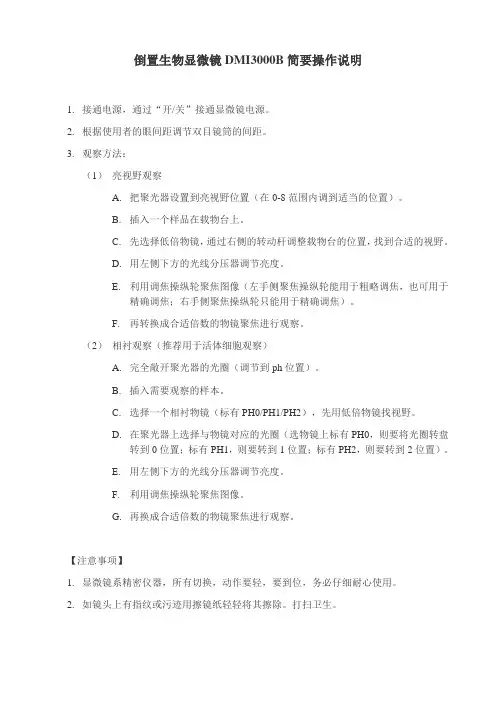
倒置生物显微镜DMI3000B简要操作说明
1.接通电源,通过“开/关”接通显微镜电源。
2.根据使用者的眼间距调节双目镜筒的间距。
3.观察方法:
(1)亮视野观察
A.把聚光器设置到亮视野位置(在0-8范围内调到适当的位置)。
B.插入一个样品在载物台上。
C.先选择低倍物镜,通过右侧的转动杆调整载物台的位置,找到合适的视野。
D.用左侧下方的光线分压器调节亮度。
E.利用调焦操纵轮聚焦图像(左手侧聚焦操纵轮能用于粗略调焦,也可用于
精确调焦;右手侧聚焦操纵轮只能用于精确调焦)。
F.再转换成合适倍数的物镜聚焦进行观察。
(2)相衬观察(推荐用于活体细胞观察)
A.完全敞开聚光器的光圈(调节到ph位置)。
B.插入需要观察的样本。
C.选择一个相衬物镜(标有PH0/PH1/PH2),先用低倍物镜找视野。
D.在聚光器上选择与物镜对应的光圈(选物镜上标有PH0,则要将光圈转盘
转到0位置;标有PH1,则要转到1位置;标有PH2,则要转到2位置)。
E.用左侧下方的光线分压器调节亮度。
F.利用调焦操纵轮聚焦图像。
G.再换成合适倍数的物镜聚焦进行观察。
【注意事项】
1.显微镜系精密仪器,所有切换,动作要轻,要到位,务必仔细耐心使用。
2.如镜头上有指纹或污迹用擦镜纸轻轻将其擦除。
打扫卫生。
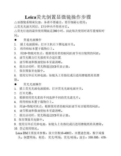
Leica荧光倒置显微镜操作步骤△显微镜系精密仪器,务请不要碰击,要仔细耐心使用。
△荧光光源关闭后,15分钟内不得再开启。
△荧光灯泡的最佳使用期限是200小时,因此每次使用时请尽量缩短时间。
⚫普通光源操作1. 插上电源插座,打开主机右下侧电源开关。
2. 将待检标本置于载物台上。
3. 用10×物镜对焦点,根据使用者的眼间距调节双目镜筒的间距。
4. 调节光圈及灯光强度至合适位置.5.调节粗调和微调使标本至最清晰。
6.拔出活动杆,使光路通过CCD至显示器。
7.保存图象至电脑中。
8.使用完毕后关掉电源,如镜头上有指纹或污迹用擦镜纸将其擦除。
⚫荧光光源操作1.插上荧光光源电源插座,打开荧光光源电源开关。
2.打开光栅。
3.根据使用荧光素的不同选择不同的荧光滤光片。
4.将待检标本置于载物台上。
5.用10×物镜对焦点,根据使用者的眼间距调节双目镜筒的间距。
6.调节粗调和微调使标本至最清晰。
7.拔出活动杆,使光路通过CCD至显示器。
8. 保存图象至电脑中。
9. 使用完毕后关掉电源,如镜头上有指纹或污迹用擦镜纸将其擦除。
10. 登记使用情况。
Leica DM主要技术参数:放大倍数10-400倍,内置滤色镜,数字成像头。
倒置明场、相差、荧光/明场、荧光/暗场。
波长:330-380,450-490,510-560。
主电90~250V。
频率:50~60HZ。
功率消耗:最大160W。
周围环境温度:10~36℃。
相对湿度:0~80%。
污染级别:2。
应用范围:倒置明场、相差及荧光,主要用于细胞及细菌等的观察及荧光摄影。
Leica DM IL LED倒置显微镜操作规程一、显微镜的用途由于微生物体积微小,远远低于肉眼的观察极限。
常以微米(um)或纳米(nm)来描述其大小。
因此,要对它们进行观察必须借助显微镜放大系统。
显微镜能将微小物体或物体的微细部分高倍放大。
二、显微镜的操作步骤1、去掉防尘罩,插上电源插座(电源插座一般是不拔的),打开电源开关。
2、将待检标本置于载物台上(放置之前要检查物镜是否调的太高,要确保其低于载物台垫片圆孔中心)。
3、用10×物镜对焦点,根据不同倍数的物镜,选择相应的滤光片,再根据使用者的眼间距调节双目镜筒的间距。
4、调节显微镜灯光至适当强度,然后调节粗调旋钮至看到清晰图像后,再改用微调旋钮调节标本图像至最清晰。
5、低倍镜下观察完标本的整体状况后,可改用高倍镜进一步细致的观察标本的局部情况,一般用40×物镜。
(注意:转换物镜前,要先旋转粗调旋钮使物镜稍微往下调低,再进行高倍和低倍物镜之间的转换。
这样是为了防止物镜碰到载物台的下缘损伤物镜。
)6、观察结束后,先将显微镜灯光调至最暗,再关闭显微镜电源,最后一定要用防尘罩盖住显微镜。
三、Leica Application suite V3软件的使用1、打开电脑,运行Leica Application suite V3软件。
2、在观察到所需的清晰图像后,拔出活动杆(此时目镜中就无法看到图像,只能在电脑显示器中看到图像),使光路通过CCD(电荷耦合元件)至显示器。
3、点击Leica Application suite V3软件菜单栏中的“获取”菜单,则可以观察到图像。
此时若感觉显示器中的图像不是很清楚,可以通过微调旋钮调节至最清楚。
4、当图像调节至最清晰时,在获取界面点击左下角的“获取图像”,则自动转换到了“浏览”菜单的界面。
此时点击该界面左下角的“采集”图像会自动保存在该软件的默认文件夹下,但是这种情况数据容易丢失。
我们可以在“我的电脑”中建立自己的文件夹,然后点击该界面右上角的“导出”图标,然后根据自己的习惯重命名,还可以选择图像的类型,保存至自己的文件夹随时备用。
徕卡Leica倒置式工业显微镜DMi8在竞争中保持领先是您事业前进的动力。
无论您从事金相学、医疗设备制造还是微电子领域,速度都显得至关重要。
您可根据自身需求定制徕卡这款高度模块化的倒置式显微镜。
它将徕卡优异的光学品质、丰富的对比度模式以及直观易用的软件集于一身,有助于加速您的工作流程。
徕卡倒置式显微镜助您加速工作流程徕卡倒置式显微镜可以为您节省时间和资金。
区别于正立式显微镜,您可直接将样品置于载物台对其表面对焦一次,然后在所有放大倍率下对焦,并对后续样品进行操作。
由于物镜位于载物台下方,与样品发生碰撞的危险大大降低。
节省时间∙您切换样品的速度将加快四倍∙您可获得更宽敞的工作空间,轻松放置大而重的样品∙可观察重量最高30 kg 的样品细致入微地观察样品您需要观察大尺寸样品并进行详尽分析吗?徕卡显微系统(Leica Microsystems)独家提供的宏观*物镜可令您在更短的时间内观察更多样品细节。
只需单击一下,即可从宏观模式 (35 mm) 切换到纳米模式 (200 nm)。
∙徕卡宏观物镜可提供4 倍于标准物镜的视场 (以0.7 倍放大倍率观察35.7 mm 样品的概况)。
∙您可在不同的放大倍率间切换:– 0.7x – 5x – 10x – 20x- 50x – 100x –,并从不同角度照亮样品。
∙ 3 档调焦驱动器可在不同放大倍率下实现灵敏可调的对焦,从而提高您的样品检测效率。
您可针对自己的工作需要打造完美工具对系统进行量身定制,使其符合您的预算、应用需求和偏好。
现在就为您工作所需的特性进行投资–并为未来需要做好准备。
更大的自由度∙您可升级自己的系统,以满足未来所需∙在手动、编码和自动功能之间进行选择∙您可使用丰富的对比度模式,满足所有应用需求一次击键即可轻松完成再现和存档Leica Application Suite (LAS) 软件提供一系列专家级模块以减轻您的工作负担,例如:∙Steel Expert∙Phase Expert∙Grain Expert定期更新和升级 LAS 软件可确保您始终走在竞争前列。
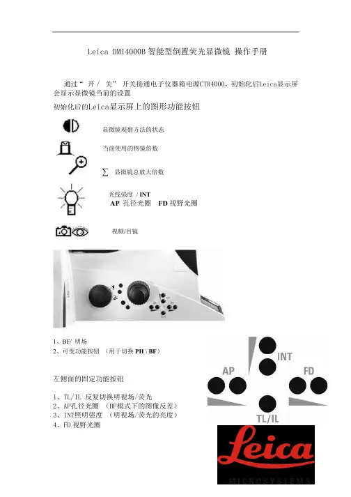
1. 目的:为了规范 LEICA DMi1型倒置显微镜标准操作规程的使用和维护保养,特制订本标准操作规程。
2. 适用范围:本操作规程适用于LEICA DMi1型倒置显微镜的所有操作和维护保养。
3. 责任:3.1仪器操作人员:负责仪器的正确使用、清洁及基本维护。
3.2仪器负责人:负责监督和培训其他人员对仪器的正确使用;负责对仪器进行定期维护和保养;仪器出现故障时,负责及时联系工程师进行维修,并跟踪维修进度。
4.内容:4.1徕卡Leica显微镜DMi1的结构图解Leica DMi1 支架右侧图解1 双目观察镜筒上半段2 目镜3 目镜镜筒4 镜筒下半段5 主电源开关6 相衬插板7 LED灯箱8 滤镜插孔,Ø 43mm9 聚光镜 10 透射光照明系统弯臂 11 聚光镜高度调节限位滚花螺钉 12 粗调和微调调焦手轮 13 物镜转换器与物镜14 照相模块保护罩 15 照相模块Leica DMi1 stand 支架左侧图解1 亮度控制器2 LED灯箱3 孔径光阑4 聚光镜5 粗调和微调调焦手轮6 载物台7 移动尺插板8 移动尺9 红外接收窗 10 红外接收窗目镜可以产生一个由物镜透射的中间实像的放大虚像。
在使用过程中,目镜所起的作用相当于一个放大镜。
Leica DMi1 正视图图解1 双目观察镜筒上半段2 目镜Leica DMi1 后视图解1 整合式照相模块接口2 支架电源3 接地连接4 照相模块USB电源连接口5 身份标签6 相机模块保护罩固定位7 红外发射窗AM Scope MU900显微采集器4.2徕卡leica显微镜DMI1的使用方法步骤:4.2.1准备阶段:首先打开LED透射光照明系统的电源,接通电源开关。
用旋钮调节亮度。
此时LED 灯会快速闪动3次,表示已激活。
若LED 灯闪动两次并保持较长时间,表示未激活。
注意:将亮度控制旋钮从零点旋转到最大亮度位置然后再旋回(在接通Leica DMI1显微镜电源后3秒钟内)一次,可以激活或禁用LED的自动关闭功能。
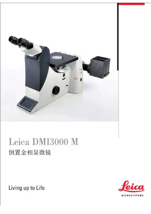
LeicaDM ILM倒置金相显微镜Leica DM ILM适用于来料、生产控制检查,检查样品制备过程和金相教学。
为您提供最优质的光学器件和最高效的显微镜系统是我们的责任。
您需要一台能快速准确显示结果的材料显微镜系统。
徕卡显微镜系统旨在减少您观察的时间,降低您的操作难度,同时提供最优化的结果。
- 徕卡品牌意味着高标准的光学性能。
HC (和谐系统) 光学系统的开发将光学显微镜标准Array提到了一个新的高度。
宽广的物镜选择范围保证了材料显微镜惊人的成像质量。
高性价比的HI Plan EPI 新物镜系列将明亮高反差与一流分辨率和优化的坚固的,耐腐蚀铸铝机身,漆面清洁,表面光滑。
显微镜的T型机身提供高稳定性,即使在高放大倍率下放置较重的样品,也能保证整体的稳健。
充足手部动作的空间是您操作灵活。
6 V 35 W照明此外,可另配外置的12V100W卤素灯反射光照明器或透射光。
根据要求,两个灯也可以同时安装开启,为明场和荧光工作。
新的灯光系统新的入射光照明系统插拔式3位反射棱镜组,很容易交换入射光及反射光照明。
滤光片组两个固定在显微镜内32mm直径的过滤器,以及一个可选的50厘米直径的中间过滤器优化观察图像。
3层机械移动载物台载物台适合不同形状和大小的样本。
几乎可以接受所有的样本量,并允许非破坏性的大型部件显微镜检查。
247X230mm 大载物台可以放置宽、高以及重的样品,内有150X150mm 的矩形台板插入可以完全移除。
小样本放在有80mm ,40mm ,30mm 和20mm 的内孔空白宽度。
同时有旋钮转动样本。
高承载能力 - 宽调节范围双层载物台支持高达样品负有重8Kg 。
XY 方向的调节范围在60 X40mm ,可迅速扫描试样。
矩形台板有4个垂直调整位,防止因制样不平对聚焦产生影响。
4新的照明系统可接纳不同类型的光源(灯泡灯丝或电弧放电),确保最佳的光强度和均匀性光通量。
外置和内置都按照克勒照明原理。
leica dmi8倒置荧光显微镜工作原理Leica DMI8倒置荧光显微镜是一种高级显微镜,主要应用于生物学和材料科学领域。
它具有极高的分辨率和成像质量,可以观察细胞、组织和材料的微观结构和功能。
下面将详细介绍Leica DMI8倒置荧光显微镜的工作原理。
Leica DMI8倒置荧光显微镜采用了倒置设计,与传统的直立显微镜不同。
正因为如此,它可以观察那些无法放置在玻片上的大型、复杂和活体样品。
在倒置显微镜中,目标在光源上方,而物镜和透镜安装在样品下方。
通过这种设计,Leica DMI8倒置显微镜可以提供更大的工作距离和更好的稳定性,同时也使得样品在观察过程中更容易调整。
Leica DMI8倒置荧光显微镜的工作原理可以分为以下几个关键步骤:1.光源Leica DMI8倒置荧光显微镜使用高亮度的荧光灯作为光源。
荧光灯产生的白光在准直镜筒中经过调整后,通过反射镜进入物镜。
2.过滤器在Leica DMI8倒置显微镜中,滤光片起到非常重要的作用。
滤光片的选择决定了可见光和荧光光谱的过滤和分离。
这对于获取高质量的显微镜影像是至关重要的。
3.物镜物镜是显微镜中最重要的组件之一,它决定了成像的分辨率和清晰度。
Leica DMI8倒置显微镜通常配备了多个物镜,包括低倍率的物镜用于观察整个样品,高倍率的物镜用于观察细节。
物镜通过仔细的设计和制造,能够提供高度的放大倍率和出色的透视效果。
4.荧光标记在观察细胞和组织的时候,研究者通常会使用荧光标记来标记特定的分子或细胞器。
Leica DMI8倒置显微镜使用荧光染料来激发样品中的荧光。
这些荧光染料吸收入射光,然后发射出恒定的波长。
这种荧光现象可以被荧光滤光片所捕捉,同时其他波长的光被过滤掉。
5.成像系统Leica DMI8倒置显微镜配备了高度灵敏的CCD相机,用于捕捉荧光图像。
荧光图像的质量很大程度上取决于相机的性能。
CCD相机能够以高速捕捉图像,并具有出色的噪声抑制能力。
leica dmi8倒置荧光显微镜工作原理Leica DMI8倒置荧光显微镜是一种高端的显微镜,具有先进的技术和优越的性能。
它采用倒置设计,即镜头和光源安装在样品底部,而样品放置在顶部,这种设计可以观察大尺寸的生物样品,例如细胞培养皿、培养皿或培养箱内的样品。
本文将介绍Leica DMI8倒置荧光显微镜的工作原理,并探讨其在生命科学领域的应用。
Leica DMI8倒置荧光显微镜的工作原理主要包括光源系统、物镜系统、检测系统和成像系统。
首先是光源系统,Leica DMI8采用了先进的多通道LED光源,可以提供高亮度和均匀度的光源,满足不同荧光染料的激发需求。
其次是物镜系统,Leica DMI8倒置荧光显微镜配备了高清晰度的物镜,可以通过调节物镜的倍率来观察不同尺寸和层次的样品。
检测系统则采用了高灵敏度的CCD相机,可以捕捉到微弱的荧光信号并转换成电信号。
最后是成像系统,Leica DMI8倒置荧光显微镜通过集成的成像软件可以实现实时观察、图像采集、图像处理和数据分析等功能,可以帮助科研人员快速获取高质量的荧光图像。
Leica DMI8倒置荧光显微镜在生命科学领域具有广泛的应用,例如细胞培养皿中的细胞观察和追踪、蛋白质表达和定位研究、细胞凋亡和增殖实验、细胞内信号转导研究等。
倒置设计可以让科研人员在不破坏样品的情况下观察细胞的生长情况和细胞内结构,特别适合活细胞的观察。
同时,Leica DMI8倒置荧光显微镜的高灵敏度和高分辨率的成像系统可以提供清晰的细胞结构和荧光信号,有助于研究者获取准确的数据和结论。
总之,Leica DMI8倒置荧光显微镜通过先进的技术和优越的性能,为生命科学领域的研究工作提供了强大的工具。
倒置设计可以观察大尺寸的样品,高灵敏度的检测系统可以捕捉微弱的信号,成像软件可以帮助科研人员进行数据处理和分析,使Leica DMI8倒置荧光显微镜成为生命科学领域不可或缺的设备。
相信随着科技的不断进步,Leica DMI8倒置荧光显微镜将在生物医学研究中发挥越来越重要的作用。
Leica
DM ILM
倒置金相显微镜
Leica DM ILM
适用于来料、生产控制检查,检查样品制备
过程和金相教学。
为您提供最优质的光学器件和最高效的显微
镜系统是我们的责任。
您需要一台能快速准
确显示结果的材料显微镜系统。
徕卡显微镜
系统旨在减少您观察的时间,降低您的操作
难度,同时提供最优化的结果。
- 徕卡品牌意味着高标准的光学性能。
HC (和
谐系统) 光学系统的开发将光学显微镜标准Array提到了一个新的高度。
宽广的物镜选择范围保
证了材料显微镜惊人的成像质量。
高性价比的
HI Plan EPI 新物镜系列将明亮高反差与一流
分辨率和优化的
坚固的,耐腐蚀铸铝机身,漆面清洁,表面光滑。
显微镜的T型机身提供高稳定性,即使在高放大倍率下放置较重的样品,也能保证整体的稳健。
充足手部动作的空间是您操作灵活。
6 V 35 W照明
此外,可另配外置的12V100W卤素灯反射光照明器或透射
光。
根据要求,两个灯也可以同时安装开启,为明场和荧
光工作。
新的灯光系统
新的入射光照明系统
插拔式3位反射棱镜组,很容易交换入射光及反射光照明。
滤光片组
两个固定在显微镜内32mm直径的过滤器,以及一个可选的50厘米直径的中间过滤器优化观察图像。
3层机械移动载物台
载物台适合不同形状和大小的样本。
几乎可以接受所有的样本量,并允许非破坏性的大型部件显微镜检查。
247X230mm 大载物台可以放置宽、高以及重的样品,内有150X150mm 的矩形台板插入可以完全移除。
小样本放在有80mm ,40mm ,30mm 和20mm 的内孔空白宽度。
同时有旋钮转动样本。
高承载能力 - 宽调节范围
双层载物台支持高达样品负有重8Kg 。
XY 方向的调节范围在60 X40mm ,可迅速扫描试样。
矩形台板有4个垂直调整位,防止因制样不平对聚焦产生影响。
4
新的照明系统可接纳不同类型的光源(灯泡灯丝或电弧放电),确保最佳的光强度和均匀性光通量。
外置和内置都按照克勒照明原理。
设置对中心的孔径光阑,提高分辨率,对比度和景深。
物镜
Aluminium, Pol contrast
Microhardness indentations
物镜的质量是显微镜成像的核心。
对反射光的明视场,对比度和荧光偏振设计,徕卡DM ILM 显微镜兼容所有无限远系统及徕卡独有的M25mm 大孔径透光物镜。
更早的徕卡类型(徕兹)RMS物镜应该可在DM ILM 上使用。
N PLAN 2.5x/0.07 11.2 mm N PLAN 5x/0.12 14.0 mm N PLAN 10x/0.25 5.8 mm N PLAN 20x/0.40 1.1 mm N PLAN 50x/0.75 0.37 mm N PLAN 100x/0.90 0.27 mm PL FLUOTAR 1.6x/0.05 1.54 mm PL FLUOTAR 2.5x/0.07 9.2 mm HC PL FLUOTAR 5x/0.15 12.0 mm HC PL FLUOTAR 10x/0.30 11.0 mm HC PL FLUOTAR 20x/0.50 1.27 mm HC PL FLUOTAR 50x/0.80 0.5 mm HC PL FLUOTAR 100x/0.90 0.3 mm HC PL
FLUOTAR 100x/1.30 OIL ∞/0 0.13 mm N PLAN 系列
PL APO 50x/0.90 0.28 mm PL APO 100x/0.95 0.16 mm PL APO 150x/0.95 0.20 mm
PL APO 250x/0.95 0.24 mm with spacer ring 25/RMS PL APO 系列
PL FLUOTAR L 50x/0.55 8.0 mm
PL FLUOTAR L 100x/0.75 4.7 mm with spacer ring 25/RMS PLAN H 20x/0.40 12.6 mm PLAN H 40x/0.60 7.1 mm
长工作距离
M (直线刻度) HCL 系列:30 mm 直径目镜HC PLAN10x/20
同样兼容其它倍率目镜:12.5X,16X,25X
M (方格)目镜10x/20 和目镜10x/20
视度补偿目镜10x/18 和目镜10x/18 PL FLUOTAR 系列
目镜观察筒定义了边缘锋利和标准倍率图像:ILB和ILT观察筒所配直径23.2mm 的目镜:
Objective series N PLAN and PL FLUOTAR
目镜观察筒
观察与照相
45°观察角,目镜筒直径 23.2 mm 三目镜筒 HC ILT
Trinocular tube HC ILT with MPS30
Trinocular tube HC L V1T with TV camera
DM ILM 具有多种观察筒供选择,具备0° -35 °视角调整。
所有的观察筒都配备了一个1X 无限远管镜。
通过360°旋转,使显微镜也可以从侧面使用,方便用户调整试样位置。
双目镜筒 HC ILB
45°观察角,目镜筒直径 23.2 mm ,通光量100 %目镜观察/100 % 照相输出。
照相输出端位于目镜侧端,它的好处在于不妨碍检视载物台上的样品。
其它观察筒适用于 IL/L: 双目 HC LB 和三目 HC L VB 0° – 35° 视角调整 通光量50 % 观察/50 %照相 三目镜筒 HC L1 T
通光量 100 % /100 % 或50 %/50 % 三目镜筒 HC L3 T
三目镜筒 HC L V1T 0° – 35° 视角调整,通光量50 % 观察/50 %照相
选配件
Zircon, pol contrast
Microhardness indentation with interferogram
Leica DM ILM with assymetric discussion tube
Ergo 模块
调高观察位置 30 mm.
变倍器
可选 1X, 1.5X, 2X 。
绘图器
复制比率 1:1 ,用于跟踪绘制显微镜下的样品结构。
显微硬度压头 MHT 10
在倒置显微镜测量显微硬度非常简单,加载范围在0.6-400pond 。
特别适用于薄件细小组织硬度。
CCD 适配器
我们提供顶级图像质量的CCD 适配器,适用数字或模拟摄像头。
选择适配器应考虑打印图像的标准倍率,及尽可能大的视野面积。
单芯片摄像头
C -mount 0.35x HC 1/
C-mount 0. 5x HC 1/ C-mount 0.63x HC 1/ C-mount1x HC 1/ 3芯片摄像头 C-mount 1X B-mount 1X B-mount 1. 25X F-mount 1X F-mount1.25X
测量和对比
目镜测微尺以优良的、高精度模式用于长度与粒径测量。
两种目镜(23.2和30mm直径)都可安装测微尺。
规格有
10mm=100直线刻度,方格刻度,及满足ASTM-E112标
准的晶粒度格尺。
Leica DM MFK2
在电视屏幕上显示格尺标记,用于长度,角度,圆面积视
Pig iron 频测量。
技术参数
Defective strip conductor
Sensor structure
Iron material, interference colour layer
高度尺寸: CCD摄像头 390 mm 数码相机 410 mm
模拟摄像头
350 – 450 mm 显微镜尺寸:
长 650 mm 宽 320 mm
物镜孔径: M25 x 0.75 目镜视野: 23.2 mm
30 mm (HC L tubes) 光阑直径:
32 mm (50 mm optional)。