
SDS-PAGE_and_Western_blot
- 格式:ppt
- 大小:3.24 MB
- 文档页数:15
Western bolt一、实验目的通过实验了解western blot技术的原理和操作。
二、实验原理SDS-PAGE是最常用的定性分析蛋白质的电泳方式,特别是用于蛋白质纯度检测和测定蛋白质分子量。
PAGE能有效的分离蛋白质,主要依据其分子量和电荷的差异,而SDS-PAGE的分离原理则仅根据蛋白质的分子量的差异。
SDS-聚丙烯酰胺凝胶电泳,是在聚丙烯酰胺凝胶系统中引进SDS(十二烷基硫酸钠),SDS会与变性的多肽,并使蛋白带负电荷,由于多肽结合SDS的量几乎总是与多肽的分子量成正比而与其序列无关,因此SDS多肽复合物在丙稀酰胺凝胶电泳中的迁移率只与多肽的大小有关,在达到饱和的状态下,每克多肽可与1.4g去污剂结合。
当分子量在15KD到200KD之间时,蛋白质的迁移率和分子量的对数呈线性关系,符合下式:logMW=K-bX,式中:MW为分子量,X为迁移率,k、b均为常数,若将已知分子量的标准蛋白质的迁移率对分子量对数作图,可获得一条标准曲线,未知蛋白质在相同条件下进行电泳,根据它的电泳迁移率即可在标准曲线上求得分子量。
硝酸纤维素膜(nitrocellulose filter membrane,简称NC膜),在胶体金试纸中用做C/T线的承载体,同时也是免疫反应的发生处。
NC膜是生物学试验中最重要的耗材之一。
第一抗体就是能和特异性抗原特异性结合的蛋白。
第二抗体是能和抗体结合,即抗体的抗体,其主要作用是检测抗体的存在,放大一抗的信号。
化学发光HRP底物(辣根过氧化物酶底物,也常被称为ECL试剂)是目前Western blot检测中最为灵敏的试剂。
显影液的A液主要成分为鲁米诺(Luminol)及发光增强剂,B液主要成分为过氧化物溶液。
二抗上含有HRP(辣根过氧化物酶),可以催化A液和B液反应发光。
三、实验器材1.电泳槽,胶板架子2.转膜仪3.NC膜4.电泳电源5.滤纸6.摇床7.X射线摄影暗匣8.X射线胶片9.塑料薄膜四、实验试剂1.runningbuffer5xrunningbuffer(1L)Tris 15.1gGlycine 94.0gSDS 5.0gddH2O 定容至1L使用前稀释5倍2.Transfer buffer10×Transfer buffer(1L)Tris 30.3gGlycine 144.0gddH2O 定容至1L使用前稀释10倍,加入1/4体积的甲醇3.TBS10×TBS(500mL)Tris 12.1gNacl 40.0gddH2O 定容至500mL用HCl调节溶液的pH值至7.64.TBST(1L)1×TBS:Tween-20=1000:1ddH2O定容至1L5.5%的脱脂奶粉溶液(50ml)脱脂奶粉 2.5gTBST 定容至50ml6.一抗溶液7.二抗溶液8.显影液现配现用,溶液A:溶液B=1:1配置。

蛋白质检验方法
蛋白质是生物体内一类十分重要的有机物质,它们参与了生物体内的几乎所有生命活动。
因此,对蛋白质的检验方法显得尤为重要。
本文将介绍几种常见的蛋白质检验方法,以供参考。
首先,最常见的蛋白质检验方法之一是SDS-PAGE。
SDS-PAGE是一种聚丙烯酰胺凝胶电泳技术,通过电泳分离蛋白质,可以根据蛋白质的大小进行分析。
这种方法操作简单,结果准确,因此被广泛应用于蛋白质的检验和分析。
其次,免疫印迹法(Western blot)也是一种常用的蛋白质检验方法。
通过Western blot,可以检测特定蛋白质在混合蛋白质中的存在情况,并且可以定量分析目标蛋白质的表达水平。
这种方法对于研究特定蛋白质在细胞信号转导、疾病诊断等方面具有重要意义。
另外,酶联免疫吸附试验(ELISA)也是一种常见的蛋白质检验方法。
ELISA通过酶标记的抗体与待测蛋白质结合,再通过底物的反应产生可定量测定的色素来检测蛋白质的存在。
这种方法具有高灵敏度、高特异性和高通量的特点,被广泛应用于医学诊断、生物
学研究等领域。
此外,质谱法也是一种重要的蛋白质检验方法。
通过质谱法,
可以对蛋白质的氨基酸序列、翻译后修饰等进行全面的分析。
质谱
法具有高分辨率、高灵敏度和高通量的特点,对于蛋白质的结构和
功能研究具有重要意义。
综上所述,蛋白质的检验方法多种多样,每种方法都有其特定
的应用领域和优势。
在实际应用中,可以根据实验的具体要求和条
件选择合适的蛋白质检验方法,以确保实验结果的准确性和可靠性。
希望本文所介绍的蛋白质检验方法对您有所帮助。
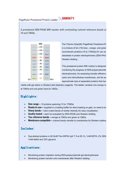
SM0671PageRuler Prestained Protein LadderA prestained SDS-PAGE MW marker with contrasting colored reference bands at10 and 70kDa.The Thermo Scientific PageRuler Prestained Proteis a mixture of ten (10) blue-, orange- and green-srecombinant proteins (10 to 170kDa) for use as sizstandards in protein electrophoresis (SDS-PAGE)Western blotting.This prestained protein MW marker is designed fomonitoring the progress of SDS-polyacrylamide geelectrophoresis, for assessing transfer efficiency onylon and nitrocellulose membranes, and for estimapproximate size of separated proteins that have b visible with gel stains or Western blot detection reagents. The ladder contains one orange refer at 70kDa and one green band at 10kDa.Highlights:•Size range– 10 proteins spanning 10 to 170kDa•Ready-to-use– supplied in a loading buffer for direct loading on gels; no need to boil •Sharp bands– color-coded bands of similar intensity for easy visualization•Quality tested – each lot evaluated by SDS-PAGE and Western blotting•Two reference bands– orange at 70kDa and green at 10kDa•Membrane-compatible– colored bands transfer to membranes for Western blottingIncludes:•Dye-stained proteins in 62.5mM Tris-H3PO4 (pH 7.5 at 25°C), 1mM EDTA, 2% SDS, 1 1mM NaN3 and 33% glycerol.Applications:•Monitoring protein migration during SDS-polyacrylamide gel electrophoresis•Monitoring protein transfer onto membranes after Western blotting•Sizing of proteins on SDS-polyacrylamide gels and Western blotsProduct Details:SDS-PAGE band profile of the Thermo ScientificPageRuler Prestained Protein Ladder. Images arefrom a 4-20% Tris-glycine gel (SDS-PAGE) andsubsequent transfer to membrane.Sm1841Spectra Multicolor Broad Range Protein LadderA prestained, 4-color, SDS-PAGE MW marker especially for proteins between 10 and 260kDa.The Thermo Scientific Spectra Multicolor Broad RangeProtein Ladder is a 4-color protein standard containing 10prestained recombinant prokaryotic proteins (10 to 260kDa)for use as size standards in gel electrophoresis and Westernblotting.This prestained protein MW marker is designed formonitoring the progress of SDS-polyacrylamide gelelectrophoresis, for assessing transfer efficiency onto PVDF,nylon and nitrocellulose membranes, and for estimating theapproximate size of separated proteins that have been made visible with gel stains or Western blot detection reagents. Four different chromophores (blue, orange, green, pink) are bound to the different component proteins, producing a brightly colored ladder with an easy-to-remember pattern.Highlights:•Size range– 10 proteins spanning 10 to 260kDa•Multicolor– four different colors for unambiguous band-size assignment•Ready-to-use– supplied in a loading buffer for direct loading on gels; no need to boil•Sharp bands– color-coded bands of similar intensity for easy visualization•Quality tested – each lot evaluated by SDS-PAGE and Western blotting•Membrane-compatible– colored bands transfer to membranes for Western blotting Includes:•Dye-stained proteins in 62.5mM Tris-H3PO4 (pH 7.5 at 25°C), 1mM EDTA, 2% SDS, 10mM DTT, 1mM NaN3 and 33% glycerol.Applications:•Monitoring protein migration during SDS-polyacrylamide gel electrophoresis•Monitoring protein transfer onto membranes after Western blotting•Sizing of proteins on SDS-polyacrylamide gels and Western blotsProduct Details:SDS-PAGE band profile of the Thermo ScientificSpectra Multicolor Broad Range Protein Ladder.Images are from a 4-20% Tris-glycine gel(SDS-PAGE) and subsequent transfer to membrane.S m1811PageRuler Plus Prestained Protein LadderA prestained SDS-PAGE MW marker with contrasting colored reference bands at 10, 25 and 70kDa.The Thermo Scientific PageRuler Plus Prestained ProteinLadder is a mixture of nine (9) blue-, orange- andgreen-stained recombinant proteins (10 to 250kDa) for useas size standards in protein electrophoresis (SDS-PAGE)and Western blotting.This prestained protein MW marker is designed formonitoring the progress of SDS-polyacrylamide gelelectrophoresis, for assessing transfer efficiency onto PVDF,nylon and nitrocellulose membranes, and for estimating theapproximate size of separated proteins that have been made visible with gel stains or Western blot detection reagents. A blue chromophore is bound to all proteins, except proteins of two reference bands of 70kDa and 25kDa that are colored with an orange dye and one green reference band of 10kDa. PageRuler Plus Prestained Protein Ladder is ready to use: no heating, further dilution or addition of a reducing agent is required before loading onto a gel.Highlights:•Size range– nine proteins spanning 10 to 250kDa•Ready-to-use– supplied in a loading buffer for direct loading on gels; no need to boil•Sharp bands– color-coded bands of similar intesity for easy visualization•Quality tested – each lot evaluated by SDS-PAGE and Western blotting•Bright reference bands– orange at 70 and 25kDa, and green at 10kDa•Membrane-compatible– colored bands transfer to membranes for Western blotting Includes:•Dye-stained proteins in 62.5mM Tris-H3PO4 (pH 7.5 at 25°C), 1mM EDTA, 2% SDS, 10mM DTT, 1mM NaN3 and 33% glycerol.Applications:•Monitoring protein migration during SDS-polyacrylamide gel electrophoresis•Monitoring protein transfer onto membranes after Western blotting•Sizing of proteins on SDS-polyacrylamide gels and Western blotsProduct Details:SDS-PAGE band profile of the Thermo Scientific PageRuler Plus Prestained Protein Ladder. Images are from a 4-20% Tris-glycine gel(SDS-PAGE) and subsequent transfer to membrane.。

实验原理蛋白质印迹是把电泳分离的蛋白质转移到固定基质上,然后利用抗原抗体反应来检测特异性的蛋白分子的技术,包括三个部分:SDS—聚丙烯酰胺凝胶电泳,蛋白质的电泳转移,免疫印迹分析。
SDS—聚丙烯酰胺凝胶电泳主要用于测定蛋白质相对分子质量,SDS是阴离子去污剂,能断裂蛋白质分子内和分子间的氢键,使分子去折叠,破坏其高级结构。
SDS与大多数蛋白质的结合比为1.4:1,由于SDS带有大量的负电荷,与蛋白质结合时掩盖了不同种类蛋白质间原有的电荷差别,使各种蛋白质带有相同密度的负电荷,形似长椭圆棒,蛋白迁移率与蛋白质相对分子质量的对数呈线性关系。
因此,利用相对分子质量标准蛋白所作的标准曲线,可以求得未知蛋白的相对分子质量。
电泳后蛋白质分子嵌在凝胶介质中,探针分子很难通过凝胶孔,将蛋白质从凝胶转移到固定基质上可以对蛋白质进行免疫检测分析。
方法有两种:①水平半干式转移即将凝胶和固定基质似三明治样夹在缓冲液浸湿的滤纸中间,通电10~30min可完成②垂直湿式转移即将凝胶和固定基质夹在滤纸中间,浸在转移装置的缓冲液中,通电2~4h或过夜可完成。
固定基质通常有硝酸纤维素膜、聚偏二氟乙烯膜和尼龙膜。
蛋白质转移到固定化膜上之后,通过蛋白质染料如丽春红S检测膜上的总蛋白,或用考马斯亮蓝检测凝胶上的蛋白剩余量,以验证转移是否成功。
用抗体作为探针进行特异性的免疫反应检测抗原蛋白,分为4步:①用非特异性、非反应活性分子封阻固定化膜上未吸附蛋白的自由结合区,以防止作为探针的抗体结合到膜上,出现检测时的高背景②固定化膜用专一性的一抗温育,使一抗与膜上的抗原蛋白分子特异性结合③酶标二抗与一抗特异结合④加入酶底物,适当保温,膜上便可见到颜色反应,检测出抗原蛋白区带。
主要溶液10%分离胶水 3.3mL、30% 丙烯酰胺混合液 4.0mL、1.0mol/L Tris(pH8.8)2.5mL、10% SDS 0.1mL、10%过硫酸铵0.1mL、TEMED 0.004mL5%浓缩胶水 2.7mL、30%丙烯酰胺混合液0.67mL、1.0mol/LTris0.5mL、10%SDS0.04Ml/10%过硫酸铵0.04mL、TEMED0.004mL1×Tris –甘氨酸电泳缓冲液Tris碱3.03g、甘氨酸18.77g、SDS 1g,用去离子水定容至1L2×SDS凝胶加样缓冲液Tris-HCl(pH6.8) 100mmol/L,β-巯基乙醇10%,10%甘油,0.01%溴酚蓝,10%SDS 转移缓冲液Tris 2.45g,甘氨酸11.25g,甲醇100mL,加去离子水至1LTBSTTris 1.21g NaCl 8.77g,Tween-20 1mL,加去离子水至1LStripping1.3mL Tris (pH 6.8),4mL10%SDS,140µlβ-巯基乙醇,用水定容到20mL实验步骤1 SDS—聚丙烯酰胺凝胶电泳⑴凝胶配置①分离胶的配置:将配置好的分离胶液混匀后迅速倒入胶槽中,至距离短玻璃板顶端约2cm 处,停止灌胶。