英文病例汇报模板
- 格式:ppt
- 大小:1.24 MB
- 文档页数:17
医学病例报告英语作文Title: Medical Case Report: Management of Chronic Hypertension in a Middle-aged Female Patient。
Abstract:This case report discusses the presentation, diagnosis, and management of chronic hypertension in a middle-aged female patient. The patient, Mrs. X, presented with a history of hypertension and was experiencing persistent elevated blood pressure despite lifestyle modifications and medication adherence. Through a comprehensive assessment, including medical history, physical examination, and diagnostic tests, the patient was diagnosed with chronic hypertension. The management approach involved pharmacological intervention, lifestyle modifications, and regular monitoring. This case highlights the importance of tailored treatment strategies and multidisciplinary care in managing chronic hypertension effectively.Introduction:Chronic hypertension, characterized by persistently elevated blood pressure levels, is a significant public health concern globally. It predisposes individuals to various cardiovascular complications, including stroke, heart failure, and renal dysfunction. This case report focuses on the management of chronic hypertension in a middle-aged female patient, emphasizing the importance of individualized treatment plans to achieve optimal blood pressure control and reduce the risk of associated complications.Case Presentation:Mrs. X, a 55-year-old female, presented to the clinic with a chief complaint of persistently elevated blood pressure readings despite adherence to antihypertensive medication. She reported a history of hypertension for the past ten years and a family history of cardiovascular diseases. On physical examination, her blood pressure was consistently elevated, averaging around 160/100 mmHgdespite being on a combination therapy of angiotensin-converting enzyme (ACE) inhibitor and diuretic.Diagnostic Assessment:Given the patient's history and physical examination findings, further diagnostic workup was pursued to assess the extent of target organ damage and potential secondary causes of hypertension. Laboratory investigations,including renal function tests, lipid profile, and electrolyte levels, were within normal limits. An electrocardiogram (ECG) revealed left ventricular hypertrophy, indicative of long-standing hypertension. Additionally, a renal ultrasound ruled out renal artery stenosis as a secondary cause of hypertension.Diagnosis:Based on the clinical presentation, diagnostic findings, and exclusion of secondary causes, Mrs. X was diagnosedwith chronic primary hypertension. The diagnosis was supported by her longstanding history of hypertension,family history of cardiovascular diseases, and evidence of target organ damage on ECG.Management:The management approach for Mrs. X's chronic hypertension involved a combination of pharmacological therapy and lifestyle modifications. Considering her persistent elevation in blood pressure despite the current medication regimen, the treatment plan was adjusted. A calcium channel blocker (amlodipine) was added to her existing therapy to achieve better blood pressure control. Furthermore, Mrs. X was counseled on dietary modifications, including a low-sodium diet and increased consumption of fruits and vegetables. She was also encouraged to engage in regular physical activity and weight management.Follow-up and Monitoring:Mrs. X was scheduled for regular follow-up visits to monitor her blood pressure response to the adjusted treatment regimen and assess for any adverse effects ofmedication. Additionally, she was advised to monitor her blood pressure at home using a digital blood pressure monitor and maintain a record for review during follow-up visits. Laboratory investigations, including renal function tests and electrolyte levels, were scheduled periodically to monitor for potential medication-related complications.Outcome:With the adjusted treatment regimen and adherence to lifestyle modifications, Mrs. X demonstrated significant improvement in blood pressure control. Subsequent follow-up visits showed a gradual reduction in her blood pressure readings, with values consistently below 140/90 mmHg. Repeat ECG performed six months later showed regression of left ventricular hypertrophy, indicating improvement in cardiac function. Mrs. X reported improved quality of life and compliance with the treatment plan.Discussion:This case illustrates the challenges encountered inmanaging chronic hypertension, particularly in patientswith resistant hypertension despite medication adherence.It underscores the importance of a comprehensive diagnostic approach to identify underlying causes and assess target organ damage. Individualized treatment strategies,including pharmacological therapy tailored to the patient's needs and preferences, are essential in achieving optimal blood pressure control. Furthermore, lifestylemodifications play a crucial role in hypertension management and should be integrated into the treatment plan. Multidisciplinary collaboration involving physicians, nurses, pharmacists, and allied healthcare professionals is vital in providing holistic care to patients with chronic hypertension.Conclusion:Effective management of chronic hypertension requires a multidimensional approach involving pharmacological therapy, lifestyle modifications, and regular monitoring. This case report highlights the successful management of chronic hypertension in a middle-aged female patient throughtailored treatment strategies and collaborative care. By addressing individual patient needs and optimizing blood pressure control, healthcare providers can mitigate the risk of cardiovascular complications and improve patient outcomes in individuals with chronic hypertension.。
英语病历报告作文格式English:A medical record report in English typically follows a specific format to ensure clear and organized documentation of a patient's medical history, diagnosis, treatment, and progress. The report usually starts with the patient's demographic information, including their name, age, gender, address, and contact information. This is followed by the reason for the patient's visit or hospitalization, including the chief complaint and any relevant medical history. The report then details the physical examination findings, laboratory and imaging test results, diagnosis, treatment plan, and prognosis. It is important to use clear and concise language, medical terminology, and abbreviations to accurately convey the patient's medical information. Additionally, proper grammar, spelling, and punctuation should be used to ensure professionalism and accuracy in the medical record report.中文翻译:英文病历报告通常遵循特定的格式,以确保清晰有序地记录患者的病史、诊断、治疗和进展情况。
英语病例报告范文Case Report: A Rare Case of Acute Myeloid Leukemia with Unusual Presentation。
Introduction:Acute Myeloid Leukemia (AML) is a malignant disorder characterized by the proliferation of abnormal myeloid cells in the bone marrow. It typically presents with symptoms such as fatigue, fever, and easy bruising. However, in some rare cases, AML can manifest with atypical symptoms, leading to diagnostic challenges. This report presents a unique case of AML with an unusual presentation.Case Presentation:A 45-year-old male presented to the emergency department with complaints of severe headaches, dizziness, and blurred vision. He had a history of chronic migraines and was initially treated for a severe migraine attack. However, his symptoms did not improve with standard migraine medications. On further evaluation, the patient was found to have profound anemia, with a hemoglobin level of 6 g/dL.Investigations:A complete blood count revealed severe pancytopenia, with a white blood cell count of 1.2 × 10^9/L, hemoglobin of 6 g/dL, and platelet count of 50 × 10^9/L. Peripheral blood smear examination showed blasts comprising 70% of the total nucleated cells, suggesting the possibility of acute leukemia. Bone marrow aspiration and biopsy were performed to confirm the diagnosis.Diagnosis:The bone marrow examination revealed hypercellularity with infiltration of blasts comprising more than 80% of the nucleated cells. Flow cytometry analysis showed expression of myeloid markers (CD13, CD33) and absence of lymphoid markers,confirming the diagnosis of acute myeloid leukemia. Cytogenetic analysis revealed the presence of a complex karyotype, which is associated with a poor prognosis.Treatment and Outcome:The patient was promptly started on induction chemotherapy with a combination of cytarabine and daunorubicin. He experienced severe myelosuppression and required supportive care, including red blood cell and platelet transfusions. Despite the initial response to chemotherapy, the patient developed refractory disease and relapsed within six months of completing consolidation therapy. Salvage chemotherapy and allogeneic stem cell transplantation were considered, but the patient declined further treatment due to poor prognosis and opted for palliative care.Discussion:This case highlights the importance of considering acute leukemia in the differential diagnosis of atypical presentations, even in the absence of classic symptoms. The unusual symptoms of severe headaches, dizziness, and blurred vision initially misled the clinicians to suspect migraine as the primary cause. However, the presence of profound anemia and pancytopenia raised suspicion of an underlying hematological disorder. Timely evaluation and appropriate diagnostic tests, including bone marrow examination, were crucial in establishing the correct diagnosis.Conclusion:This case report emphasizes the need for a high index of suspicion for acute leukemia, especially in patients presenting with unusual symptoms. Prompt diagnosis and initiation of appropriate treatment are essential for improving patient outcomes. Further research is warranted to better understand the underlying mechanisms of atypical presentations in AML and to develop targeted therapies for patients with poor prognostic factors.。

Divisi on: _________ Ward: __________ Bed: _________ Case No. ___________Name: _____________ Sex: __________ Age: ___________ Natio n: ___________ Birth Place: ________________________________ Marital Status: _____________ Work-orga nizatio n & Occupatio n: _____________________________________ Livi ng Address & Tel: _________________________________________________ Date of admissio n: ______ D ate of history taken: ______ Informant: __________Chief Complaint: ___________________________________________History of Present Illness:Past History:General Health Status: 1.good 2.moderate 3.poorDisease history:(if any, please write down the date of onset, brief diagnosticand therapeutic course, and the results.)Respiratory system:1. None2.Repeated pharyngeal pain3.chronic cough4.expectoration:5. Hemoptysis6.asthma7.dyspnea8.chest painCirculatory system:1.None2.Palpitation3.exertional dyspnea4..cyanosis5.hemoptysis6.Edema of lower extremities7.chest pain8.syncope9.hypertension Digestive system:1.None2.Anorexia3.dysphagia4.sour regurgitation5.eructation6.nausea7.Emesis8.melena9.abdominal pain 10.diarrhea11.hematemesis 12.Hematochezia 13.jaundiceUrinary system:1.None2.Lumbar pain3.urinary frequency4.urinary urgency5.dysuria6.oliguria7.polyuria 8.retention of urine 9.incontinence of urine 10.hematuria11.Pyuria 12.nocturia 13.puffy faceHematopoietic system:1.None2.Fatigue3.dizziness4.gingival hemorrhage5.epistaxis6.subcutaneous hemorrhageMetabolic and endocrine system:1.None2.Bulimia3.anorexia4.hot intolerance5.cold intolerance6.hyperhidrosis7.Polydipsia8.amenorrhea9.tremor of hands 10.character change 11.Marked obesity12.marked emaciation 13.hirsutism 14.alopecia15.Hyperpigmentation 16.sexual function changeNeurological system:1.None2.Dizziness3.headache4.paresthesia5.hypomnesis6. Visual disturbance7.Insomnia8.somnolence9.syncope 10.convulsion 11.Disturbance of consciousness12.paralysis 13. vertigoReproductive system:1.None2.othersMusculoskeletal system:1.None2.Migrating arthralgia3.arthralgia4.artrcocele5.arthremia6.Dysarthrosis7.myalgia8.muscular atrophyInfectious Disease:1.None2.Typhoid fever3.Dysentery4.Malaria 4.Schistosomiasis4. Leptospirosis 7.Tuberculosis 8.Epidemic hemorrhagic fever9.othersVaccine inoculation:1.None2.Yes3.Not clearVaccine detail _________________________________________ Trauma and/or operation history: Operations:1.None2.YesOperation details: _______________________________________ Traumas:1.None2.YesTrauma details: _________________________________________ Blood transfusion history:1.None2.Yes ( 1.Whole blood 2.Plasma3.Ingredient transfusion) Blood type:Transfusion time: ______Transfusion reaction1.None2.YesClinic manifestation: ____________________________ Allergic history:1.None2.Yes3.Not clear allergen: __________________________________clinical manifestation: _____________________________________Personal history:Custom living address_: __________________________________________Resident history in endemic disease are_a_: _________________________Smoking: 1.No 2.YesAverage ___pieces per day; about___yearsGiving-up 1.No 2.Yes (Time: _______________________ ) Drinking: 1.No 2.YesAverage ___grams per day; about ___yearsGiving-up 1.No 2.Yes(Time: _________________________ ) Drug abuse:1.No 2.YesDrug names: _______________________________________Marital and obstetrical history:Married age: _________ years old Pregnancy ______________ timesLabor _______________ times(〔.Natural labor: _____ times 2.0perative labor: __________ times3. __________________ Natural abortion: _______________ times4.Artificial abortion: ____ times5. _______________________ P remature labor: ________ times6.stillbirth________________________ t imes)Health status of the Mate:1.Well2.Not fineDetails: _______________________________________________Menstrual history:Menarchal age: ______ Duration ________ d ay Interval ________ daysLast menstrual period: ___________ Menopausal age: _____ years oldAmount of flow: 1.small 2. moderate 3. large Dysmenorrheal: 1. prese nee2.abse nc M enstrual irregularity 1. No 2.YesFamily history: (especially pay atte ntio n to the in fectious and hereditary diseaserelated to the present illness)Father: l.healthy 2.ill: ________ 3.deceased cause: ____________________ Mother:1.healthy 2.ill: ________ 3.deceased cause: ____________________ Others: ________________________________________________________The an terior stateme nt was agreed by the in forma nt.Sig nature of in forma nt: Datetime:Physical ExaminationVital signs:Temperature:0C Blood pressure: / mmHg Pulse: _________ bpm (l.regular 2.irregular ) Respirati on: _______ bpm (l.regular 2.irregular ) General conditions:Development:I.Normal 2.Hypoplasia 3.HyperplasiaNutrition: l.good 2.moderate 3.poor 4.cachexiaFacial expression 1. no rmal 2.acute 3.chro nic other __________________Habitus: l.asthenic type 2.sthenic type 3.ortho-thenic typePosition: l.active 2.positive pulsive 4.other ______________________ Consciousness l .clear 2.somnolence 3.confusion 4.stupor 5.slight coma6. mediate coma7.deep coma8.deliriumCooperation: 1Yes 2.No Gait: l.normal 2.abnormal ______Skin and mucosa:Color: 1.normal 2.pale 3.redness 4.cyanosis 5.jaundice 6.pigmentationSkin eruption:1.No 2.Yes( type: _________ distribution: __________________ ) Subcutaneous bleeding1: .no 2.yes (type: ____ distribution: ______________ )Edema:1. no 2.yes ( location and degree _______________________________ ) Hair: 1.normal 2.abnormal(details _____________________________________ ) Temperature and moisture:normal cold warm dry moist dehydration Liverpalmar : 1.no 2.yes Spider angioma(location: __________________________ ) Others: __________________________________________________________Lymph nodes: enlargement of superficial lymph node:1. no2.yesDescription: _______________________________________________Head:Skull size:1.normal 2.abnormal (description: ___________________________ ) Skull shape:1.normal 2.abnormal(description: __________________________ ) Hair distribution :1.normal 2.abnormal(description: ______________________ ) Others: ___________________________________________________________ Eye: exophthalmos: __________ e yelid: ___________ conjunctiva: _________ sclera: ________________ C ornea: ______________________Pupil: 1.equally round and in size 2.unequal (R _____ mm L _______ mm)Pupil reflex: 1.normal 2.delayed (R___s L___s ) 3.absent (R___L___) others:__________________________________________________________ Ear: Auricle 1.normal 2.desformation (description: _____________________ ) Discharge of external auditory canal:1.no 2.yes (1.left 2.right quality: ___ )Mastoid tenderness 1.no 2.yes (1.left 2.right quality: ________________ )Disturbance of auditory acuity:1.no 2.yes(1.left 2.right description: _____ ) Nose:Flaring of alae nasi :1.no 2.yes Stuffy discharge 1.no 2.yes(quality _____ ) Tenderness over paranasal sinuses:1.no 2.yes (location: ______________ ) Mouth: Lip _____________ Mucosa ____________ T ongue _______________ Teeth:1.normal 2.Agomphiasis 3. Eurodontia 4.others: _____________________Gum :1.normal 2.abnormal (Description __________________________ )Tonsil: __________________________ Pharynx: _____________________Sound: 1.normal 2.hoarseness 3.others: ____________________________ Neck:Neck rigidity 1.no 2.yes ( _____________ transvers fingers)Carotid artery: 1.normal pulsation 2.increased pulsation 3.marked distentionTrachea location:1.middle 2.deviation (1.leftward ________ 2.rightward ____ ) Hepatojugular vein reflux: 1. negative 2.positiveThyroid: 1.normal 2.enlarged ______ 3.bruit (1.no 2.yes _______________ )Chest:Chest wall: 1.normal 2.barrel chest 3.prominence or retraction: (left _______ right ___________ P recordial prominence _____________________ ) Percussion pain over sternum1.No 2.YesBreast: 1.Normal 2.ab no rmal _____________________________________Lung: Inspection: respiratory movement 1.normal 2.abnormal ___________ Palpation: vocal tactile fremitus:1. no rmal 2.ab no rmal ____________pleural rubb ing sen sati on :1. no 2.yes ___________________Subcuta neous crepitus sen sati on :1. no 2.yes _____________ Percussion:1resonance 2. Hyperresonance &location _____________3 Flatness&location ________________________________4. dulln ess & location: _____________________________5. tympa ny &location: _____________________________lower border of lung: (detailed percussi on in respiratory disease)midclavicular line : R: ____ i n tercostae L: ____ in tercostaemidaxillary line: R: _______ i n tercostae L: ____ in tercostaescapular li ne: R: ________ i n tercostae L: ____ in tercostaemoveme nt of lower borders:R: ______ cmL: _________ c m Auscultation: Breath ing sound : 1.no rmal 2.ab no rmal ___________Rales:1. no 2.yes _________________________________ Heart: lnspection:Apical pulsation: 1.normal 2.unseen 3.increase 4.diffuseSubxiphoid pulsation: 1.no 2.yesLocati on of apex beat: 1. no rmal 2.shift ( _____in tercosta,dista nee away from left MCL ____ cm) Palpation:Apical pulsation:1. normal 2.lifting apex impulse 3.negative pulsationThrill:1. no 2.yes(location: __________ phase: ________________ )Percussion relative dullness border: 1.normal 2.abnormal(Dista nee betwee n An terior Medli ne and left MCL ____ cm) Auscultation: Heart rate: __bpm Rhythm:1.regular 2.irregular ______Heart sound: 1.no rmal 2.abnormal ______________________Extra sound: 1.no 2.S3 3.® 4. opening snapP2 ____________ A _________ Pericardial frictio n soun d:1. no 2.yesMurmur: 1.no 2.yes (location ___________ phase ___________quality _____ i ntensity ________ tran smissio n _________effects of position ________________________________effects of respiration _____________________________Peripheral vascular signs1.None2.paradoxical pulse3.pulsus alternans4. Water hammer pulse5.capillary pulsation6.pulse deficit7.Pistol shot sound8.DuroziezsignAbdomen:Inspection:Shape: 1.normal 2.protuberance 3.scaphoid 4.frog-belly Gastricpattern 1.no 2.yes Intestinal pattern 1.no 2.yesAbdominal vein varicosis 1.no 2.yes(direction: _____________ )Operation scar1.no 2.yes _______________________________ Palpation: 1.soft 2. tensive (location:_____________________________ )Tenderness: 1.no 2.yes(location: _____________________ )Rebound tenderness:1.no 2.yes(location: _______________ )Fluctuation: 1.present 2.abscentSuccussion splash: 1.negative 2.positiveLiver: ______________________________________________Gallbladder:____________________ ______ M urphy sign: ___________Spleen:____________________Kidneys: _____________Abdominal mass: ______Others: _____________________________________________ Percussion:Liver dullness border: 1.normal 2.decreased 3.absentUpper hepatic border:Right Midclavicular Line _______ IntercostaShift dullness:1.negative 2.positive Ascites: _____________ degreePain on percussion in costovertebral area: 1.negative 2.positve ___ Auscultation: Bowel sounds : 1.normal 2.hyperperistalsis 3.hypoperistalsis4.absence Gurgling sound:1.no 2.yesVascular bruit 1.no 2.yes (location ___________________ ) Genital organ: 1.unexamined 2.normal 3.abnormalAnus and rectum: 1.unexamined 2.normal 3.abnormalSpine and extremities:Spine: 1.normal 2.deformity (1.kyphosis 2.lordosis 3.scoliosis)3.Tenderness(location _____________________________ )Extremities: 1.normal 2.arthremia & arthrocele (location _________________ )3.Ankylosis (location __________ )4.Aropachy: 1.no 2.yes5.Muscular atrophy (location ______________________ ) Neurological system1:.normal 2.abnormal ______________________________Important examination results before hospitalized Summary of the history: _____________________________________Initial diagnosis: ____________________________________________Recorder:Corrector:。
英语病例报告作文Title: A Case Report: Management of Infectious Mononucleosis in a Young Adult。
Abstract:Infectious mononucleosis (IM), caused by the Epstein-Barr virus (EBV), is a common viral illness characterized by fever, pharyngitis, lymphadenopathy, and fatigue. This case report discusses the presentation, diagnosis, and management of IM in a 22-year-old female.Case Presentation:A 22-year-old female presented to the outpatient clinic with complaints of fever, sore throat, and fatigue for the past week. She reported a recent history of close contact with a friend who had been diagnosed with IM. On examination, she had cervical lymphadenopathy and pharyngeal erythema with exudates. The monospot test waspositive, confirming the diagnosis of IM.Management:The patient was counseled on the nature of IM and advised to rest, maintain hydration, and avoid contact sports due to the risk of splenic rupture. Symptomatic management with acetaminophen for fever and analgesia was recommended. Corticosteroids were not initiated due to the absence of severe complications such as airway obstruction or hemolytic anemia. The patient was educated on the importance of good hand hygiene to prevent transmission of the virus to others.Follow-up:The patient was followed up in the clinic after two weeks. By this time, her fever had resolved, and she reported improvement in sore throat and fatigue. Repeat monospot test was negative, indicating resolution of acute EBV infection. She was advised to gradually resume normal activities but to avoid strenuous exercise for another fewweeks to prevent relapse.Discussion:IM typically presents with a triad of fever, pharyngitis, and lymphadenopathy, often accompanied by fatigue. Diagnosis is confirmed by a positive monospot test or Epstein-Barr virus-specific serology. Management is largely supportive, focusing on symptom relief and prevention of complications. While corticosteroids may be considered in severe cases, they are not routinely recommended due to potential adverse effects and lack of conclusive evidence of benefit. Patients should be educated about the self-limiting nature of the disease and the importance of rest and hydration. Close follow-up is essential to monitor for complications and ensureresolution of symptoms.Conclusion:Infectious mononucleosis is a common viral illness that predominantly affects young adults. Prompt recognition andappropriate management are essential to alleviate symptoms and prevent complications. Clinicians should be familiar with the typical presentation of IM and be prepared to provide supportive care while ensuring patient education and follow-up.。
英语简要病历报告作文Title: Patient Medical Report: A Case of Respiratory Infection。
Date: April 16, 2024。
Patient Information:Name: [Patient Name]Age: [Age]Gender: [Gender]Date of Admission: [Date]Admitting Physician: Dr. [Physician Name]Chief Complaint:The patient presents with symptoms of cough, fever, shortness of breath, and fatigue.History of Present Illness:The patient, [Patient Name], a [Age]-year-old [Gender], presented to the emergency department with complaints of cough, fever, shortness of breath, and fatigue for the past five days. The cough was productive, with yellowish-green sputum. The fever was intermittent and associated with chills. The patient also reported experiencing mild chest pain exacerbated by coughing.Past Medical History:The patient has a past medical history significant for asthma, for which they use an inhaler as needed. There are no known allergies to medications.Medications:The patient takes [Medication Name] for asthma asneeded.Social History:The patient is a non-smoker and denies any history of alcohol or illicit drug use. They work as a [Occupation] and have no recent travel history.Family History:There is no significant family history of respiratory illnesses.Physical Examination:On physical examination, the patient was febrile with a temperature of [Temperature], tachycardic with a heart rate of [Heart Rate], and tachypneic with a respiratory rate of [Respiratory Rate]. Oxygen saturation was [Oxygen Saturation]% on room air. Lung auscultation revealed coarse crackles in the lower lung fields bilaterally. There was no evidence of cyanosis, clubbing, or peripheral edema. 。
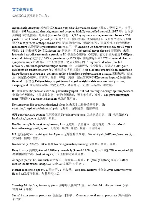
英文病历文章病例写作是医生日常的工作。
Associated symptoms 相关症状 Nausea, vomiting*2, sweating, dizzy(恶心、呕吐2次、出汗、眩晕) 1997:external chest tightness and dyspnea initially controlled atenolol. 1997年:出现胸外疼痛与呼吸困难,最终经服atenolol控制。
4/12 symptoms worse, exercise tolerance 200 yards on flat, limited by chest pain 4月12日,症状加重,受胸痛限制,仅耐受平地行走200码 No rest pain, no orthopnoea, no PND 无静息时疼痛,无端坐呼吸、无阵发性夜间呼吸困难Risk factors危险因素 Hypertension-no高血压:无 Smoking-20 cigarettes per day for 16 years 吸烟:16年来每天20支 Diabetes-no糖尿病:无 Cholesterol-never checked胆固醇:未查Ischemic heart disease-angina, previous MI缺血性心脏病:心绞痛、有心肌梗死病史 PMH(past medical history)过去史 1963: appendectomy 1963年:阑尾切除手术 1972: duodenal ulcer, no symptoms since1972年:十二指肠溃疡,之后无症状 1986: myocardial infarction, full recovery / No subsequent investigation1986年:心肌梗死,完全恢复,无随访 1989: gout quiescent on treatment1989年:痛风治疗期间症状静止 No diabetes, hypertension, rheumatic heart disease, tuberculosis, epilepsy, asthma, jaundice, cerebrovascular disease.无糖尿病、高血压、风湿性心脏病、结核病、癫痫、哮喘、黄疸、脑血管疾病 S/E(systems inquiry)系统回顾General 一般情况 Fatigue lately, appetite unchanged, weight stable, no sweats or pruritus, sleeping well 最近有疲劳感,食欲无改变,体重稳定,无出汗或骚痒,睡眠佳。
英文病例报告作文范文Case Report: Unusual Stomach Trouble.Man, 45, keeps saying his stomach hurts and he vomits sometimes. He's been like this for half a year, even after trying different meds. He hasn't changed his eating or lifestyle, so what's the problem? When we checked him out, his stomach area was a bit tender, but nothing else unusual.The lab tests showed his liver enzymes were up and he had a bit of anemia. The ultrasound of his belly showed his stomach wall was thicker than normal, like he might have chronic gastritis. But when we did an endoscopy, we saw something weird: there were little sores all over his stomach lining. It didn't look like a regular ulcer.We thought of a few other possibilities: maybe it's Crohn's disease or an autoimmune problem with his stomach. Or could it be eosinophilic gastroenteritis? Since his symptoms were so unusual and the regular treatments didn'twork, we decided to do a biopsy. And guess what? It showed there were a lot of eosinophils in his stomach lining, which means he has eosinophilic gastroenteritis.After starting him on corticosteroids, he felt much better in just two weeks. His stomach pain went away and he didn't vomit as much. When we checked his stomach again three months later, all the sores had healed up. It was a relief to see the treatment worked so well.This case really shows you can't always trust first impressions. Even if a patient's symptoms seem like a common problem, they might be something else entirely. It's always worth doing a thorough investigation to get theright diagnosis and the right treatment.。
病例报告英语作文Introduction:This case report presents the clinical findings, management strategies, and outcomes of a 52-year-old male patient diagnosed with Type 2 Diabetes Mellitus (T2DM). The objective is to illustrate the importance of comprehensive patient evaluation and individualized treatment plans in managing diabetes.Patient Profile:Mr. John Smith, a 52-year-old Caucasian male, was referred to the clinic for the management of T2DM. He had a history of hypertension, hyperlipidemia, and a family history of diabetes. His occupation as a truck driver involved longhours of sedentary work, and he reported a diet high in processed foods.Clinical Findings:Upon initial examination, Mr. Smith's body mass index (BMI) was 34.5 kg/m², indicating obesity. His hemoglobin A1c(HbA1c) level was 9.2%, which is above the target range for diabetes management. His fasting plasma glucose (FPG) was 180 mg/dL, and he exhibited signs of peripheral neuropathy.Diagnosis:Based on the American Diabetes Association (ADA) criteria, Mr. Smith's elevated HbA1c and FPG levels confirmed the diagnosis of T2DM.Treatment Plan:1. Lifestyle Modification: Mr. Smith was advised to adopt a balanced diet with reduced carbohydrates and increased fiber intake. A structured exercise program was initiated, including 30 minutes of moderate-intensity activity five times a week.2. Pharmacological Therapy: Metformin was prescribed as the first-line medication to improve glycemic control. Additionally, a statin was added to manage hyperlipidemia, and an ACE inhibitor for his hypertension.3. Regular Monitoring: Mr. Smith was scheduled for regular follow-ups to monitor his blood glucose levels, renal function, and lipid profile.Outcomes:After six months of intervention, Mr. Smith demonstrated significant improvements. His BMI reduced to 31.2 kg/m², HbA1c decreased to 7.5%, and FPG to 140 mg/dL. He reported reduced fatigue and an improvement in his peripheral neuropathy symptoms.Discussion:The case highlights the multifactorial approach to T2DM management, emphasizing the need for lifestyle changes and targeted pharmacological interventions. Regular monitoring and patient education are crucial for achieving and maintaining glycemic control.Conclusion:Mr. Smith's case report underscores the effectiveness of a personalized treatment plan in managing T2DM. It also serves as a reminder of the importance of early diagnosis and comprehensive care in preventing diabetes-related complications.。

医学病例报告英语作文Title: Medical Case Report A Rare Neurological Disorder。
Introduction:This case report discusses a rare neurological disorder called Moyamoya disease. The patient, a 32-year-old female, presented with recurring episodes of transient ischemic attacks (TIAs) and was diagnosed with this condition. This report aims to provide a comprehensive understanding of the disease, its clinical presentation, diagnostic procedures, treatment options, and prognosis.Clinical Presentation:The patient initially complained of frequent headaches, dizziness, and difficulty speaking. She also experienced weakness and numbness on the right side of her body. The symptoms worsened over time, leading to multiple episodesof TIAs. The patient's medical history revealed nosignificant risk factors for stroke, such as hypertension or diabetes.Diagnostic Procedures:The diagnostic workup included a detailed medical history, physical examination, and various imaging studies. Magnetic resonance imaging (MRI) and magnetic resonance angiography (MRA) revealed bilateral stenosis of the distal internal carotid arteries and the proximal anterior cerebral arteries. These findings were consistent with Moyamoya disease.Treatment:The treatment plan for the patient involved a multidisciplinary approach. Medications, including antiplatelet agents and anticoagulants, were prescribed to prevent further TIAs and stroke. Additionally, the patient underwent surgical revascularization to improve blood flow to the affected areas of the brain. Direct and indirect bypass procedures were performed to establish collateralcirculation.Prognosis:The prognosis for patients with Moyamoya disease varies depending on several factors, including the severity of symptoms, age at diagnosis, and promptness of treatment. Early diagnosis and intervention can significantly improve outcomes and reduce the risk of stroke. Regular follow-up visits and close monitoring of the patient's condition are essential to manage any potential complications and ensure optimal long-term outcomes.Discussion:Moyamoya disease is a rare cerebrovascular disorder characterized by progressive narrowing of the internal carotid arteries and the development of collateral blood vessels. It primarily affects children and young adults, with a higher prevalence in individuals of Asian descent. The exact cause of the disease remains unknown, although genetic and environmental factors are believed to play arole.Conclusion:Moyamoya disease is a rare neurological disorder that requires early recognition and appropriate management to prevent potentially devastating complications such as stroke. This case report highlights the importance of a comprehensive diagnostic approach and multidisciplinary treatment strategies. Further research is needed to better understand the pathogenesis and develop novel therapeutic interventions for this challenging condition.。

英语病例报告作文Medical Case Report: A Rare Case of Acute Myeloid Leukemia in a Young Adult。
Abstract:Acute myeloid leukemia (AML) is a rare and aggressive form of cancer that affects the blood and bone marrow. Itis characterized by the rapid growth of abnormal white blood cells, which can interfere with the normal production of red blood cells, platelets, and other white blood cells. This case report presents the clinical and laboratory findings of a 25-year-old male patient who was diagnosed with AML. The patient's symptoms, diagnostic tests, treatment, and outcomes are discussed in detail.Introduction:AML is a relatively rare form of leukemia, accounting for approximately 1% of all cancer diagnoses. It typicallyoccurs in older adults, with a median age at diagnosis of 68 years. However, AML can also affect younger individuals, as demonstrated by the case of the 25-year-old male patient described in this report. The incidence of AML in young adults is low, and the disease is often more aggressive and difficult to treat in this population.Case Presentation:The patient presented to the hospital with complaints of fatigue, shortness of breath, and easy bruising. He reported a 3-month history of progressively worsening symptoms, including fever, night sweats, and weight loss. On physical examination, the patient appeared pale and had petechiae and ecchymoses on his skin. Laboratory tests revealed pancytopenia, with a white blood cell count of 1.2 × 10⁹/L, hemoglobin level of 8.5 g/dL, and platelet count of 45 × 109/L. A peripheral blood smear showed the presence of blast cells, which raised suspicion for leukemia. Further testing, including bone marrow aspiration and biopsy, confirmed the diagnosis of AML.Treatment and Outcome:The patient was started on induction chemotherapy with a regimen of cytarabine and daunorubicin. He experienced severe myelosuppression and required supportive care with transfusions of red blood cells and platelets. Despite initial improvement in his blood counts, the patient developed febrile neutropenia and was found to have a bloodstream infection with a multidrug-resistant organism. He was treated with broad-spectrum antibiotics and antifungal agents, but his condition continued to deteriorate. Unfortunately, the patient succumbed to complications of his leukemia and infection, and he passed away 2 months after his initial presentation.Discussion:This case highlights the challenges of managing AML in young adults. The disease is often more aggressive and refractory to treatment in this population, leading to poor outcomes. In addition, the patient's susceptibility to infections due to severe myelosuppression furthercomplicated his clinical course. This case underscores the need for more effective and less toxic therapies for AML, particularly in young patients who may be more resilient to aggressive treatment approaches.Conclusion:Acute myeloid leukemia is a rare and aggressive form of cancer that can affect individuals of all ages. This case report illustrates the challenges of diagnosing andtreating AML in a young adult, and it emphasizes the needfor further research to improve outcomes in this population. Healthcare providers should be aware of the unique clinical characteristics of AML in young patients and strive to develop more effective and less toxic therapies for this challenging disease.。
脑梗死取栓病例汇报范文英文回答:Case Report: Mechanical Thrombectomy for Acute Ischemic Stroke.Patient Presentation:A 65-year-old male presented to the emergency department with sudden-onset left-sided weakness, slurred speech, and right facial droop.National Institutes of Health Stroke Scale (NIHSS) score: 17。
Imaging Studies:Computed tomography (CT) angiogram: Left middle cerebral artery (MCA) M1 segment occlusion.Magnetic resonance imaging (MRI) diffusion-weighted imaging (DWI): Ischemic changes in the left MCA territory.Treatment:The patient was immediately transferred to the interventional neuroradiology suite.Under general anesthesia, a right transfemoral approach was used for arterial access.A 5-Fr guiding catheter (Envoy, Codman Neurovascular) was navigated into the left internal carotid artery.Using a Solitaire FR guidewire (Medtronic), the thrombus was successfully retrieved from the left MCA M1 segment.Post-procedural CT angiogram confirmed complete recanalization of the affected artery.Outcome:The patient's NIHSS score improved to 2 within 24 hours after the procedure.At discharge, the patient was able to ambulate independently and had minimal residual left-sided weakness.Conclusion:Mechanical thrombectomy is an effective treatment for acute ischemic stroke due to large vessel occlusion. Inthis case, the timely intervention led to significant improvement in neurological function and a favorable outcome.中文回答:脑梗死取栓病例汇报。
病例报告英语作文Title: A Case Report: The Diagnosis and Treatment of a Rare Medical Condition。
Abstract:This case report presents a rare medical condition of a 45-year-old patient who presented with a unique set of symptoms. The aim of this report is to describe the diagnostic process, treatment plan, and patient outcome. The case highlights the importance of a multidisciplinary approach and thorough investigation in diagnosing and managing rare medical conditions.Introduction:Rare medical conditions pose significant challenges in terms of diagnosis and treatment due to their limited prevalence and diverse clinical presentations. This case report focuses on a patient with an unusual set of symptomsthat required a comprehensive evaluation to determine the underlying cause.Case Presentation:A 45-year-old male patient presented with a three-month history of fatigue, weight loss, and intermittent fevers. The patient reported no significant medical history or family history of similar symptoms. Initial physical examination revealed enlarged lymph nodes in the neck and groin, along with hepatomegaly. Laboratory investigations showed elevated inflammatory markers and abnormal liver function tests.Diagnostic Assessment:Given the patient's symptoms and physical examination findings, a wide range of potential diagnoses were considered, including infectious, autoimmune, and neoplastic diseases. The patient underwent a series of diagnostic tests, including blood cultures, serology for infectious diseases, imaging studies, and a lymph nodebiopsy.Results:Blood cultures were negative for any bacterial orfungal growth. Serology tests ruled out common infectious diseases such as tuberculosis and HIV. Imaging studies revealed multiple enlarged lymph nodes in various regionsof the body. A lymph node biopsy was performed, and histopathological examination showed features consistentwith Castleman disease, a rare lymphoproliferative disorder. Treatment and Outcome:The patient was referred to a multidisciplinary team consisting of hematologists, oncologists, and infectious disease specialists for further management. The treatment plan included the administration of corticosteroids to alleviate symptoms and reduce inflammation. Additionally,the patient received targeted therapy with rituximab, a monoclonal antibody, to target the abnormal lymphocytes.Over the course of several months, the patient showed significant improvement in symptoms, with a reduction in lymph node size and normalization of liver function tests. Regular follow-up visits were scheduled to monitor the patient's progress and adjust the treatment plan as necessary.Discussion:Castleman disease is a rare disorder characterized by abnormal lymph node enlargement and systemic symptoms. The diagnosis of this condition requires a combination of clinical suspicion, thorough investigation, and histopathological examination. Treatment options vary depending on the subtype of Castleman disease and may include surgery, chemotherapy, or targeted therapy.Conclusion:This case report highlights the importance of a multidisciplinary approach in diagnosing and managing rare medical conditions. The successful diagnosis and treatmentof Castleman disease in this patient demonstrate the significance of thorough investigation, collaboration among healthcare professionals, and individualized treatment plans. Further research is needed to enhance our understanding of rare medical conditions and improvepatient outcomes.。