人基质金属蛋白酶-13 MMP-13 ELISA试剂盒 北京驰明瑞
- 格式:pdf
- 大小:1.32 MB
- 文档页数:5

山东医药2022年第62卷第35期冠心病患者冠状动脉支架置入术后电针刺激双侧内关穴、郄门穴预防支架内再狭窄效果观察刘芙君1,陈黎明2,王勇11山东第一医科大学附属省立医院推拿科,济南250021;2山东第一医科大学附属省立医院心内科摘要:目的观察冠心病患者冠状动脉支架置入术(PCI)后电针刺激双侧内关穴、郄门穴的临床效果。
方法选取冠心病PCI后患者348例,随机分为电针组167例、非电针组155例。
两组均接受标准的药物治疗,电针组给予电针刺激双侧内关穴、郄门穴,每周治疗3次,共24周。
两组治疗后行冠脉造影,记录支架内再狭窄(ISR)发生情况,ELISA法检测血清S100A4。
治疗后随访至少12个月或以主要心血管不良事件(MACE)为随访终点,比较两组的生存时间及MACE发生情况。
结果电针组、非电针组分别发生ISR15例(8.98%)、39例(25.16%),两组比较P<0.05。
治疗前后电针组血清S100A4水平分别为(515.06±13.24)、(412.07±14.56)ng/mL,非电针组分别为(513.06±14.24)、(510.91±13.01)ng/mL;与治疗前比较,治疗后电针组血清S100A4水平降低,且低于非电针组治疗后(P均<0.05)。
电针组MACE发生率为10.18%,非电针组为25.81%,两组比较P<0.05。
电针组生存时间为(10.11±0.87)月,非电针组生存时间为(7.55±0.64)月;两组比较P<0.05。
结论电针刺激双侧内关穴、郄门穴可以有效降低冠心病患者PCI后ISR的发生率、同时有助于改善患者的预后,可能与其降低血清S100A4水平有关。
关键词:支架内再狭窄;冠状动脉支架置入术;电针刺激;内关穴;郄门穴;冠心病doi:10.3969/j.issn.1002-266X.2022.35.018中图分类号:R541.4文献标志码:A文章编号:1002-266X(2022)35-0075-03冠状动脉支架置入术(PCI)可极大地改善冠心病患者的预后及生活质量,明显减少心肌梗死导致的死亡,但术后支架内再狭窄(ISR)可增加主要心血管不良事件(MACE)的发生率,是影响患者近期和远期疗效及预后的主要因素[1-2]。

Catalog Number EK1M02-48EK1M02-96定量检测血清、血浆和细胞培养上清中的人基质金属蛋白酶2(MMP-2)浓度。
本产品仅用于科学研究,非诊断试剂,不能用于临床诊断。
1.背景介绍基质金属蛋白酶2(MMP-2),又名72kDa 的IV 型胶原酶和明胶酶A ,在人类中由MMP2基因编码。
激活MMP-2需要蛋白水解加工。
MMP-2在子宫内膜破裂、调节血管形成和炎症应答中起作用。
MMP-2参与许多事件,如癌症进程、癌细胞浸润、细胞信号转导、新血管形成和淋巴管生成。
MMP2基因突变与Torg-Winchester 综合征、多发性骨溶解、关节炎综合征有关,可能与瘢痕疙瘩有关。
非囊性纤维化支气管扩张中,相比于呼吸道感染铜绿假单胞菌的患者,感染流感嗜血杆菌患者的MMP-2浓度是升高的。
2.检测原理本试剂盒采用双抗体夹心酶联免疫吸附检测技术。
特异性抗人MMP-2抗体预包被在高亲和力的酶标板上。
酶标板孔中加入标准品、待测样本和生物素化的检测抗体,经过孵育,样本中存在的MMP-2与固相抗体和检测抗体结合。
洗涤去除未结合的物质后,加入辣根过氧化物酶标记的链霉亲和素(Streptavidin-HRP)。
洗涤后,加入显色底物TMB ,避光显色。
颜色反应的深浅与样本中MMP-2的浓度成正比。
加入终止液终止反应,在450nm 波长(参考波长570-630nm)测定吸光度值。
3.试剂盒检测的局限1)请在本试剂盒标示的有效期内使用。
2)试剂盒的试剂不能与其他批号的试剂或其他来源的试剂混合使用。
3)任何标准品稀释、操作人员、移液技术、洗涤技术、孵育温度、试剂盒保存时间的改变,都将影响结合反应。
4)本试剂盒在设计上去除或降低了生物学样本中的一些内源性干扰因素,并非所有可能的影响因素都已经去除。
Human MMP-2ELISA Kit 检测试剂盒(酶联免疫吸附法)EK1M02-01二、基本信息1.2.未提供的材料设备1)能够检测450nm吸光度的酶标仪,参考波长570nm或630nm2)移液器及枪头、加样槽3)准备试剂用的试管、离心管、量筒等4)蒸馏水或去离子水5)涡旋振荡器、微孔板振荡器3.贮存试剂盒保存于2-8℃,有效期标注于标签上。

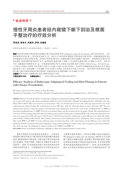
慢性牙周炎患者经内窥镜下龈下刮治及根面平整治疗的疗效分析贺高美贺高美,,郭丽云郭丽云,,刘建华刘建华,,李婷李婷,,吴梅爱深圳市龙岗区第二人民医院口腔科,广东深圳518112摘要目的研究慢性牙周炎患者经内窥镜下龈下刮治及根面平整(subgingival scaling and root planning,SRP)治疗的疗效。
方法选择深圳市龙岗区第二人民医院从2019年3月—2021年3月收治的83例慢性牙周炎患者为研究对象。
以随机抽签法将其分作内窥镜组41例及对照组42例。
对照组开展单纯SRP治疗,内窥镜组则开展经内窥镜下SRP治疗。
分析两组治疗前后牙周状况指标,血清及龈沟液炎症因子水平,血清基质金属蛋白酶-1(MMP-1)以及基质金属蛋白酶-13(MMP-13)水平。
结果治疗后,内窥镜组牙周附着水平(AL)、牙周袋探诊深度(PD)以及探诊出血指数(BOP)分别为(3.58±1.01)mm、(2.71±0.32)mm、(1.35±0.12)均低于对照组,差异有统计学意义(t=3.628、17.640、16.356,P<0.05)。
且治疗后内窥镜组血清炎症因子水平、龈沟液炎症因子水平、血清MMP-1以及MMP-13水平均低于对照组,差异有统计学意义(P<0.05)。
结论慢性牙周炎患者经内窥镜下龈下刮治及根面平整治疗的疗效较佳,可减轻炎症反应,降低MMP-1以及MMP-13水平。
关键词慢性牙周炎;内窥镜;龈下刮治;根面平整治疗;疗效中图分类号R781文献标志码A doi10.11966/j.issn.2095-994X.2022.08.09.33Efficacy Analysis of Endoscopic Subgingival Scaling and Root Planing in Patients with Chronic PeriodontitisHE Gaomei,GUO Liyun,LIU Jianhua,LI Ting,WU MeiaiDepartment of Stomatology,Second People´s Hospital of Longgang District,Shenzhen,Guangdong Province,518112ChinaAbstract Objective To study the efficacy of endoscopic subgingival scaling and root planing(SRP)in patients with chronic periodontitis.Meth‐ods A total of83patients with chronic periodontitis admitted to the Second People´s Hospital of Longgang District of Shenzhen City from March2019to March2021were selected as research objects.They were divided into41cases in the endoscopy group and42cases in the control group by random drawing.The control group was given SRP alone,while the endoscopic group was given endoscopic SRP.The peri‐odontal status indicators,serum and gingival crevicular fluid serum inflammatory factor levels,serum matrix metalloproteinase-1(MMP-1) and matrix metalloproteinase-13(MMP-13)levels were analyzed before and after treatment in the two groups.Results The periodontal attach‐ment level(AL),the probing depth(PD)of the periodontal pocket and the bleeding index on probing(BOP)in the endoscopic group after treat‐ment were respectively(3.58±1.01)mm,(2.71±0.32)mm,(1.35±0.12),lower than the control group,and the difference was statistically signifi‐cant(t=3.628,17.640,16.356,P<0.05).After treatment,the levels of serum inflammatory factors,inflammatory factors in gingival crevicular fluid,serum MMP-1and MMP-13in the endoscopy group were lower than those in the control group,and the differences were statistically significant(P<0.05).Conclusion Endoscopic subgingival scaling and root planing have better curative effect in patients with chronic periodon‐titis,which can reduce the inflammatory response and the levels of MMP-1and MMP-13.Key words Chronic periodontitis;Endoscopy;Subgingival scaling;Root planing treatment;Curative effect慢性牙周炎作为牙科临床常见疾病之一,主要是由菌斑微生物及其相关产物共同作用导致的慢性感染性疾病,随着病情的不断进展,可能引起牙周支持组织的进行性破坏,导致牙周膜、牙龈以及牙槽骨等出现不可逆损害[1]。

膝关节骨关节炎ute序列磁共振炎性面积与关节液MMP-13指标的相关性分析金鹏;陈俊杰;庄汝杰【摘要】目的分析膝关节骨关节炎(KOA) ute序列磁共振炎性面积与关节液MMP-13的相关性.方法收集2014年11月至2015年2月就诊且符合KOA诊断标准的患者24例,对患侧膝关节行矢状位ute序列磁共振扫描以获取影像学膝软骨炎性面积,同日收集患者膝关节液标本,应用酶联免疫吸附剂测定法(ELISA)检测关节液基质金属蛋白酶13 (MMP-13)指标,最后采用Pearson相关分析探讨磁共振影像学炎性面积与MMP-13指标的相关性.结果 24例KOA患者关节液MMP-13指标为(0.179±0.03) pg/ml,磁共振炎性面积为(144.23±91.95) mm2,Pearson相关性分析显示两组数据呈正相关(r=0.920,P<0.01).结论 KOA ute序列磁共振炎性面积与关节液MMP-13指标呈正相关,MMP-13是KOA炎症发展的重要指标.【期刊名称】《浙江临床医学》【年(卷),期】2016(018)004【总页数】2页(P659-660)【关键词】膝关节骨关节炎;磁共振成像;超短回波时间;MMP-13【作者】金鹏;陈俊杰;庄汝杰【作者单位】310053 浙江中医药大学;310006 浙江省中医院;310006 浙江省中医院【正文语种】中文膝关节骨关节炎(KOA)是一种以关节软骨退化、骨质增生为主要特点的慢性退行性关节病,常发生于中老年人群[1],是在力学及生物学因素的共同参与下,软骨细胞、细胞外基质及软骨下骨三者之间分解和合成失衡、代谢紊乱的结果。
基质金属蛋白酶13(MMP-13)因可以优先降解其主要成分为II 型胶原软骨基质而在骨关节炎(OA)生物学发病机制中所发挥的作用备受关注。
本文旨在分析KOA ute序列磁共振炎性面积与关节液MMP-13的相关性,为MMP-13作为KOA炎症发展的一项重要指标提供研究支持。

基质金属蛋白酶在骨关节炎症性疾病中的研究进展发布时间:2022-04-28T04:50:17.945Z 来源:《世界复合医学》2022年2期作者:戴炯华,刘芳通信作者[导读] 基质金属蛋白酶(Matrix metalloproteinases,MMPs)是一类锌依赖性蛋白酶家族,其可通过降解细胞外基质、调控相应信号通路来参与细胞增殖、迁移、和分化等多种生物学过程[1]。
戴炯华,刘芳通信作者南华大学衡阳医学院岳阳市人民医院研究生协作培养基地,湖南衡阳421001【摘要】基质金属蛋白酶(Matrix metalloproteinases,MMPs)是一类锌依赖性蛋白酶家族,其可通过降解细胞外基质、调控相应信号通路来参与细胞增殖、迁移、和分化等多种生物学过程[1]。
MMPs在正常关节组织中低水平表达,而在关节炎症病理状态下表达明显增高,其参与调节炎症反应的各个方面,在骨关节炎、痛风性关节炎、类风湿性关节炎等发挥重要作用,本文对基质金属蛋白酶与骨关节常见炎症性疾病相关性研究进展做一综述,为科研提供便利。
【关键词】基质金属蛋白酶;骨关节炎;痛风性关节炎;类风湿性关节炎Research progress of matrix metalloproteinases in inflammatory diseases of bone and joint[Abstract]Matrix metalloproteinases(MMPs)are a family of zinc-dependent proteases,which can participate in various biological processes such as cell proliferation,migration and differentiation by degrading extracellular Matrix and regulating corresponding signal pathways[1].MMPs low level expression in normal joint tissues,and expressed in joint inflammation pathological condition significantly increased,to participate in the aspects of regulating the inflammatory response,in osteoarthritis,rheumatoid arthritis,gouty arthritis,etc play an important role,in this paper,matrix metalloproteinases and common inflammatory joint disease correlation research progress.[Key words]matrix metalloproteinase;osteoarthritis;gout arthritis;rheumatoid arthritis1.基质金属蛋白酶的概述人体正常的生长发育过程离不开细胞外基质(Extracellular matrix,ECM)适时的降解,ECM是一种大分子网络,其中胶原是ECM中最丰富的蛋白质,MMPs是唯一能够降解胶原的酶。

人基质金属蛋白酶-9(MMP-9)酶联免疫分析(ELISA)试剂盒使用说明书厦门慧嘉生物科技有限公司本试剂仅供研究使用目的:本试剂盒用于测定人血清,血浆及相关液体样本中基质金属蛋白酶-9(MMP-9)含量。
实验原理:本试剂盒应用双抗体夹心法测定标本中人基质金属蛋白酶9(MMP-9)水平。
用纯化的人基质金属蛋白酶9(MMP-9)抗体包被微孔板,制成固相抗体,往包被单抗的微孔中依次加入人基质金属蛋白酶9(MMP-9),再与HRP标记的基质金属蛋白酶9(MMP-9)抗体结合,形成抗体-抗原-酶标抗体复合物,经过彻底洗涤后加底物TMB显色。
TMB在HRP酶的催化下转化成蓝色,并在酸的作用下转化成最终的黄色。
颜色的深浅和样品中的人基质金属蛋白酶9(MMP-9)呈正相关。
用酶标仪在450nm波长下测定吸光度(OD值),通过标准曲线计算样品中人基质金属蛋白酶-9(MMP-9)浓度。
试剂盒组成:样本处理及要求:1. 血清:室温血液自然凝固10-20分钟,离心20分钟左右(2000-3000转/分)。
仔细收集上清,保存过程中如出现沉淀,应再次离心。
2. 血浆:应根据标本的要求选择EDTA或柠檬酸钠作为抗凝剂,混合10-20分钟后,离心20分钟左右(2000-3000转/分)。
仔细收集上清,保存过程中如有沉淀形成,应该再次离心。
3. 尿液:用无菌管收集,离心20分钟左右(2000-3000转/分)。
仔细收集上清,保存过程中如有沉淀形成,应再次离心。
胸腹水、脑脊液参照实行。
4. 细胞培养上清:检测分泌性的成份时,用无菌管收集。
离心20分钟左右(2000-3000转/分)。
仔细收集上清。
检测细胞内的成份时,用PBS(PH7.2-7.4)稀释细胞悬液,细胞浓度达到100万/ml左右。
通过反复冻融,以使细胞破坏并放出细胞内成份。
离心20分钟左右(2000-3000转/分)。
仔细收集上清。
保存过程中如有沉淀形成,应再次离心。
化妆品功效评价(Ⅳ)——延缓皮肤衰老功效宣称的科学支持李诚桐;赵华【摘要】概述了皮肤的衰老表现和衰老机制学说,介绍了生物化学法、细胞生物学法、三维重组皮肤模型替代法、动物实验法、主观评估、客观仪器评价法等延缓皮肤衰老化妆品功效评价方法,提供了延缓皮肤衰老功效评价的新思路,展望了延缓衰老化妆品功效评价方法的未来发展方向.【期刊名称】《日用化学工业》【年(卷),期】2018(048)004【总页数】8页(P188-195)【关键词】延缓皮肤衰老化妆品;皮肤衰老;功效评价【作者】李诚桐;赵华【作者单位】北京工商大学理学院化妆品系,北京100048;北京市植物资源研究开发重点实验室,北京100048;北京工商大学理学院化妆品系,北京100048;北京市植物资源研究开发重点实验室,北京100048【正文语种】中文【中图分类】TQ658伴随着人们对精神生活的追求不断提高,消费者更加注重皮肤的保养,并希望通过使用化妆品来延缓皮肤衰老。
市场的需求促使化妆品企业相继推出多种延缓皮肤衰老宣称的产品,而产品是否能够达到宣称目的,以及如何对延缓皮肤衰老宣称提供科学支持,也成为生产企业、消费群体以及市场监管部门所共同关注的话题。
1 皮肤的衰老人的一生中要经历生长期、成熟期和老年期(衰老期)3个阶段。
衰老是一个复杂的、涉及多种不同器官和系统的系列变化过程,是生命的基本特征之一,它是生物个体在生命周期中必然发生的、难以逆转的退行性变化。
在人体衰老阶段,各器官、组织和系统都会发生变化,但从外观上最容易观察到的是皮肤及其附属器官的变化[1]。
皮肤覆盖在身体表面,由表皮、真皮、皮下组织构成,呈多层次分布。
皮肤的衰老是一个多维立体的过程[2]。
皮肤老化在表皮、真皮、皮下组织和附属器官的主要表现有以下几个方面。
1)表皮。
表皮老化主要表现为皮肤变薄,角质形成细胞增大且部分角化不全,其中比较明显的指标为皮肤水分含量的丢失。
由于水合能力降低,皮肤组织细胞的水分减少,细胞皱缩,出现细小皱纹。
MMP-33)酶联免疫分析(ELISA)人基质金属蛋白酶-3(MMP-试剂盒使用说明书本试剂仅供研究使用目的:本试剂盒用于测定人血清,血浆及相关液体样本中基质金属蛋白酶-3(MMP-3)的含量。
实验原理:本试剂盒应用双抗体夹心法测定标本中人基质金属蛋白酶-3(MMP-3)水平。
用纯化的人基质金属蛋白酶-3(MMP-3)抗体包被微孔板,制成固相抗体,往包被单抗的微孔中依次加入人基质金属蛋白酶-3(MMP-3),再与HRP标记的羊抗人抗体结合,形成抗体-抗原-酶标抗体复合物,经过彻底洗涤后加底物TMB显色。
TMB在HRP酶的催化下转化成蓝色,并在酸的作用下转化成最终的黄色。
颜色的深浅和样品中的人基质金属蛋白酶-3(MMP-3)呈正相关。
用酶标仪在450nm波长下测定吸光度(OD值),通过标准曲线计算样品中人基质金属蛋白酶-3(MMP-3)浓度。
试剂盒组成:试剂盒组成48孔配置96孔配置保存说明书1份1份封板膜2片(48)2片(96)密封袋1个1个酶标包被板1×481×962-8℃保存标准品:225μg/L0.5ml×1瓶0.5ml×1瓶2-8℃保存标准品稀释液 1.5ml×1瓶 1.5ml×1瓶2-8℃保存酶标试剂3ml×1瓶6ml×1瓶2-8℃保存样品稀释液3ml×1瓶6ml×1瓶2-8℃保存显色剂A液3ml×1瓶6ml×1瓶2-8℃保存显色剂B液3ml×1瓶6ml×1瓶2-8℃保存终止液3ml×1瓶6ml×1瓶2-8℃保存浓缩洗涤液(20ml×20倍)×1瓶(20ml×30倍)×1瓶2-8℃保存样本处理及要求:1.血清:室温血液自然凝固10-20分钟,离心20分钟左右(2000-3000转/分)。
仔细收集上清,保存过程中如出现沉淀,应再次离心。
胶原蛋白酶2,9检测试剂盒(MMP Zymography assay kit) P1700描述:基质金属蛋白酶(matrix metalloproteinase, MMP)以无活性的酶原形式分泌,活化后能降解细胞外基质的胶原蛋白,又称胶原酶。
胶原酶通过影响细胞外基质参与胚胎发育、形态发生、组织重塑等过程,并参与炎症、肿瘤、心血管、神经等许多疾病过程。
血浆和组织中的MMP-2、MMP-9参与各种生理与病理过程,其在临床诊断和治疗中的意义日益受到重视1。
本试剂盒采用酶谱法(zymography)检测MMP-2、MMP-9活性。
Zymography是一种广为使用的、基于SDS-PAGE电泳和反相凝胶染色的蛋白酶检测方法,其灵敏度可达1 nM,高于ELISA方法2,其基本原理和程序是:制备加有胶原酶底物的SDS-PAGE凝胶,含蛋白酶的样品在此凝胶中进行电泳,电泳结束后取出凝胶与酶反应Buffer孵育,凝胶染色与脱色。
凝胶中由于含底物蛋白而被深染,形成深色背景,但在有蛋白酶条带的位置,蛋白酶降解凝胶中的底物蛋白,因而不被染色而形成透亮区域,从而能同时指示MMP-2、MMP-9的大小位置(酶谱)及活性。
本试剂盒提供MMP-2、MMP-9特异蛋白底物及酶学反应缓冲液,可检测少至0.1~0.5 µl血液中的MMP-2、MMP-9酶谱及活性,检测灵敏度约为1 nM。
试剂盒可进行50次标准小胶检测,如果小胶加样孔为10~15个,则总计可检测500~750个样品。
参考文献:1.Nagase H and Woessner JF. Matrix Metalloproteinases. J Biol Chem, 1999, 274, 21491-214942.Heussen C, and Dowdle EB. Electrophoretic analysis of plasminogen activators in polyacrylamide gels containing sodiumdodecyl sulfate and copolymerized substrates. Anal. Biochem,1980, 102, 196–202适用: 检测MMP-2、MMP-9酶谱及活性组成与储存(50 assays):12个月有效1. 2 × SDS-PAGE non-reducing buffer 1.5 ml , −20 ºC 储存2. 10 × Substrate G 50 ml −20 ºC 储存3. 10 × Buffer A 50 ml 4 ºC 储存使用时用蒸馏水稀释4. 10 × Buffer B 50 ml 4 ºC 储存使用时用蒸馏水稀释5. SDS-PAGE凝胶蛋白质考马斯亮蓝染色液(货号P1501) 4 ºC储存操作步骤:1.制备含MMP底物蛋白的SDS-PAGE凝胶:推荐分离胶浓度为8%。
人基质金属蛋白酶-13 MMP-13使用说明书产品编号:本试剂盒仅供科研使用,不得用于临床及诊断使用!操作步骤1.取出试剂盒,于室温(20-25℃)放置15-30分钟。
实验过程应在室温(20-25℃)内进行。
2.取出酶标板,按照标准品的次序分别加入100μl的标准品溶液于空白微孔中。
3.空白微孔中加入100μl的样品,空白对照加入100μl的蒸馏水;4.在各孔中加入50μl的酶标记溶液;(不含空白对照孔)5.将酶标板用封口胶密封后,37℃孵育反应1小时;(在孵育箱中保持稳定的温度与湿度)6.充分清洗酶标板3-5次,保持各孔有充足的水压;(浓缩洗涤液以1:100的比例与蒸馏水稀释)7.酶标板洗涤后用吸水纸彻底拍干;8.各孔加入显色剂A、B液各50μl;(不含空白对照孔)9.20-25℃下避光反应15分钟;10.各孔加入50μl终止液,终止反应;结果判断1.30分钟内在波长450nm的酶标仪上读取各孔的OD值;2.百分结合率计算:设S0管计数为B0,各标准管或样品管计数为B,非特异管计数为NSB,则百分结合率计算公式如下:B/ B0=(B-NSB)/( B0-NSB)×100%3.logit计算:各标准点或样品管的logit值计算公式如下:logit=ln(B/ B0)/(1-B/ B0)4.将标准品的OD均值与标准品0点的OD均相除,为标准点的百分结合率,在log-logit坐标纸上绘图。
5.Log-logit双对数标准曲线:坐标纸上横轴从左至右第一个1-9表示为第一个10进位,第二个1-9表示为第二个10进位。
第三个1-9表示为第三个10进位。
坐标纸纵轴为百分比(1-99),即各标准吸光值的百分结合率。
取一条通过各点的直线。
要求尽可能多的点在线上,同时剩余的点均匀分布在直线的两边。
样品也同样由吸光值计算百分结合率,再从纵轴上的相应结合率找到直线上的点,此点对应的横坐标浓度即为样品的浓度,无须换算。
6.人工处理:以标准浓度取log值为横坐标,对应的logit值为纵坐标在普通坐标纸上或以标准浓度为横坐标,对应的B/B0为纵坐标在logit-log坐标纸上画出标准曲线(理想化时是一条直线)。
根据待测样品的B/B0可以从坐标纸上查出样品的浓度值。
如果使用普通坐标纸,查出的数值应取反对数才是最后的浓度值。
7.自动处理:使用logit-log或四参数数据处理模式,由电脑自动计算得出结果。
8.敏感度:1.0ng/ml;9.图例Human matrix metalloproteinase 13 ELISA Kit96 TestsCatalogue Number:Store all reagents at 2-8°CFOR LABORATORY RESEARCH USE ONLY. NOT FOR THERAPEUTIC OR DIAGNOSTIC APPLICATIONS!PLEASE READ THROUGH ENTIRE PROCEDURE BEFORE BEGINNING!INTENDED USEThis BOGOO MMP-13 ELISA kit is intended Laboratory for Research use only and is not for use in diagnostic or therapeutic procedures.The Stop Solution changes the color from blue to yellow and the intensity of the color is measured at 450 nm using a spectrophotometer. In order to measure the concentration of MMP-13 in the sample, this MMP-13 ELISA Kit includes a set of calibration standards. The calibration standards are assayed at the same time as the samples and allow the operator to produce a standard curve of Optical Density versus MMP-13 concentration. The concentration of MMP-13 in the samples is then determined by comparing the O.D. of the samples to the standard curve.PRINCIPLE OF THE ASSAYThe coated well immunoenzymatic assay for the quantitative measurement of serum MMP-13 utilizes a monoclonal anti-MMP-13 and a MMP-13-HRP conjugate. The assay asample and buffer are incubated together with anti-MMP-13 antibody coated plate for sixty and washed. The diluted MMP-13-HRP conjugate is then added to each well and incubated. After the incubation period, the wells are decanted and washed three times. The wells are then incubated with a substrate for the enzyme. The product of the enzyme-substrate reaction forms a blue colored complex. Finally, a stopping solution is added to stop the reaction, which will then turn the solution yellow.The intensity of color is measured spectrophotometrically at 450nm in a microplate reader. The intensity of the color is inversely proportional to the MMP-13 concentration since MMP-13 from samples and MMP-13-HRP conjugate compete for the anti-MMP-13 antibody binding site. Since the number of sites is limited, as more sites are occupied by MMP-13 from the sample, fewer sites are left to bind MMP-13-HRP conjugate.Standards of known MMP-13 concentrations are run concurrently with the samples being assayed and a standard curve is plotted relating the intensity of the color (Optical Density) to the concentration of MMP-13. The unknown MMP-13 concentration in each sample is interpolated from this curve.REAGENTS PROVIDEDAll reagents provided are stored at 2-8° C. Refer to the expiration date on the label.1.MICROTITER PLATE 96 wells2.ENZYME CONJUGATE 6.0 mL 1 vial3.STANDARD.1 0ng/ml 1 vial4.STANDARD.2 50ng/ml 1 vial5.STANDARD.3 100ng/ml 1 vial6.STANDARD.4 250ng/ml 1 vial7.STANDARD.5 500ng/ml 1 vial8.STANDARD.6 1000ng/ml 1 vial9.SUBSTRATE A 6.0 mL 1 vial10.SUBSTRATE B 6.0 mL 1 vial11.STOP SOLUTION 6.0 mL 1 vial12.WASH SOLUTION x100 10 mL 1 vial13.Instruction 1MATERIALS REQUIRED BUT NOT SUPPLIED1.Microplate reader capable of measuring absorbance at 450 nm.2.Precision pipettes to deliver 2 ml to 1 ml volumes.3.Adjustable 10ml -100ml pipettes for reagent preparation.4.Adjustable 10ml -100ml pipettes for reagent preparation.5.100 ml and 1 liter graduated cylinders.6.Calibrated adjustable precision pipettes, preferably with disposable plastic tips. (A manifold multi-channelpipette is desirable for large assays.)7.Absorbent paper.8.37°C incubator.9.Distilled or deionized water.10.Data analysis and graphing software. Graph paper: linear (Cartesian), log-log or semi-log, or log-logit asdesired.11.Tubes to prepare standard or sample dilutions.ASSAY PROCEDURE1. 1 .Prepare all Standards before starting assay procedure (see Preparation Reagents). It is recommended thatall Standards and Samples be added in duplicate to the Microtiter Plate.Standards or Samples to2.First, secure the desired number of coated wells in the holder, then add 100 μL ofthe appropriate well of the antibody pre-coated Microtiter Plate.3.Add 50 μL of Conjugate to each well. Mix well. Complete mixing in this step is important. Cover andincubate for 1 hours at 37°C.4.Prepare Substrate Solution no more than 15 minutes before end of incubation (see Preparation of Reagents).5.Wash the Microtiter Plate using one of the specified methods indicated below:6.Manual Washing: Remove incubation mixture by aspirating contents of the plate into a sink or proper wastecontainer. Using a squirt bottle, fill each well completely with distilled or de-ionized water, then aspiratecontents of the plate into a sink or proper waste container. Repeat this procedure four more times for a totalof FIVE washes. After final wash, invert plate, and blot dry by hitting plate onto absorbent paper or papertowels until no moisture appears. Note: Hold the sides of the plate frame firmly hen washing the plate toassure that all strips remain securely in frame.7.Automated Washing: Aspirate all wells, then wash plate FIVE times using distilled or de-ionized water.Always adjust your washer to aspirate as much liquid as possible and set fill volume at 350 μL/well/wash(range: 350-400 μL). After final wash, invert plate, and blot dry by hitting plate onto absorbent paper paper towels until no moisture appears. It is recommended that the washer be set for a soaking time of 10seconds or shaking time of 5 seconds between washes.8.Add 50 μL Substrate A&B to each well. Cover and incubate for 15 minutes at 20-25°C.9.Add 50 μL of Stop Solution to each well. Mix well.10.Read the Optical Density (O.D.) at 450 nm using a microtiter plate reader within 30 minutes.BOGOO CALCULATION OF RESULTS1.Calculate the mean absorbance value A450 for each set of reference standards and samples.2.Divide the average A450 value for each standard, control and test sample by the average A450 of standard0and multiply by 100 to obtain %B/B0 for each sample.3.Prepare a standard curve by plotting the average absorbance of each standard versus the correspondingconcentrations of the standards on linear-log graph paper or the %B/B0 value for each standard versus the corresponding concentration of the standard on linear-log or logit-log graph paper. logit=ln(B/ B0)/(1-B/ B0)4.Any values obtained for diluted samples must be further converted by applying the appropriate dilution factorin the calculations.5.The standard density is a X, the B/ B0 is a Y, sitting to mark the paper in the logit- log up draw a standardcurve.According to the B/ B0 that need to be measured the sample can from sit to mark the density value that the paper looks up the sample up.6.The sensitivity by this assay is 1.0ng/ml7.The standard curve presented here is an example of the data typically produced with this kit; however, yourresults will not be identical to these.8.Standard curve。