BSA-binding properties and anti-proliferative effects of amino acids functionalized
- 格式:pdf
- 大小:2.27 MB
- 文档页数:9

2021255vitro and in vivo models :a systematic review [J/OL ].Nutrients ,2019,11(12):11122875.DOI :10.3390/nu11122875.[10]余天奉,韩泽平,黎毓光,等.Hsa -miR -145联合槲皮素对人前列腺癌LNCaP 细胞侵袭、迁移的影响[J ].现代肿瘤医学,2017,25(15):2387-2391.[11]Sheng J ,Zou X ,Cheng Z ,et al.Recent Advances in HerbalMedicines for Digestive System Malignancies [J ].Front Pharma‐col ,2018,9:01249.DOI :10.3389/fphar.2018.01249.[12]YAN Z ,LAI Z ,LIN J.Anticancer properties of traditional Chi‐nese medicine [J ].Comb Chem High Throughput Screen ,2017,20(5):423-429.[13]MAURYA SK ,SHADAB G ,SIDDIQUE HR.Chemosensitizationof therapy resistant tumors :targeting multiple cell signaling path‐ways by lupeol ,a pentacyclic triterpene [J ].Curr Pharm Des ,2020,26(4):455-465.[14]MAGAR RT ,SOHNG JK.A review on structure ,modificationsand structure -activity relation of quercetin and its derivatives [J ].J Microbiol Biotechnol ,2020,30(1):11-20.[15]DANG JH ,JIN ZJ ,LIU XJ ,et al.Metformin in combination withcisplatin inhibits cell viability and induces apoptosis of human ovarian cancer cells by inactivating ERK 1/2[J ].Oncol Lett ,2017,14(6):7557-7564.[16]MAO Y ,XI L ,LI Q ,et bination of PI3K/Akt pathway in‐hibition and Plk1depletion can enhance chemosensitivity to gem‐citabine in pancreatic carcinoma [J ].Transl Oncol ,2018,11(4):852-863.[17]LU TX ,ROTHENBERG ME.MicroRNA [J ].J Allergy Clin Im‐munol ,2018,141(4):1202-1207.[18]XU WX ,LIU Z ,DENG F ,et al.MiR -145:a potential biomarkerof cancer migration and invasion [J ].Am J Transl Res ,2019,11(11):6739-6753.[19]ZHANG H ,JIANG M ,LIU Q ,et al.miR -145-5p inhibits the pro‐liferation and migration of bladder cancer cells by targeting TAGLN2[J ].Oncol Lett ,2018,16(5):6355-6360.[20]张雪峰,张雪琦,吴翠翠,等.miRNA -145与膀胱癌侵袭和转移的关系[J ].标记免疫分析与临床,2019,26(9):1552-1555,1582.[21]LIU X ,WU Y ,ZHOU Z ,et al.Celecoxib inhibits the epithelial -to -mesenchymal transition in bladder cancer via the miRNA -145/TGFBR2/Smad3axis [J ].Int J Mol Med ,2019,44(2):683-693.[22]WANG J ,ZHANG H ,SITU J.KCNQ1OT1aggravates cell prolif‐eration and migration in bladder cancer through modulating miR -145-5p/PCBP2axis [J ].Cancer Cell Int ,2019,19(1):325.[23]YE D ,SHEN Z ,ZHOU S.Function of microRNA -145and mech‐anisms underlying its role in malignant tumor diagnosis and treat‐ment [J ].Cancer Manag Res ,2019,11:969-979.(收稿日期:2020-03-12,修回日期:2020-04-21)紫花前胡苷抗宫颈癌细胞作用研究付琼,殷金凤,颜爱华作者单位:成都市第二人民医院妇产科,四川成都610017通信作者:颜爱华,女,副主任医师,研究方向为妇科肿瘤,Email :摘要:目的探究紫花前胡苷抗宫颈癌HeLa 细胞的作用效果及相关信号通路。

1.1.1多肽合成按照多肽合成常规操作,合成来源于上海楚肽生物科技有限公司(Apeptide CO.,Ltd.) HPLC检测纯度为95%。
多肽C端增加的半胱氨酸残基用于以其巯基连接于偶联剂上。
1.1.2 合成多肽与载体的偶联载体蛋白选择BSA(Roche公司),用SPDP(PIERCE公司)连接法将合成的多肽与BSA进行偶联:4.6mgSPDP溶解于740ulDMSO,终浓度为20mM。
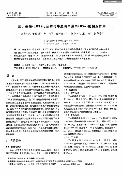
0.1008gBSA溶解于2ml PBS-EDTA溶液中,室温静置1h。
HiTrap TM Deaslting column脱盐柱洗脱多余的SPDP。
4mg多肽加入偶联好的BSA-SPDP体系中室温过夜。
1.1.3 抗多肽抗体的制备选体重1. 5~2 kg的健康雄性新西兰大白兔,按常规操作卡介苗活化其免疫系统。
将偶联的BSA-多肽过滤除菌,100mg(2ml)加等体积的不完全佐剂,充分混悬。
在新西兰大白兔背部多点皮下注射,4周后加强免疫,其后每隔2周进行1次加强免疫,注射途径与初次免疫相同,共两次后,检测效价。
耳源静脉采血、分离血清。
1.1.4 多肽抗体的纯化Protein G柱分离纯化IgG抗体:PBSpH7.4平衡Protein G亲和柱,以0.1M Gly-HCl pH3.0洗脱,PBSpH7.4透析,分装,-20℃保存。
1mM预冷的HCl充分膨胀Sepharose 4FF(GE公司)后,15个体积的1mMHCl清洗去残留的蔗糖,每次清洗2min。
偶联缓冲液0.1MNaHCO3 pH8.3 0.5M NaCl平衡2次。
多肽溶于偶联缓冲液,调节pH值到pH8.3后,加入凝胶,室温混旋3h。
加入1M预冷的乙醇胺混旋2h阻断未偶联上的活化位点。
50mM Tris-HCl pH8.0,1MNaCl 溶液与50mM Gly-HCl pH3.5,1M NaCl 交替清洗8次。
PBS缓冲液平衡10倍体积。
装柱,平衡,上样后以0.1MGly-HCl pH2.2洗脱,PBS透析,分装,-20℃冻存。

FAPα靶向标记化合物131I-FAPI-03的合成及初步体内外实验研究马 欢1,2, 廖家莉2, 杨远友2, 刘 宁2, 李飞泽2(1. 四川省医学科学院 四川省人民医院(电子科技大学附属医院) 核医学科,成都 610072;2. 四川大学 原子核科学技术研究所,辐射物理及技术教育部重点实验室,成都 610064)摘要:本研究以4-喹啉基-甘氨基-2-氰基吡咯烷为骨架,对连接基团进行碳链延长及羟基修饰成功合成FAPI 衍生物ATE-FAPI-03;通过亲电取代反应实现其131I 标记,并对标记化合物131I-FAPI-03的脂水分配比、体外稳定性等进行分析;开展细胞结合、内吞、流出等实验以评价131I-FAPI-03的体外动力学特征;并考察了131I-FAPI-03在荷胶质瘤小鼠体内的分布情况。
结果表明:131I-FAPI-03为亲脂性小分子,并具有良好的体外稳定性;与FAPα阳性细胞U87MG 孵育10 min 时的结合率为(22.00 ± 0.35)%,且随着孵育时间的延长结合率有明显的上升趋势,而与FAPα阴性细胞MCF-7的结合率始终处于较低水平;通过竞争结合实验测得131I-FAPI-03的IC 50值为45.5 nM ,表明其对FAPα具有较高的亲和力;大部分与U87MG 细胞结合的131I-FAPI-03可被细胞内吞,但其在细胞中的滞留能力偏低。
131I-FAPI-03在荷胶质瘤小鼠体内具有快速的肿瘤靶向能力:经尾静脉注射5 min 后,肿瘤组织对131I-FAPI-03的放射性摄取值为(14.90 ±3.21)% ID/g ,注射2 h 后,肿瘤/肌肉的放射性摄取比值达到(43.7 ± 16.7)。
上述结果为新型FAPα靶向药物的研发提供了重要的参考。
关键词:FAPα;131I ;FAPI ;胶质瘤中图分类号:TL92+3 文献标志码:A 文章编号:1000-7512(2024)02-0097-09doi :10.7538/tws.2024.37.02.0097Synthesis and Preliminary Evaluation of FAPα Targeted Tracer 131I-FAPI-03MA Huan 1,2, LIAO Jiali 2, YANG Yuanyou 2, LIU Ning 2, LI Feize2(1. Department of Nuclear Medicine , Sichuan Provincial People’s Hospital ,University of Electronic Science and Technology of China , Chengdu 610072, China ;2. Key Laboratory of Radiation Physics and Technology of the Ministry of Education ,Institute of Nuclear Science and Technology , Sichuan University , Chengdu 610064, China )Abstract: Using N-(4-quinolinoyl)-Gly-(2-cyanopyrrolidine) as scaffold ,we prolonged the linker with serine to obtain a FAPI derivative ATE-FAPI-03,which was subsequently labeled with131I byelectrophilic substitution. Then the in vitro stability ,Log P value ,binding affinity ,targeting properties and biodistribution behavior of 131I-FAPI-03 was evaluated. Results show that 131I-FAPI-03 was lipophilic and stable in vitro ,capable of specifically binding to FAPα-positive U87MG cells收稿日期:2023-12-04;修回日期:2024-01-18基金项目:中央高校基本科研业务费专项资金资助(2023SCU12132);四川省医学科学院.四川省人民医院青年人才基金(2022QN32)通信作者:李飞泽第37卷 第2期同 位 素Vol. 37 No. 22024年 4 月Journal of IsotopesApr. 2024fast with a major proportion trapped intracellularly. After 10 min of incubation,131I-FAPI-03 showed a specific binding rate of (22.00 ± 0.35)%,and the binding rate increased with the incubation time,to a peak of (37.5 ± 0.83)% at 180 min. However,the FAPα-negative MCF-7 cells exhibited very low uptake of 131I-FAPI-03 at any time point. The IC50 measured by the competition assay indicated significant binding property of 131I-FAPI-03. Biodistribution studies revealed that 131I-FAPI-03 could rapidly accumulate in tumor sites with an uptake of (14.90 ± 3.21)% ID/g at 5 min post injection. At 2 h post injection,131I-FAPI-03 displayed the highest tumor-to-muscle ratio of 43.7 ± 16.7. All above results provided important reference for the development of novel FAPα-targeting tracers.Key words: FAPα; 131I; FAPI; glioma肿瘤相关成纤维细胞(CAF)作为肿瘤微环境的主要组成部分,积极参与细胞外基质结构的重塑、肿瘤侵袭与转移、免疫抑制和耐药生成过程[1-3]。

Product InformationMonoclonal ANTI-FLAG® M2, Clone M2 Produced in Mouse, Affinity Isolated AntibodyF1804Storage Temperature −20 °CProduct DescriptionMonoclonal ANTI-FLAG® M2 is a mouse-derived, affinity-purified IgG1 monoclonal antibody that binds to fusion proteins containing a FLAG® peptide sequence.1 The M2 antibody will recognize a FLAG® peptide sequence at the N-terminus,Met-N-terminus, C-terminus, or internal sites of a fusion protein. Binding of the M2 antibody is not dependent on calcium.Monoclonal ANTI-FLAG® M2 is useful for detection, identification, and capture of fusion proteins that contain a FLAG® peptide sequence by common immunological procedures, such as Western blotting, immunofluorescence, and immunoprecipitation. ReagentSupplied at a concentration of ∼ 1 mg of protein per mL of solution, and formulated in 50% glycerol,10 mM sodium phosphate, and 150 mM NaCl,pH 7.4. The formulation contains no antimicrobial preservatives.Antigenic binding siteN-Asp-Tyr-Lys-Asp-Asp-Asp-Asp-Lys-C SpecificityThe monoclonal antibody detects only the target protein band(s) on a Western blot from an E. coli, plant or mammalian crude cell lysate.SensitivityThe monoclonal antibody detects as little as 2 ng of target protein by dot blot. The Western blot is tested down to 10 ng, but may detect lower using the procedure detailed below. Precautions and DisclaimerFor R&D use only. Not for drug, household,or other uses. Please consult the Safety Data Sheet for information regarding hazards and safe handling practices.Preparation InstructionsImmediately prior to use, dilute the monoclonal antibody in Tris-buffered saline (TBS), pH 8.0, with 3% nonfat milk (Cat. No. T8793). Dilutions in the described procedures are provided as guidelines. Adjust the antibody concentration to maximize detection sensitivity and minimize background. StorageStore the undiluted antibody at −20 °C in working aliquots. The product, as formulated, will not freeze when stored at the recommended temperature. Note: Over time, small amounts of purified antibodies can precipitate due to intermolecular hydrophobic interactions. If precipitate is observed in this product, briefly centrifuge the vial to pellet the precipitate. Withdraw the desired volume of antibody solution from the clear supernatant for use. This should not alter the performance of the purified antibody in most applications.ProceduresWestern Blot Immunostaining2-5Note: this procedure is based on chemiluminescent detection using Chemiluminescent Peroxidase Substrate-1 (Cat. No. CPS160). Dilutions of both primary and secondary antibodies may require optimization when using other substratesor conditions.1.Separate fusion proteins containing aFLAG® peptide sequence from sample lysatesusing a standard SDS-PAGE protocol. Load2.5–10 µg of total protein lysate per lane.2.Transfer proteins from the gel to a nitrocellulosemembrane, Immobilon®-P, or otherpolyvinylidene difluoride (PVDF) membrane. ThePVDF membrane may provide greaterdownstream sensitivity.3.Wash the blot in at least 0.5 mL/cm2 of purifiedwater for 2–3 minutes employing gentle agitation (50–60 rpm).4.Block the blot with at least 0.5 mL/cm2 ofTris Buffered Saline (TBS), pH 8.0, with 3%nonfat milk for 30 minutes at room temperature, employing gentle agitation.5.Remove the blocking agent and wash once with0.5 mL/cm2 of Tris buffered saline, pH 8.0(Cat. No. T6664).6.Add the desired concentration of monoclonalantibody to the blot. A final antibodyconcentration of 1 µg/mL (1:1,000 dilution of the antibody as supplied) in at least 0.5 mL/cm2 ofTBS with 3% nonfat milk is suggested. Incubateat room temperature for 30 minutes employinggentle agitation.Note: Dilutions must be optimized for differentsubstrates and systems.7.Decant off the Monoclonal ANTI-FLAG® M2solution and wash once with at least 0.5 mL/cm2of TBS, pH 8.0.8.Add the secondary antibody in the form ofAnti-Mouse IgG-Peroxidase (Cat. No. A9044) orequivalent. The concentration of secondaryantibody must be optimized based on thesubstrate being used. For detection usingChemiluminescent Peroxidase Substrate-1, a final secondary antibody dilution of 1:30,000 should be employed. Specifically, it is suggested theantibody as supplied be diluted in at least0.5 mL/cm2 of TBS with 3% nonfat milk. Incubatethe blot employing gentle agitation at roomtemperature for 30 minutes. 9.Wash the blot at least three times for a total of15 minutes (5 minutes per wash) in TBS with0.05% TWEEN® 20, pH 8.0 (Cat. No. T9039).Agitate gently, employing at least 0.5 mL/cm2 ofwash solution.10.Develop the blot using ChemiluminescentPeroxidase Substrate-1 or an equivalent reagentfor 5 minutes. Do not agitate the blot during this incubation step. Drain briefly and wrap inplastic film.11.Expose BioMax® Light film to the blot for a rangeof times from several seconds up to 10 minutes.It is recommended that a quick exposure of10–30 seconds be performed to determine theoptimal exposure time needed. If the signal is too intense even at the short exposure times, allowthe signal to decay over a 1–8 hour period(or longer if necessary), and then re-exposethe film.Indirect ImmunofluorescentCytochemical StainingMonoclonal ANTI-FLAG® M2 may be utilized in immunocytochemical staining procedures when used in conjunction with a labeled secondary antibody (indirect).6 A generic procedure for adherent cell staining is described, using immunofluorescence, employing an Anti-Mouse IgG-FITC conjugate asthe label.1.Grow and transfect cells on coverslips.2.Fix the cells by incubation with phosphatebuffered saline, pH 7.4 (Cat. No. P3813),containing 4% paraformaldehyde (Cat. No.P6148), and 4% sucrose (Cat. No. S1888) for15 minutes at room temperature.3.Wash the fixed cells with PBS for 5 minutes.Repeat once.4.Permeabilize the cells by incubation with 0.25%TRITON™ X-100 (Cat. No. T9284) in PBS for5 minutes.5.Wash the cells with PBS for 5 minutes.Repeat once.6.Block by incubation with 10% bovine serumalbumin (Cat. No. A9647) in PBS (10% BSA/PBS) for 30 minutes at 37 °C.7.Incubate with Monoclonal ANTI-FLAG® M2 dilutedin the range of 1:500 to 1:2,000 in 3% BSA/PBSfor 2 hours at 37 °C.8.Wash with PBS for 5 minutes. Repeat twice.9.Incubate with the secondary antibody, Anti-MouseIgG- FITC (Cat. No. F9137) at a 1:1,000 dilutionin 3% BSA/PBS for 45 minutes at 37 °C.10.Wash with PBS for 5 minutes. Repeat twice.11.Mount coverslips with cells side down on glassslides using a small drop of mounting mediumsuch as polyvinyl alcohol for semi-permanentmounting. The inclusion of an anti-fading agentlike DABCO® in the mounting medium(25–100 mg/mL, for example Cat. No. 10981) isstrongly recommended. Seal coverslips to glassslides (with nail polish, for instance).12.Examine by fluorescence microscopy. FITC has anabsorption maximum at 492 nm with an emission maximum at 520 nm. Immunoprecipitation (IP)Monoclonal ANTI-FLAG® M2 may be used in IP procedures when used in conjunction with an insoluble carrier matrix, such as a Protein G resin. Alternatively, EZview™ Red Protein G Affinity Gel (Cat. No. E3403) or the Protein G Immunoprecipitation Kit (Cat. No. IP50) may be used. EZview Red ANTI-FLAG® M2 Affinity Gel(Cat. No. F2426) or ANTI-FLAG® M2 Affinity Gel (Cat. No. A2220) may be utilized directly for IP. See reference 5 for general protocols.Enzyme Immunoassay (EIA)Monoclonal ANTI-FLAG® M2 may be used in EIA procedures. Typically, a fusion protein containing a FLAG® peptide sequence is directly adsorbed (or otherwise presented) within the wells of a multiwell polystyrene plate. The Monoclonal ANTI-FLAG® M2 antibody may be diluted up to 1:50,000 for subsequent incubation within the plate wells. Detection may be accomplished using Anti-Mouse IgG-Peroxidase (Cat. No. A9044) or equivalent, diluted 1:10,000, followed by an appropriate substrate for visualization.We also offer the ANTI-FLAG® High Sensitivity, M2 coated 96-well plate (Cat. No. P2983) for EIA-based screening applications. References1.Brizzard, B.L., et al., Immunoaffinity purificationof FLAG® epitope-tagged bacterial alkalinephosphatase using a novel monoclonal antibodyand peptide elution. BioTechniques, 16(4):730-735 (1994).2.Bjerrum, O.J., and Heegaard. N.H.H., CRCHandbook of Immunoblotting of Proteins, Volume I, Technical Descriptions. CRC Press(Boca Raton, FL), pp. 229-236 (1988).3.Dunbar, B.S. (ed.), Protein Blotting: A PracticalApproach. IRL Press (New York, NY),pp. 67-70 (1994).4.Fortin, A., et al., A 56- to 54-kilodalton non gratasignal in immunoblot analysis using thehorseradish peroxidase chemiluminescencesystem. Biochem. Cell Biol., 72(5-6):239-243 (1994).5.Harlow, E., and Lane, D., Antibodies: ALaboratory Manual. Cold Spring Harbor Laboratory Press (Cold Spring Harbor, NY) (1988).6.Ciaccia, A.V., and Price, E.M., IBI FLAG Epitope,1: 4-5 (1992)..Troubleshooting Guide (Western Blot Immunostaining Procedure) Problem Possible Cause SolutionFusion protein is not detected. Protein is notexpressed.Verify nucleic acid sequence and reading frame of the FLAG® fusionprotein in vector construct. If sequence is present, attempt tooptimize expression.Target proteinpoorly representedin sample.Positive controls (10 ng/lane recommended) should always beincluded. If the positive control works, the sample may not containthe FLAG® fusion protein of interest, or it may be present atconcentrations too low to detect. Immuno-precipitation withANTI-FLAG® M2 Affinity Gel (Cat. No. A2220) may be required forlow concentrations of FLAG fusion proteins.Positive controls we have available:•Amino-terminal FLAG-BAP™ Fusion Protein (Cat. No. P7582).•Carboxy-terminal FLAG-BAP™ Fusion Protein (Cat. No. P7457).•Amino-terminal Met-FLAG-BAP™ Fusion Protein (Cat. No. P5975).Defective detectionreagentsRun appropriate controls to ensure performance. Use 10 ng/lane of acontrol FLAG-BAP™-fusion protein as a positive control. If no signal isobtained with the control, repeat the procedure using a fresh lot ofsecondary antibody-HRP conjugate, along with freshly preparedreagents.Inadequateexposure timeusingchemiluminescentsystem.If no signal is observed on the film, expose for longer times. It isrecommended to try exposure times ranging from about 5 seconds toas long as 10 minutes.Inappropriate film Switch to film designated for chemiluminescent detection such asBioMax® Light.No target proteinpresent onmembrane.Verify transfer onto the membrane by visualizing proteins usingPonceau S solution (Cat. No. P7170). If possible, a positive controlshould always be run to ensure that the detection systemcomponents are functioning normally. Pre-stained protein markers(for example, Cat. No. C1992 or C4861) may also be used to verifycomplete transfer of proteins from gel to membrane.Antigen is coveredby blockingreagent, due tooverblocking.Masking of a signal can occur if the blocking reagent (such as caseinor gelatin containing blocking buffers) is used at an excessively highconcentration. A dilution ranging from 1:1 to 1:3 may be performedto decrease the concentration of blocking reagent. If the problempersists, use TBS with 3% nonfat milk (Cat. No. T8793).Antibodyconcentration isnot optimal.Determine the optimal working dilution for the MonoclonalANTI-FLAG® M2 antibody via titration. Consider using a higherconcentration of antibody, if no signal or a weak signal is detected.Also, antibody used at an excessively high concentration can causesignal inhibition, especially in chemiluminescent detection systems.The life science business of Merck operatesas MilliporeSigma in the U.S. and Canada.Merck and Sigma-Aldrich are trademarks of Merck KGaA, Darmstadt, Germany or its affiliates.All other trademarks are the property of their respective owners. Detailed information on trademarks is available via publicly accessible resources.Problem Possible Cause SolutionHigh non-specific background Cellular extract concentration istoo high.2.5–10 µg of total lysate protein perlane is usually enough to obtain agood signal. Load less cellularextract or serially dilute the cellularextract to determine the optimalsignal to noise ratio.Monoclonal ANTI-FLAG® M2antibody concentration is too high.Dilute the Monoclonal ANTI-FLAG®M2 antibody to a concentrationranging from 0.1–0.5 µg/mL. UseTBS with 3% nonfat milk asthe diluent.Secondary antibody cross-reactivity.For the secondary antibody, it isrecommended that users initiallyemploy dilutions of 1:30,000.Higher dilutions may be necessary,or a more specific secondaryantibody should be used.Monoclonal ANTI-FLAG® M2antibody cross-reacts with naturallyoccurring FLAG®-like epitopes.Increasing the temperature to37 °C during the blocking, binding,and wash steps may reducecross-reactivity. Lysates frommock-transfected controls(transfected with plasmid lackinginsert DNA) will help distinguish theFLAG®-fusion proteins from othercross-reacting proteins.NoticeWe provide information and advice to our customers on application technologies and regulatory matters to the best of our knowledge and ability, but without obligation or liability. Existing laws and regulations are to be observed in all cases by our customers. This also applies in respect to any rights of third parties. Our information and advice do not relieve our customers of their own responsibility for checking the suitability of our products for the envisaged purpose.The information in this document is subject to change without notice and should not be construed as a commitment by the manufacturing or selling entity, or an affiliate. We assume no responsibility for any errors that may appear in this document.Technical AssistanceVisit the tech service page at /techservice.Standard WarrantyThe applicable warranty for the products listed in this publication may be found at /terms. Contact InformationFor the location of the office nearest you, go to /offices.。

重组蛋白疫苗其它翻译后修饰(氧化、脱酰胺)分析重组蛋白疫苗是一类不包含完整病原体,并由异源表达系统中生产的特定蛋白质抗原配制而成的一类疫苗。
蛋白质翻译后修饰(PTMs)是指蛋白质在翻译中或翻译后的化学修饰过程,其在细胞分化、蛋白质降解、信号传导和调节过程、基因表达调节以及蛋白质相互作用等许多细胞过程中起着关键作用。
氧化是一种较为常见的重组蛋白疫苗翻译后修饰,主要发生在含有硫的氨基酸上,如半胱氨酸等,这种氧化修饰可能导致重组蛋白疫苗的结构和功能改变,从而影响其治疗效果。
重组蛋白疫苗中天冬酰胺和谷氨酰胺残基的非酶催化脱酰胺作为一种常见的翻译后修饰也时常发生,这种修饰可能会影响蛋白质的结构、稳定性、折叠和聚集等,从而影响重组蛋白疫苗的安全性和有效性,因而也成为重组蛋白疫苗的关键质量属性。
如何测定重组蛋白疫苗的氧化和脱酰胺等翻译后修饰也成为药物质控过程的重要步骤。
对于重组蛋白疫苗的氧化修饰而言,通常通过高效液相色谱(HPLC)和质谱(MS)等分析技术进行检测。
通过比较处理前后的重组蛋白疫苗样本,用HPLC和MS分析含有硫的氨基酸含量的变化,从而确定氧化的程度。
脱酰胺分析方法也类似,质谱分析(LC-MS或LC-MS/MS)具有更高的灵敏度,是检测PTM的“金标准”。
脱酰胺反应导致质量增加,这很容易通过MS检测到。
此外,还可通过2D-毛细管电泳、RP-HPLC等方法检测。
生物制品表征其他翻译后修饰(氧化、脱酰胺)分析示意图。
百泰派克生物科技(BTP),采用CNAS和ISO9001双重认证质量控制体系管理实验室,为客户提供符合全球药政法规的药物质量研究服务。
我们基于Thermo Fisher 的Q ExactiveHF质谱平台,Orbitrap Fusion质谱平台,Orbitrap Fusion Lumos 质谱平台结合Nano-LC,为广大科研工作者提供重组蛋白质药物氧化修饰分析以及脱酰胺修饰分析服务,可以确定重组蛋白样品是否发生氧化修饰以及脱酰胺修饰以及发生修饰的氨基酸位点,并提供包括方法学建立、验证和转让的完整服务。
Kit ContentsFlowComp ™ Dynabeads ® contains ~1 × 10⁹ (~10 mg) beads/mL in phosphate buffered saline (PBS), pH 7.4, with 0.1% bovine serum albumin (BSA) and 0.02% sodium azide as a preservative. FlowComp ™ Human CD3 Antibody contains monoclonal CD3 antibody in PBS with 0.5% BSA and 0.02% sodium azide. FlowComp ™ Release Buffer contains modified biotin in 0.1% BSA and 2 mM EDTA.Caution: Sodium azide may react with lead and copper plumbing to form highly explosive metal azides.Product DescriptionFlowComp ™ Human CD3 is intended for positive magnetic isolation of CD3+ T cells from human peripheral bloodmononuclear cells (PBMCs). The isolated cells are highly pure, viable, and bead-free (fig. 1). In the first step, FlowComp ™ Human CD3 Antibody is added and binds to the target cells. In the second step, CD3+ T cells, that have bound the specific antibodies, are captured by the FlowComp ™ Dynabeads ®. In the third and last step, the cells are released from the FlowComp ™ Dynabeads ®.Dynabeads ® FlowComp ™ Human CD3Isolation from PBMCFor research use only. Not for human or animal therapeutic or diagnostic use.Catalog no. 11365D Store at 2˚C to 8˚CRev. Date: February 2012 (Rev. 001)Downstream ApplicationsIsolated cells are bead-free and may be used directly in anydownstream application including flow cytometry. The cells readily proliferate in response toDynabeads ® Human T Activator CD3/CD28 and can be measured by incorporating EdU or in a CFSE assay.Required Materials• Magnet (DynaMag ™ portfolio).See /magnets for recommendations.• Mixer allowing tiltingand rotation of tubes (e.g. HulaMixer ® Sample Mixer).• Isolation Buffer:Ca 2+ and Mg 2+ free PBSsupplemented with 0.1% BSA and 2 mM EDTA.Note: BSA can be replaced by human serum albumin (HSA) or 2% FBS/FCS.• Optional: Flow cytometry antibodies. We recommend using anti-CD3clone UCHT-1 from Caltag Medsystems as primary fluorescent antibody for flow staining of cells after isolation. Optional clones: OKT3, HITa3.• Optional: For viability analysis, SYTOX ® Red is recommended.General Guidelines• Visit /cellisolation and follow ourQuickLinks for recommended sample preparation procedures.• Use a mixer that provides tilting and rotation of the tubes to ensure thatbeads do not settle in the tube.• This product should not be used with the MPC ™-1 magnet(Cat. no. 12001D).• Avoid air bubbles (foaming) during pipetting.• Never use less than the recommended volume of beads.• Carefully follow the recommended pipetting volumes and incubationtimes.• Keep all buffers cold.• To avoid unspecific labeling of cells during flow staining, werecommend using gammaglobulin prior to staining with primary fluorescent antibody.• For better purity, repeat the washing step once or transfer the bead-bound cells to a new tube before adding the FlowComp ™ Release Buffer.• All incubations at room temperature can also be performed at2°C to 8°C.ProtocolThis protocol is intended for isolation of bead-free CD14+ monocytes, starting with 5 × 10⁷ PBMC/test where typically 10–20% are CD14+ monocytes. When working with higher cell numbers/number of tests, scale up all volumes accordingly, as shown in Table 1.Wash the BeadsSee Table 1 for volume recommendations.1. Resuspend the beads in the vial (i.e. vortex for >30 sec, or tilt and rotatefor 5 min).2. Transfer the desired volume of beads to a tube.3. Add the same volume of Isolation Buffer, or at least 1 mL, andresuspend.4. Place the tube in a magnet for 1 min and discard the supernatant.5. Remove the tube from the magnet and resuspend the washed beadsin the same volume of Isolation Buffer as the initial volume of beads (step 2).Prepare Cells• Prepare a PBMC suspension according to “General Guidelines”.Resuspend the cells at 1 × 10⁸ cells/mL in Isolation Buffer. • Prepare approximately 10 mL of Isolation Buffer per 5 × 10⁷cells.PBMC: ~2 × 10⁹Figure 1: Purity of human CD3+ cells isolated from PBMC using Dynabeads ® FlowComp ™Human CD3.For support visit /support or *****************************©2012 Life Technologies Corporation. All rights reserved. The trademarks mentioned herein are the property of Life Technologies Corporation or their respective owners, except where otherwise stated. LIFE TECHNOLOGIES AND/OR ITS AFFILIATE(S) DISCLAIM ALL WARRANTIES WITH RESPECT TO THIS DOCUMENT, EXPRESSED OR IMPLIED, INCLUDING BUT NOT LIMITED TO THOSE OF MERCHANTABILITY OR FITNESS FOR A PARTICULAR PURPOSE. IN NO EVENT SHALL LIFE TECHNOLOGIES AND/OR ITS AFFILIATE(S) BE LIABLE, WHETHER IN CONTRACT, TORT, WARRANTY, OR UNDER ANY STATUTE OR ON ANY OTHER BASIS FOR SPECIAL, INCIDENTAL, INDIRECT, PUNITIVE, MULTIPLE OR CONSEQUENTIAL DAMAGES IN CONNECTION WITH OR ARISING FROM THIS DOCUMENT, INCLUDING BUT NOT LIMITED TO THE USE THEREOF .SPEC-06682on labels is the symbol for catalog number.REF Table 1: Volumes for human CD3+ T cells. This protocol is scalable from 1 × 10⁷ to 5 × 10⁸ PBMC. larger tube than recommended in step 14 to successfully remove the biotin in the sample.** When incubating, tilt and rotate the vial so the cells and beads are kept in the bottom of the tube. Do not perform end-over-end mixing if the volume is small relative to the tube size.Description of MaterialsFlowComp ™ Dynabeads ® are uniform, superparamagnetic polystyrene beads (2.8 μm in diameter) coated with modified streptavidin. FlowComp ™ Human CD3 Antibody contains a DSB-X conjugated monoclonal mouse anti-human CD3. FlowComp ™ Release Buffer contains a modified biotin that displaces the modified biotin on the antibody to release cells from the beads.Limited Use Label LicenseThe purchase of this product conveys to the purchaser the limited, nontransferable right to use the purchased amount of the product only to perform internal research for the sole benefit of the purchaser. No right to resell this product or any of itscomponents is conveyed expressly, by implication, or by estoppel. This product is for internal research purposes only and is not for use in commercial applications of any kind, including, without limitation, quality control and commercial services such as reporting the results of purchaser’s activities for a fee or other form of consideration. For information on obtaining additional rights, please contact outlicensing@ or Out Licensing, Life Technologies, 5791 Van Allen Way, Carlsbad, California 92008.Manufactured by Life Technologies AS, Norway. Life Technologies AS complies with the Quality System Standards ISO 9001:2008 and ISO 13485:2003.Limited Product WarrantyLife Technologies Corporation and/or its affiliate(s) warrant their products as set forth in the Life Technologies' General Terms and Conditions of Sale found on Life Technologies' website at /termsandconditions . If you have any questions, please contact Life Technologies at /support .Isolate CellsThis protocol is based on 5 × 10⁷PBMC, but is directly scalable from 1 × 10⁷to 5 × 10⁸ cells, according to Table 1.1. Transfer 500 μL (5 × 10⁷ cells) prepared cells to a tube and add 25 μLFlowComp ™ Human CD3 antibody. 2. Mix well and incubate for 10 min at 2°C to 8°C.3. Wash by adding 2 mL Isolation Buffer and centrifuge for 8 min at350 × g.4. Remove the supernatant and resuspend in 1 mL Isolation Buffer.5. Add 75 μL washed FlowComp ™ Dynabeads ® and mix well(e.g. vortex 2–3 seconds).6. Incubate for 15 min at room temperature under rolling and tilting.7. Add 1 mL isolation buffer, pipet 2–3 times (or vortex 2–3 seconds) andplace the tube in a magnet for 2 min.8. While the tube is still in the magnet, carefully remove and discard thesupernatant containing the CD3 negative cells.9. Repeat steps 7–8 to wash the bead-bound CD3+ cells. These steps arecritical to obtain a high purity of isolated cells.Release Cells10. Resuspend the bead-bound cells in 1 mL Release Buffer.11. Incubate for 10 min with rolling and tilting at room temperature.12. Pipet 10 times to efficiently release the cells and place in a magnet for2 min. Avoid foaming.13. Transfer the supernatant containing the bead-free CD3+ cells to a newtube, and again place on the magnet for 1 min to remove any residual beads. Transfer again the supernatant containing the bead-free cells to a new tube.14. Add 2 mL Isolation Buffer followed by centrifugation for 8 min at350 × g. Discard the supernatant and resuspend the cell pellet in preferred medium.Keep the cells on 2°C to 8°C until further use in downstream applications.Related Products。
bsa封闭原理免疫荧光## BSA Blocking Principle in Immunofluorescence.### Introduction.Immunofluorescence is a powerful technique used in biology and medicine to visualize and localize specific proteins within cells and tissues. It involves the use of fluorescently labeled antibodies that bind to targetproteins of interest, allowing for their detection and analysis. However, nonspecific binding of antibodies toother molecules in the sample can lead to background fluorescence and interfere with accurate signal interpretation. To prevent nonspecific binding, a blocking step is typically employed using bovine serum albumin (BSA).### Mechanism of BSA Blocking.BSA is a large, inert protein that binds to hydrophobic surfaces on other proteins and molecules. When added to asample, BSA effectively coats these surfaces, blocking them from interacting with antibodies. This prevents nonspecific binding of antibodies to non-target molecules, thereby reducing background fluorescence and improving the signal-to-noise ratio in immunofluorescence assays.### Procedure for BSA Blocking.The procedure for BSA blocking in immunofluorescence typically involves the following steps:1. Prepare a blocking solution by dissolving BSA in a suitable buffer (e.g., PBS). The optimal concentration of BSA varies depending on the experimental conditions, but a typical range is 1-5%.2. Incubate the sample with the blocking solution for a period of time (e.g., 30 minutes to 1 hour) at room temperature or on ice. This allows the BSA to bind to and block nonspecific binding sites.3. Remove the blocking solution by washing the samplemultiple times with the appropriate buffer.4. Proceed with the immunofluorescence staining procedure, adding the primary antibody specific to the target protein.### Advantages of BSA Blocking.The use of BSA blocking in immunofluorescence offers several advantages:Reduces background fluorescence by preventing nonspecific antibody binding.Improves the signal-to-noise ratio, enhancing the accuracy of signal interpretation.Facilitates the specific detection and localization of target proteins.Reduces the likelihood of false-positive or false-negative results.### Other Blocking Agents.While BSA is a commonly used blocking agent in immunofluorescence, other proteins and molecules can also be employed for this purpose. Some alternatives include:Non-fat dry milk.Casein.Gelatin.Fish skin gelatin.The choice of blocking agent depends on factors such as the specific experimental conditions, the target protein, and the availability of reagents.### Conclusion.BSA blocking is a crucial step in immunofluorescenceassays, as it effectively reduces nonspecific binding of antibodies and improves the specificity and accuracy of signal interpretation. By following proper blocking procedures, researchers can enhance the quality of their immunofluorescence data and obtain more reliable insights into the localization and expression of target proteins.## BSA封闭原理在免疫荧光的应用。
实验四蛋白质印迹分析【实验目的】了解蛋白质印迹法的基本原理及其操作和应用。
【实验原理】蛋白质印迹法又称为免疫印迹法,这是一种可以检测固定在固相载体上蛋白质的免疫化学技术方法。
待测蛋白既可以是粗提物也可以经过一定的分离和纯化, 另外这项技术的应用需要利用待测蛋白的单克隆或多克隆抗体进行识别。
如图所示,可溶性抗原,也就是待测蛋白首先要根据其性质,如分子量,分子大小,电荷以及其等电点等采用不同的电泳方法进行分离;通过电流将凝胶中的蛋白质转移到聚偏二氟乙烯膜上;利用抗体(一抗) 与抗原发生特异性结合的原理,以抗体作为探针钓取目的蛋白。
值得注意的是在加入一抗前应首先加入非特异性蛋白,如牛血清白蛋白对膜进行“封阻” 而防止抗体与膜的非特异性结合。
经电泳分离后的蛋白往往需再利用电泳方法将蛋白质转移到固相载体上, 我们把这个过程称为电泳印迹。
常用的两种电转移方法分别为:1.半干法: 凝胶和固相载体被夹在用缓冲溶液浸湿的滤纸之间,通电时间为10 分钟~30 分钟。
2. 湿法:凝胶和固相载体夹心浸放在转移缓冲溶液中,转移时间可从45 分钟延长到过夜进行。
由于湿法的使用弹性更大并且没有明显浪费更多的时间和原料,因此我们在这里只描述湿法的基本操作过程。
对于目的蛋白的识别需要采用能够识别一抗的第二抗体。
该抗体往往是购买的成品,已经被结合或标记了特定的试剂,如辣根过氧化物酶。
这种标记是利用辣根过氧化物酶所催化的一个比色反应,该反应的产物有特定的颜色且固定在固相载体上,容易鉴别。
因此可通过对二抗的识别而识别一抗,进而判断出目标蛋白所在的位置。
其他的识别系统包括碱性磷酸酶系统和125I 标记系统。
【实验材料】1. 实验器材SDS/PAGE实验相关材料;电转移装置;供电设备;PVDF膜 ( Millipore Immobion-P #IPVH 000 10 ); Whatman 3MM纸;其他工具:镊子、海绵垫、剪子、手套、小塑料或玻璃容器、浅盘。
BSA改性标记生物素BSA-Biotin生物素化BSA生物素-牛血清白蛋白BSA-Biotin;BSA-BIO牛血清白蛋白偶联生物素生物素标记牛血清白蛋白牛血清白蛋白改性标记生物素生物素化牛血清白蛋白生物素化BSA产品详情:中文名称:牛血清白蛋白修饰生物素英文名称:Bovine serum albumin Biotin;BSA-Biotin;BSA-BIO规格:ml纯度:≥95%保存时间:1 year用途:仅供科研用产地:西安保存条件:-20℃或以下温度保存。
勿反复冻融或于4℃以上久置。
产品描述:是用原装进口生物素(Biotin)与载体蛋白-牛血清白蛋白(BSA)经化学交联获得的偶联物。
本品溶于PBS(pH7.4)中,蛋白浓度≥1mg/ml,可作为抗原免疫动物用于抗体的制备,或包被于固相载体用于生物素及其抗体的检测。
本产品经稀释后可用于ELISA检测,效价≥1:10000。
生物素(Biotin)又称维生素H、辅酶R,是水溶性维生素,也属于B族维生素。
生物素是一种维持机体自然生长和正常机能所必须的水溶性维生素,参与维生素C的合成,是脂肪和蛋白质正常代谢不可或缺的物质。
生物素为无色长针状结晶,具有尿素与噻吩相结合的骈环,并带有戊酸侧链;微溶于水和乙醇,不溶于其它常见的有机溶剂。
在中等强度的酸及中性溶液中可稳定数日,在碱性溶液中稳定性较差。
在普通温度下相当稳定,但高温和氧化剂可使其丧失活性。
运输说明:极低温产品:极低温产品运输过程中加装干冰运输。
低温产品:低温产品运输过程中加装专用冰袋运输。
常温产品:常温产品运输过程中无需加冰或者特殊包装。
温馨提示:瑞禧供应产品仅用于科研,不能用于人体。
OriginalarticleBSA-binding properties and anti-proliferative effects of amino acids functionalized polyoxomolybdatesChunyan Li a ,Wen Qi a ,Hongqian Cao a ,Yanfei Qi a ,*,Shuang Zhang b ,*,Shihan Xu b ,Jiaheng Sun a ,Shuanli Guo aa School of Public Health,Jilin University,Changchun,Jilin 130021,PR ChinabState Key Laboratory Integrated Optoelectronics,College of Electronic Science and Engineering,Jinlin University,Changchun 130012,ChinaA R T I C L E I N F OArticle history:Received 29September 2015Received in revised form 29January 2016Accepted 31January 2016Keywords:PolyoxometalatesAnti-proliferative effect Bovine serum albumin In vitroA B S T R A C TGlycine decorated heteropolymolybdates,K 2Na[AsMo 6O 21(O 2CCH 2NH 3)3]Á6H 2O 1and K 2Na 2[g -Mo 8O 26(O 2CCH 2NH 3)2]Á6H 2O 2,have been synthesized and evaluated for in vitro anti-proliferative effects.The identity and high purity of compounds 1and 2were con firmed by elemental analysis,FT-IR spectrum,UV –vis spectrum,and X-ray diffraction.Crystal data for 2:triclinic,P -1,a =9.792(2)Å,b =10.077(2)Å,c =10.351(2)Å,a =83.865(4) ,b =71.110(4) ,g =62.284(3) ,V =854.3(3)Å3,Z =1,R (final)=0.0486.The inhibitory effects of 1and 2on human non-small cell lung cancer cell line A549were investigated by MTT assay,nuclear staining,and the flow cytometry.It indicated that compound 1inhibited the proliferation of A549cells in a dose-dependent and time-dependent manner,which is more effective than the positive control,5-fluorouracil (5-FU)(P <0.05).The staining and flow cytometry results showed that compound 1induced the apoptosis and necrosis of A549cells and inhibit cell proliferation,which is associated with S-phase pound 2showed a modest activity in a dose-dependent manner.In addition,the interaction between compound 1and bovine serum albumin (BSA)was evaluated by spectroscopic methods.The results showed that the compound 1effectively quenched the intrinsic fluorescence of BSA via static quenching and changed the conformation of BSA.ã2016Elsevier Masson SAS.All rights reserved.1.IntroductionPolyoxometalates (POMs),a well-de fined inorganic metal-oxide cluster compounds,have many properties that make them unmatched for applications in catalysis,magnetic,functional materials,and medicine [1–6].Since 1974Jasmin [7]firstly found that HPA-23could suppress the tumor,POMs have attracted great interest among researchers and made great progresses in the antitumor,antiviral,antibacterial fields.Recently,in order to increase the biological ef ficiency,effectively reduce the toxic side effects,improve the stability activity and selectivity of POMs-based agents,many novel POMs have been synthesized by modifying their structure,charge,polarity and composition.POMs function-alized by organic molecules,such as amino acid [8],peptides [9],bisphosphonate [10],phosphinimines [11],isocyanates,and organometal [12,13],has been proven to be an ef ficient method to prepare potentially drugs for the synergistic effects between the organic and inorganic segments [14,15].For example,Li et al.[16]reported the successful synthesis of organogermanium substituted heteropolytungstates,and found that the antitumoral activity of heteropolyanions could be increased by grafting organometallic groups onto the POMs surface.In another report,Han et al.[17]studied the antitumor activity of glycine functionalized hetero-molybdate (HGly)4[HPMo 12O 40]2Á22H 2O,which is stronger than the mother compound H 3PMo 12O 40on HeLa and Pc –3m cells in vitro .More recently,Chen et al.reported that amino acid functionalized POMs exhibited higher antitumor activities against MCF-7and HepG2in vitro [18].Despite the practical importance and relatively developed synthetic strategy,the most challenging is always to research the relationship between structure and biology activity.Therefore,more work is required to perform in different types of POMs and POMs derivatives.Furthermore,the interactions between drugs and bio-macro-molecules have been an interesting research field,which can provide signi ficant information on distribution,ef ficacy,delivery and elimination rates of the drugs [19–21].Among bio-macro-molecules,bovine serum albumin (BSA),a large molecule to store and transport endogenous and exogenous compounds in blood plasma,has been widely used as a model in evaluating the interactions of ligands with proteins.Spectroscopic techniques,*Corresponding authors.E-mail addresses:qiyanfei@ (Y.Qi),zhangs@ (S.Zhang)./10.1016/j.biopha.2016.01.0420753-3322/ã2016Elsevier Masson SAS.All rights reserved.Biomedicine &Pharmacotherapy 79(2016)78–86Availableonline atScienceDirectsuch as FTIR,CD,fluorescence,UV–vis absorption and synchronous fluorescence are commonly reliable methods utilized to analyse the binding properties of different drugs with BSA.Although the antitumor and antiviral activities of POMs have been investigated intensively,the binding actions and mechanism between POMs and BSA still remain limited.Herein,based on our previous research works,the anti-proliferative effects of two structure determined polyoxomolyb-dates functionalized by glycine,K2Na[As III Mo6O21(O2CCH2NH3)3]Á6H2O1and K2Na2[g-Mo8O26(O2CCH2NH3)2]Á6H2O2,have been investigated in vitro.Morphological changes and the apoptosis of tumoral cells were detected by confocal laser scanning micro-scope.The changes of cell cycle were detected by theflow cytometry.The MTT results indicated that the inhibitory activity of compound1on A549cell line was higher than that of the positive drug5-Fu,while the compound2showed modest anti-prolifer-ative effects.The anti-proliferative effects of compound1was attributable to the specific induction of apoptosis and cell cycle S phase arrest.In addition,the binding interactions and mechanism of compounds1and2with BSA were investigated at physiological conditions using the methods offluorescence and UV–vis absorption spectroscopy.2.Experimental2.1.MaterialsAll the chemicals were of analysis grade and used without further purification.The elemental analyses(C,H and N)in1and2 were performed on a PerkinElmer2400CHN Elemental Analyzer. Mo,As and K were determined by a Leaman inductively coupled plasma(ICP)spectrometer.Infrared spectrum was recorded in the range400–4000cmÀ1on an Alpha Centaurt FT/IR Spectropho-tometer using KBr pellets.The crystalline structure of compound1 and2was detected by X-ray diffraction on a Rigaku D/max-rA power diffractometer using Cu-K a1monochromated radiation (l=1.5418Å).2.2.Synthesis and characterization of K2Na[As III Mo6O21(O2CCH2NH3)3]Á6H2O1Compound1has been synthesized according to literature[25]. Pale green crystals of1were isolated in one day with the yield of 60%.Elemental analysis(%)calcd for1:Mo,40.51;As,5.27;K,5.50;C, 5.07;N,2.96;H,1.92(%);found;Mo,40.39;As,5.62;K,5.78;C,5.26; N,2.73;H,1.99(%).IR(KBr pellet,cmÀ1):3536,3157,2708,2639, 1611,1496,1462,1416,1332,1115,1052,905,778,652,556,425.2.3.Synthesis of K2Na2[g-Mo8O26(O2CCH2NH3)2]Á6H2O2 Compound2was accomplished by dissolving Na2MoO4ÁH2O (6.0mmol),HOOCCH2NH2(6.0mmol),and KCl(6.0mmol)in30mL distilled water with stirring.The pH of the solution was adjusted to 3.5by addition of4M HCl.Then the solution was refluxed for1h andfiltered after cooling into a50mL baker.Slow evaporation of the solvent at room temperature led to pink crystals of compound 2with the yield of65%.Elemental analysis(%)calcd for2 (C4H22K2Mo8N2Na2O36):Mo,49.01;Na,2.94;K,4.99;C,3.07;N, 1.79;H,1.42(%):found:Mo,49.29;Na,2.87;K,4.95;C,3.13;N, 1.64;H,1.49(%).IR(KBr pellet,cmÀ1):3523,3194,3003,2356, 1658,1617,1455,1391,1297,1097,1043,916,849,690,595,420.2.4.X-ray crystallographyThe structure of compound2was determined by single crystal X-ray diffraction.Data were collected on a Rigaku R-AXIS RAPID IP diffractometer with Mo-K a(l=0.71073Å)at298K.Empirical absorption corrections(c scan)were applied for compound2.The structures were solved by the direct method and refined by the Full-matrix least squares on F2using the SHELXL-97software [33,34].All of the non-hydrogen atoms were refined anisotropi-cally.Hydrogen atoms of organic ligands werefixed in ideal positions.The hydrogen atoms attached to water were not located.A summary of crystal data and structure refinements for compound2is provided in Table1.Crystallographic data for the structural analysis have been deposited with the Cambridge Crystallographic Data Center,CCDC reference number613899for 2.2.5.Cell culture and treatmentA549cells(provided by the Department of College Electronic Science and Engineering,Jilin University,PR China)were main-tained in RPMI1640medium(Gibco,USA)supplemented with10% (v/v)fetal bovine serum(FBS;Gibco),and100U/ml penicillin–streptomycin at37 C in in5%carbon dioxide atmosphere.The medium was changed every two days,and cells were digested by 0.25%trypsin.Prior to exposures to drugs,the cell viability was verified to be>85%according to the standard trypan blue exclusion test.2.6.Anti-proliferative properties of compound1The anti-proliferative properties of the compound1on the human cancer cells were determined by the MTT assay as described in literature[35].Briefly,A549cells were plated on 96-well plates at a density of1.0Â104cells/mL.After a24h in period of incubation,the dilutions of compound1at different doses(2000,1000,500,250and125m M)were added and allowed to incubate for24h.The5-Fu was used as the positive control group with the concentration of500,1000,2000m M in RPMI1640. A549cells in the negative control group were treated with the same volume of medium.The cell viability was determined by MTTTable1Crystal data and structure refinement for2.Empirical formula C2H11KMo4NNaO18Formula weight776.92Temperature296(2)KWavelength0.71073ÅCrystal system,space group Triclinic,P-1Unit cell dimensions a=9.792(2)Åa=83.865(4)b=10.077(2)Åb=71.110(4)c=10.351(2)Åg=62.284(3)Volume854.3(3)Å3Z,calculated density2,3.020Mg/m3Absorption coefficient 3.230mmÀ1F(000)732Crystal size0.28Â0.24Â0.19mmu range for data collection 2.08–28.50Limiting indicesÀ13h8,À13k12,À13l13 Reflections collected/unique6283/4340[R(int)=0.0301] Completeness to u=28.5098.8%Absorption correction EmpiricalMax.and min.transmission0.541and0.421Refinement method Full-matrix least-squares on F2Data/restraints/parameters4287/0/244Goodness-of-fit on F2 1.002Final R indices[I>2sigma(I)]a R1=0.0486,b wR2=0.1165R indices(all data)a R1=0.0765,b wR2=0.1394Largest different peak and hole 2.175andÀ1.754e.AÀ3a R1=P||F0|–|F C||/P|F0|.b wR2=P[w(F02–F C2)2]/P[w(F02)2]1/2.C.Li et al./Biomedicine&Pharmacotherapy79(2016)78–8679assay(final concentration,0.5mg/mL).The cells were incubated for another4h at37 C.Then,the supernatants were removed cautiously,the dimethyl sulfoxide(DMSO,150m M)was added to each well,and the plate was then shaken for about10min to dissolve the blue crystals adequately.The cytotoxicity was measured by the reduction of MTT observed in mitochondria at 48h and72h after the initial treatment.Measured microplate absorbance at the wavelength of490nm on a microplate reader (Biotek Co.,USA).The inhibitory rate was calculated using the following equation:inhibitory rate(%)=(OD control–OD treatment)/ OD controlÂ100%.2.7.Morphological observationThe apoptosis was morphologically measured by staining with Hoechst33342/PI as described in a previous study.A549cells were seeded in24-well plates and incubated for24h to make the cells attached.And then Cells were treated with control group and compound1in different concentrations for24h at37 C,5%CO2. The cells werefixed with methanol/acetic acid(3:1)for30min, washed three times with PBS(pH7.4)and stained with Hoechst 33342/PI solution(1:1,V/V)for10min at room temperature in the dark.The cellular morphology was observed using thefluores-cence confocal microscopy(Olympus FV1000,Japan).2.8.Flow cytometric analysis of cell cycle distributionBriefly,A549cells were seeded in a12-well plate at a density of 1Â106cells per well.After12h,the cells were exposed to compound1with the concentrations of25,50and100m M for 24h.After the designated durations,the cells were collected and centrifuged at1500rpm for5min,washed three times with PBS, The cells were incubated in300m L DNA staining solution(0.1% Triton X-100,200m g/mL DNase free RNase and50m g/mL propidium iodid)and incubated in the dark at room temperature for30min.The percentage of cells in G1,S and G2/M phases were calculated using aflow cytometer(Epics XL ADC,Beckman Coulter, Miami,FL,USA).2.9.BSA binding experiments2.9.1.UV–vis absorptionThe UV absorption spectra of6.25m M compounds1,6.25m M BSA,and the mixture of compound1and BSA with molar ratio 1:1were recorded on a Metash6100UV–vis spectrophotometer using1cm quartz cuvette in the wavelength range from190to 600nm.2.9.2.Fluorescence titration experimentsA3mL portion of a0.05m M BSA solution and PBS buffer solution were respectively added in the sample and blank pool with1cm quartz cuvette.Thefluorescence intensity of BSA was recorded.The excitation wavelength was set at300nm and emission spectra were recorded in the wavelength from310to 600nm.And then the equivalent compound1buffer was gradually added into the sample pool(cumulative titrant volume less than 0.1mL),to givefinal concentrations in the range of0–4.62Â10À5 mol/L.Thefluorescence intensities of different concentration compound1-BSA complexes were determined at310–440nm wavelength range.In the meantime,the synchronousfluorescence intensity of the mixture solution was determined at the room temperature using thefixed excitation wavelength and emission wavelength at D l values of15nm and60nm.The widths of the excitation and emission slits were set to5.0and5.0nm,respectively.2.10.Statistical analysisData were expressed as meanÆSD(standard deviation)and analyzed by SPSS16.0statistical software(significance was established at P<0.05).All experiments were performed in triplicate.Statistical significance was evaluated by the one-way analysis of variance(ANOVA)and Student’s t-test.LSD post hoc analyses of multiple comparisons were statistically analyzed.3.Results and discussion3.1.Structure description of2Single crystal X-ray crystallography shows that compound2 consists of[g-Mo8O26]4Àanions,DL-glycine,K+,Na+cations and the isolated H2O molecules.As shown in Fig.1,the octamolybdate [Mo8O26]4Àanion shows g-isomer,which is composed of eight edge-sharing MoO6octahedra.There are four crystallographically independent Mo atoms in the unit.The Mo–O distances of each MoO6octahedron can be divided into three groups:M–O t (terminal)1.702(6)–1.710(6)Å,M–O c(central) 2.47(2)–2.51(2)Åand M–O b(bridge)1.716(6)–2.448(6)Å.The valence sum calcu-lations show that the average empirical oxidation state valence of the Mo atoms is+6.The DL-glycine is coordinated to molybdenum atoms via monodentate carboxylate-oxygen atoms(Mo(3)–O(6) 2.128(6)Å).The arrangement of polyanion is similar as in the previously reported Na4[Mo8O26(alaOH)2],Na2[Mo8O26(lysOH)2] and Na4[Mo8O26(proOH)2]Á22H2O complexes where alaOH,lysOH and proOH represent alanine,lysine and proline amino acids [22–24].The space group is the centrosymmetric P-1,hence both enantiomers D-and L-glycine are present in the structure and the asymmetric unit contains half of the total molecule.The C–N distance is1.480(1)Å,and the C–O(carboxylate)distances in gly are1.283(1)Åfor C1–O6and1.240(1)Åfor C1–O18.Since the compounds were prepared at pH3.5,the glycine a-amino group is protonated(p K a9.6).Therefore,the glycine ligands are in their zwitterionic form.One crystallographically unique Na ion in the structure of compound2is coordinated by one carboxyl oxygen atom from a glycine molecule(Na1–O18,2.357(8)Å),four oxygen atoms from four water molecules(Na1–O17,2.519(10)Å,Na(1)–O(16)#7,2.422 (8)Å,Na(1)–O(16)2.770(9)Åand Na1–O15,2.350(9)Å),and one oxygen atom from the octamolybdate[Mo8O26]4Àanion(Na1–O7, 2.302(7)Å).The coordination site of K+ions is an anomalous geometry formed byfive oxygen atoms(O1,O2,O3,O4and O14) from three octamolybdate groups and two oxygen atom(O15and O16)from water molecules to complete the nine coordination environment.The K–O bond distances are in the range2.741(6)–3.274(7)Åand the O–K–O bond angles range from53.47(2)tobined ball-stick and polyhedral representation of2.The MoO6octahedra are shown in yellow and the balls represent carbon(black),nitrogen(blue),oxygen (red)and hydrogen(white).(For interpretation of the references to color in this figure legend,the reader is referred to the web version of this article.)80 C.Li et al./Biomedicine&Pharmacotherapy79(2016)78–86159.65(18) .It is notable that two Na +ions form a binuclear Na –O –Na cluster in an edge-sharing mode side by side form double-bridges which connected the neighbor [Mo 8O 26(Gly)2]to form a 1D line,and then these chains are linked together by the K +ions to form a 3D structure.3.2.XRD,FT-IR,and UV spectra of compounds 1and 2The simulated and experimental X-ray powder diffraction (XRD)patterns of compound 1are shown in Fig.2a and b.Theirpeak positions are in good agreement with each other,which indicates the phase purity of the products and the successfully synthesis of K 2Na[As III Mo 6O 21(O 2CCH 2NH 3)3]Á6H 2O according to literature [25].The simulated and experimental XRD patterns of compound 2are shown in Fig.2c and d.Their peak positions are in good agreement with each other,which indicates the phase purity of the products.The differences in intensity may be a result of the preferred orientation of the powder samples of 1and 2.Fig.2.X-ray powder diffraction patterns of compounds 1and 2.(a)Simulation and (b)experiment for 1;simulation (c)and experiment (d)for 2.Fig.3.The inhibitor effects of control group,5-Fu group,and compounds 1and 2groups on A549cells at 24h.Results represent the mean ÆSD from three independent experiments.*P <0.05for compounds 1and 2vs.5-Fu at 500m M.**P <0.05,for the compound 1vs.5-Fu at 1000m M.***P <0.05,for compound 1vs.5-Fu at 2000mM.Fig.4.The inhibitor effects of compound 1on A549cells at different times.Data are presented as the mean ÆSD of three independent experiments.Results represent the mean ÆSD from three independent experients.*P <0.05for the compound 1at different time and doses vs.control.#P <0.05for the compound 1in the same dose at different times.C.Li et al./Biomedicine &Pharmacotherapy 79(2016)78–8681The IR spectrum of the compound 1has the characteristic asymmetric stretching vibration peaks at 905,778,652,556,and 425cm À1,which are attributed to n (Mo –O terminal )and n (Mo –O bridge )of polyoxoanion,shown in Fig.S1(a).The strong absorption bands at 3029,2708,2639,1611,1496,1462,1416,1332,1115,and 1052cm À1are attributed to the characteristic peaks of the glycine groups.The peaks at 2639–3157cm À1are attributed to the vibration absorption peaks of N –H of NH 3+.The other strong features at 3536cm À1are assigned to the water molecules.The characteristic peaks of the compound 1are nearly consistent with the reported inthe literature.These results suggest that the compound 1is successfully synthesized according to literature [25].The IR spectrum of compound 2exhibits characteristic bands at 916,849,690,595and 420cm À1ascribed to n (Mo –O terminal )and n (Mo –O bridge )of [g -Mo 8O 26]4À,strong bands in the 1043–2658cm À1are characteristic of COO as and COO sym stretching frequencies of glycine ligands,as shown in Fig.S1(b).These stretching peaks were observed to shift to lower energy,indicating carbonyl oxygen coordination to the molybdenum ion.The peaks at 2356–3194cm À1are attributed to the vibrationabsorptionFig.5.Morphological changes of A549cells by Hoechst 33342staining treated with 100m M of compound 1at 24h.(Original magni fication 60Â).Fig.6.A549cells stained with Hoechst 33342/PI dye.Arrow a shows variable cells (bright blue),arrow b shows apoptotic cells (strong blue and weak red fluorescence),and arrow c shows necrotic cells (bright pink/red fluorescence).(Original magni fication 10Â).(For interpretation of the references to color in this figure legend,the reader is referred to the web version of this article.)82C.Li et al./Biomedicine &Pharmacotherapy 79(2016)78–86peaks of N–H of NH3+.The other strong features at3523cmÀ1are assigned to the water molecules.As shown in Fig.S2,the UV–vis spectra of compounds1and2 exhibit similar characteristic absorption peaks at ca.207,226nm for1,and207,231nm for2,respectively.The absorption band is corresponding to the Oterminal/bridge-Mo charge-transfer tran-sitions.3.3.The growth inhibition effect in vitro assaysThe growth of the A549cell in the presence of various concentrations of compound1and2was measured.The effects of negative control,1,2and5-Fu groups on the viability of A549cells are given in Fig.3and Table S1.The cytotoxicity of compound1and 2on A549cells showed that the50%lethal concentration(IC50)was about332.87m M at24h and180.40m M at72h for1, 1072.02m M at24h and330.18m M at72h for2.After treatment, the viability of A549cells of compound1and2groups have a significant reduction than that in the negative control and positive 5-Fu groups(P<0.05).The inhibition ratios of1and2continuously increase with the enhancement of concentration,indicating that the anti-proliferative effects of1and2are dependent on their concentrations.Because the MTT results of1are better than2, further anti-proliferative studies are concentrated on compound1.The inhibitory of the A549cells was measured in control group and compound1groups at24,48and72h,respectively,as shown in Fig.4.The inhibition ratio of compound1-treated cells with the concentration of2000m M reached the peak value90.14%at24h, 96.92%at48h and still kept about98.57%at72h.The inhibition of the A549cells treated with different concentrations of1were Fig.7.Effect of compound1on cell cycle arrest.(a)A549cells were treated with0,125,250,and500m M complex1for24h.The relative number of cells within each cell cyclewas determined byflow cytometry.(b)Statistical results of G1phase cells.Results represent the meanÆSD from three independent experiments.*P<0.05for compound1vs. control.C.Li et al./Biomedicine&Pharmacotherapy79(2016)78–8683increased with time,indicating that the anti-proliferative effect of compound1is time-dependent.3.4.Morphological analysisIn order to determine whether compound1can change the morphology in A549cells,Hoechst33342staining was performed by the confocal laser scanning microscopy.As seen in Fig.5,the control cells grew well and the membrane were integrity,while cells treated with100m M of compound1for24h were round and floated,the connection between cells became loose,and theproliferation of cell slow down.The formation of apoptotic bodies within the nucleus was clearly observed in A549cells treated with compound1,while no apoptotic cells were present in control.Furthermore,compound1induced apoptosis in A549cells was determined using a Hoechst33342/PI double staining assay.The color of cells can stained into blue by Hoechst,while the nucleus can be stained into red by PI.Therefore the normal cells were light blue,the apoptotic cells were brilliant blue and light red,and the dead cells were brilliant red.Under the confocal laser scanning microscope,as shown in Fig.6,variable cells,apoptotic cells, necrotic cells could be found in control,100m M,300m M and 500m M doses of compound1,respectively.When the dose ofcompound1was increased,cell shrinkage and the number of dead cells were increasingly appeared.The massive necrotic and apoptotic cells were found at the concentration500m M.These results showed that compound1clearly induced apoptosis in A549cells in a dose-dependent manner.3.5.Flow cytometric analysis for cell cycle distributionTo analysis the changes of cell cycle,A549cells(1Â106)were seeded and exposed to compound1with the concentrations of25, 50and100m M for12h and detected byflow cytometry.As shown in Table S2and Fig.7,the level of S phase A549cells was about 29.24%,36.78%,36.36%and36.49%for negative control and compound1with various doses.These results demonstrated that the compound1can induce S phase cell cycle arrest in A549cells compared with that of control group(P<0.05).3.6.BSA binding3.6.1.UV–vis absorptionUV–vis absorption measurement is an effective and easy way to study the structural changes of protein and ligand–protein complex formation[26].Fig.8was the absorption spectrum of compound1,free BSA and compound1-BSA system.In the spectrum,the characteristic absorption peak of isolate BSA at the dose of6.25m M in PBS(pH7.4)was found near226and277nm. And compound1was found near207nm and226nm.And then, the absorption peak of BSA was slightly increased with the addition of compound1solution.It indicated that an interaction occurred between compound1and BSA.3.6.2.Fluorescence spectroscopic experimentsBSA belongs to endogenousfluorophores with tryptophan and tyrosine residues in the molecule.Thefluorescence peak of BSA is very strong in near330nm with300nm as the excitation wavelength.Fig.9shows thefluorescence emission spectra of BSA in the presence of various concentrations of compound1at the room temperature.Thefluorescence intensity of BSA gradually decreased with increasing the concentrations of compound1.The maximum emission peak of BSA was lightly red shifts and deceased,which suggested that the microenvironment around BSA altered with the addition of compound1and the formation of compound1-BSA complexes occurred.Fluorescence quenching can be divided into dynamic and staticquenching[27].The dynamic quenching only affects luminophoremolecules of excited states,but does not change the absorptionspectrum of them.On the contrary,the static quenching oftenleads to the change of the absorption spectrum of the luminophoremolecules during the formation of new complexes.According tothe UV–vis analyses,the maximum absorbance peak of BSA isreduced by compound1.Therefore,thefluorescence quenching ofcompound1to BSA is probably static quenching.Furthermore,theStern–Volmer equation:F0/F=1+K sv C q=1+K q t0C q,which is com-monly used to identify whether the quenching is caused bydynamic quenching or formation of complex.In this equation,F0and F are thefluorescence intensities in the absence and presenceof the quencher,respectively.C q,K SV and K q are the concentrationof quencher,the Stern–Volmer quenching constant and thequenching rate constants,respectively.Maximum K q value ofvarious quenching agent on the biological macromolecules is2Â1010L molÀ1sÀ1,t0is the average lifetime offluorescent substance withoutfluorescence quencher,and thefluorescence lifetime of biological macromolecule is10À8s[27,28].The Stern–Fig.8.Effect of compound1on the absorption spectra of BSA.UV–vis spectra of(a) BSA(6.25m M),(b)compound1(6.25m M)and(c)compound1-BSA with molar ratio of1:1.Fig.9.Fluorescence spectra of BSA(0.05m M)in the presence of different concentrations of compound1(0, 6.7,13.3,19.9,26.5,33.1,39.7,46.2m M)in PBS.Insert:Lineweaver-Burk curve offluorescence titration data of BSA with different concentration of compound1(0,6.7,13.3,19.9,26.5,33.1,39.7,46.2m M)at 20 C in PBS.84 C.Li et al./Biomedicine&Pharmacotherapy79(2016)78–86。