
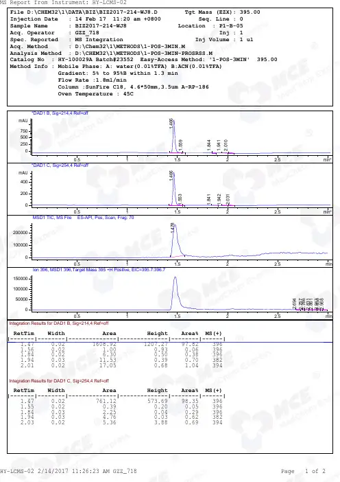
Bay_41-4109_racemate_LCMS_23552_MedChemExpress
- 格式:pdf
- 大小:173.63 KB
- 文档页数:2

LBI-38904BMaintenance ManualORION™UHFSCAN AND SYSTEMMOBILE RADIOTABLE OF CONTENTSSynthesizer/Receiver/Exciter . . . . . . . LBI-39033Power Amplifier . . . . . . . . . . . . . . LBI-39034PA Interface . . . . . . . . . . . . . . . . LBI-38994Control Logic/IF Board . . . . . . . . . . LBI-38907Control Units . . . . . . . . . . . . . . . LBI-38992Assemblies . . . . . . . . . . . . . . . . LBI-38909Service Section . . . . . . . . . . . . . . LBI-38908Ericsson Inc.Private Radio SystemsMountain View RoadLynchburg, Virginia 24502ericssonz1-800-528-7711 (Outside USA, 804-528-7711)Printed in U.S.A.Copyright© October 1993, Ericsson GE Mobile Communications Inc.SPECIFICATIONS*Frequency Range:403-440 MHz 440-470 MHz 470-512 MHzRegulatory ApprovalFCC (United States)AXATR-315-A2403-440 MHz 20/40 Watts AXATR-315-B2440-470 MHz 30/40 Watts AXATR-315-C2470-512 MHz 35 Watts AXATR-316-A2403-440 MHz 100 Watts AXATR-316-B2440-470 MHz 100 Watts AXATR-316-C2470-512 MHz 80 Watts DOC (Canada)TR-315TR-315403-440 MHz 20/40 Watts 440-470 MHz30/40 WattsBattery Drain:ReceiveSquelched 1.1 Amperes at 13.8 V oltsUnsquelched 3.0 Amperes at 13.8 V olts (15 Watts Output)Transmitter20 Watts 35/40 Watts 80/100 Watts12 Amperes at 13.2 V olts 14 Amperes at 13.6 V olts 25/28 Amperes at 13.4 V olts Frequency Stability:0.0002% depending on model Temperature Range:-30° C (-22° F) to +60° C (+140° F)Duty Cycle:100% Receive, 14% Transmit TransmitterTransmit Output Power:20W/35W/40W/80W/100W Conducted Spurious:-85 dB Modulation:±4.5 kHzAudio Sensitivity:55 to 110 millivoltsAudio Frequency Characteristics:Within +1 dB to -3 dB of a 6 dB/octave pre-emphasis 300 Hz and within +1 dB to -4.5 dB of a 6 dB/octave pre-emphasis 3000 Hz per EIA standards. Post-limiter filter per FCC and EIA.Distortion:Less than 2% (1000 Hz)Less than 5% (3000 Hz)Deviation Symmetry:0.3 kHz maximum Maximum Frequency Separation:403-440 MHz 37 MHz 440-470 MHz 30 MHz 470-512 MHz 42 MHz Microphone Load Impedance:600 OhmsPower Adjust Range:100% to 50% of rated power (U.S.A. Models)100% to 30% of rated power (Euro Models)RF Output Impedance:50 Ohms FM Noise:45 dBContinuedThis manual covers Ericsson and General Electric products manufactured and sold by Ericsson Inc.NOTICE!Repairs to this equipment should be made only by an authorized service technician or facility designated by the supplier.Any repairs, alterations or substitution of recommended parts made by the user to this equipment not approved by the manufacturer could void the user’s authority to operate the equipment in addition to the manufacturer’s warranty.NOTICE!This manual is published by Ericsson Inc., without any warranty. Improvements and changes to this manual necessitated by typographical errors, inaccuracies of current information, or improvements to programs and/or equipment, may be made by Ericsson Inc., at any time and without notice. Such changes will be incorporated into new editions of this manual. No part of this manual may be reproduced or transmitted in any form or by any means, electronic or mechanical, including photocopying and recording, for any purpose, without the express written permission of Ericsson Inc.The software contained in this device is copyrighted by the Ericsson Inc. Unpublished rights are reserved under the copyright laws of the United States.NOTICE!LBI-389041DESCRIPTIONThe synthesized ORION mobile radio combinations are completely solid-state, utilizing microcomputer technology and integrated circuits to provide high-quality, high-reliabil-ity radios. Standard combinations may be equipped with:•Microcomputer Controlled Frequency Synthesizer•Up to 16 Channels•0.0002% Frequency Stability•Other Structured OptionsThe basic radio consists of three printed wiring boards mounted in a cast aluminum frame. The three boards are:1.The Control Logic/IF board2.The Frequency Synthesizer/Receiver/Exciter board3.The Power Amplifier boardThe radio is of double-layer construction with tuning ad-justments easily accessible from the top of the radio.The Control Logic/IF Board is located on the top of theradio, while the Power Amplifier and the Synthesizer/Re-ceiver/Exciter boards are located on the bottom of the radio.SYNTHESIZER/INTERCONNECTThe synthesizer consists of a microcomputer, E lectricallyE rasable P rogrammable R ead O nly M emory (EEPROM), afrequency synthesizer IC, transmit and receive V oltage C on-trolled O scillator’s (VCO) and associated circuitry. The fre-quency synthesizer under control of the microcomputergenerates all transmit and receive Radio Frequencies (RF).The EEPROM stores binary data for all radio frequen-cies, Channel Guard tones/digital codes and the timing func-tion of the C arrier C ontrol T imer (CCT). Themicrocomputer accesses the EEPROM and provides the correctW ALSH bits to the Channel Guard circuitry to generate thecorrect Channel Guard tone or digital code on a per-channelbasis.PROGRAMMINGThe EEPROM allows the radio to be programmed or repro-grammed as needed to adapt to changing system requirements.Radio Frequencies, Channel Guard tone and digital codes andthe CCT function can be reprogrammed.The EEPROM can be reprogrammed through the radiofront connector using a personal computer. This programmerallows all information to be loaded simultaneously.Programming instructions are provided in the respectiveProgrammer Maintenance Manuals.TRANSMITTERThe transmitter consists of the exciter, frequency synthe-sizer, transmitter VCO and a Power Amplifier (PA) assembly.The PA assembly consists of a PA board mounted on a heatsink assembly. The PA board also contains an antenna switch-ing diode and a low-pass filter.Audio and Channel Guard circuitry for the transmitter is lo-cated on the Control Logic/IF Board.RECEIVERThe receiver consists of the frequency synthesizer, RXVCO, injection amplifiers, front end, IF and limiter detector.Audio, squelch and Channel Guard circuitry for the receiver islocated on the Control Logic/IF Board.SYSTEM CONTROL FUNCTIONA microprocessor on the Control Logic/IF board controlsthe frequency synthesizer, the TX ON/OFF, the decoding ofCTCSS tones, the generation of CTCSS tones,... etc. The audioprocessor circuitry of the transmitter and the receiver are lo-cated on the Control Logic/IF board. Squelch circuitry and aconnection to the digital AEGIS circuit is also located on theControl Logic/IF board.OPERATIONComplete operating instructions for the ORION Two-WayRadio are provided in Operator’s Manual LBI-38888 for thecontrol unit used.MAINTENANCEThe Service Section in maintenance manual LBI-38908contains the maintenance information to service this radio. TheService Section includes:•Dissassembly Procedures•Replacement of IC’s, chip capacitors and resistors•Alignment procedures for the transmitter and receiver•Troubleshooting Procedures and wave formsA mechanical layout for the radio is found in ORION As-semblies Maintenance Manual LBI-38909.Figure 1 - ORION Mobile RadioSPECIFICATIONS* - Cont.ReceiverAudio Output:15 Watts with less than 3% distortion(To 4.0 ohm speaker)Sensitivity:0.35 µV (STD)/0.22 µV (PRE)12 dB SINAD (IEIA method)Selectivity:-85 dB (STD)-80 dB (PRE)EIA Two-Signal Method(25 kHz Channels)Spurious Response:-100 dB (STD)/-90 (PRE)Intermodulation 25 kHz:-85 dB (STD)/-80 dB (PRE)Maximum Frequency Separation:403-440 MHz .... 37 MHz440-470 MHz .... 30 MHz470-512 MHz .... 42 MHzFrequency Response:Within +1, -3 dB of 6 dB/octave de-emphasis from 300 to 3000MHz (1000 Hz reference)RF Input Impedance:50 OhmsHum/Noise ratio:Unsquelched-50 dBSquelched-70 dBChannel Spacing:30 kHz*These specifications are intended primarily for use of the service technician. Refer to the appropriate Specifications Sheet for the complete specifications.LBI-389042SYSTEM INTERCONNECTION DIAGRAMLBI-38904 Array U.S.A. LOW POWER3SYSTEM INTERCONNECTION DIAGRAM LBI-38904U.S.A. HIGH POWER4SYSTEM INTERCONNECTION DIAGRAMLBI-38904 Array EUROPEAN5。

Pharmacol,2022,20(1):94-95.[8]㊀Luan D,Dadpey B,Zaid J,et al.Adipocyte-secreted IL-6sensitizes macrophages to IL-4signaling[J].Diabetes,2023,72(3):367-374.[9]㊀Wada T,Hoshino M,Kimura Y,et al.Both typeⅠandⅡIFN induce insulin resistance by inducing different isoformsof SOCS expression in3T3-L1adipocytes[J].Am PhysiolEndocrinol Metab,2011,300(6):1112-1123. [10]㊀Jung TW,Park HS,Choi GH,et al.β-aminoisobutyric acidattenuates LPS-induced inflammation and insulin resistancein adipocytes through AMPK-mediated pathway[J].Bi-omed Sci,2018,25(1):27.[11]㊀Nam SW,Kim MS,Han Y,et al.WJCPR11reverses theTNF-α-induced inhibition of adipocyte differentiation andglucose uptake[J].Biochem Biophys Res Commun,2021(578):150-156.[12]㊀Duan T,Du Y,Xing C,et al.Toll-like receptor signalingand its role in cell-mediated immunity[J].Front Immu-nol,2022(13):812774.[13]㊀Zhang ZD,Li HX,Gan H,et al.RNF115inhibits the post-ER trafficking of TLRs and TLRs-mediated immune re-sponses by catalyzing K11-linked ubiquitination of RAB1Aand RAB13[J].Adv Sci(Weinh),2022,9(16):2105391.[14]㊀Yim JJ,Ding L,Schaffer AA,et al.A microsatellite poly-morphism in intron2of human Toll-like receptor2gene:functional implications and racial differences[J].FEMSImmunol Med Microbiol,2004,40(2):163-9. [15]㊀Senn JJ.Toll-like receptor-2is essential for the develop-ment of palmitate-induced insulin resistance in myotubes[J].Biol Chem,2006,281(37):26865-26875. [16]㊀Itani SI,Ruderman NB,Schmieder F,et al.Lipid-inducedinsulin resistance in human muscle is associated with chan-ges in diacylglycerol,protein kinase C,and IkappaB-alpha[J].Diabetes,2002,51(7):2005-2011.ʌ文章编号ɔ1006-6233(2024)03-0411-06槐耳多糖调节SPHK1/S1P/S1PR3信号通路对宫颈癌细胞恶性生物学行为的影响李丽品,㊀马素艳,㊀安入征(河北省石家庄市平安医院肿瘤科,㊀河北㊀石家庄㊀050000)ʌ摘㊀要ɔ目的:探究槐耳多糖(HP)调节SPHK1/S1P/S1PR3信号通路对宫颈癌细胞恶性生物学行为的影响㊂方法:MTT检测槐耳多糖(0㊁25㊁50㊁100㊁200㊁400μg/mL)处理的宫颈癌细胞活力,筛选最佳药物浓度㊂实验分为对照组(Control组)㊁槐耳多糖低㊁中㊁高浓度组(HP-L㊁HP-M㊁HP-H组)和槐耳多糖高浓度+SphK1激活剂K6PC-5组(HP-H+K6PC-5组),观察细胞增殖㊁迁移和侵袭情况;west-ern blot检测SPHK1㊁S1P㊁S1PR3㊁Snail㊁N-cadherin㊁E-cadherin蛋白水平㊂结果:处理24㊁48㊁72h后,与0μg/mL比较,50μg/mL㊁100μg/mL㊁200μg/mL和400μg/mL的HP处理的细胞活力显著降低(P<0.05)㊂HP-L组㊁HP-M组和HP-H组细胞Edu阳性率㊁侵袭细胞数㊁划痕愈合率及Snail㊁N-cadherin㊁SPHK1㊁S1P㊁S1PR3水平显著低于Control组(P<0.05),E-cadherin水平显著升高(P<0.05)㊂HP-H+ K6PC-5组细胞Edu阳性率㊁侵袭细胞数㊁划痕愈合率及Snail㊁N-cadherin㊁SPHK1㊁S1P㊁S1PR3水平显著高于HP-H组(P<0.05),E-cadherin水平显著降低(P<0.05)㊂结论:HP可能通过抑制SPHK1/ S1P/S1PR3信号通路抑制宫颈癌细胞的增殖㊁侵袭和迁移㊂ʌ关键词ɔ㊀槐耳多糖;㊀SPHK1/S1P/S1PR3信号通路;㊀宫颈癌;㊀恶性生物学行为ʌ文献标识码ɔ㊀A㊀㊀㊀㊀㊀ʌdoiɔ10.3969/j.issn.1006-6233.2024.03.011Impacts of Polysaccharide of Trametes Robiniophila Murr on the Malignant Biological Behavior of Cervical Cancer Cells by Regulating theSPHK1/S1P/S1PR3Signaling PathwayLI Lipin,MA Suyan,AN Ruzheng㊃114㊃ʌ基金项目ɔ河北省中医药管理局科研计划项目,(编号:2010132)(Shijiazhuang Ping'an Hospital,Hebei Shijiazhuang050000,China)ʌAbstractɔObjective:To investigate the impacts of Huaier polysaccharide(HP)regulating the SPHK1/S1P/S1PR3signaling pathway on the malignant biological behavior of cervical cancer cells.Meth-ods:MTT was used to detect the viability of cervical cancer cells treated with Sophora auricula polysaccharides (0,25,50,100,200,400μg/mL)and screen for the optimal drug concentration.The subjects were divided into control group,low,medium,and high concentration groups of Huaier polysaccharide(HP-L,HP-M, HP-H groups),and high concentration of Huaier polysaccharide+SphK1activator K6PC-5group(HP-H+ K6PC-5group).The cell proliferation,migration and invasion were observed;Western blot was used to de-tect the protein levels of SPHK1,S1P,S1PR3,Snail,N-cadherin,and E-cadherin.Results:After treat-ment for24,48and72h,HP treatments of50μg/mL,100μg/mL,200μg/mL and400μg/mL significantly decreased cell viability compared with0μg/mL(P<0.05),Edu positive rate,number of invasive cells, scratch healing rate and Snail,N-cadherin,SPHK1,S1P and S1PR3levels in HP-L,HP-M and HP-H groups were obviously lower than those in Control group(P<0.05),E-cadherin levels significantly increase (P<0.05).The Edu positive rate,number of invasive cells,scratch healing rate and Snail,N-cadherin, SPHK1,S1P and S1PR3levels in HP-H+K6PC-5group were obviously higher than those in HP-H group (P<0.05),E-cadherin levels significantly decrease(P<0.05).Conclusion:HP may inhibit the prolifera-tion,invasion and migration of cervical cancer cells by inhibiting SPHK1/S1P/S1PR3signaling pathway.ʌKey wordsɔ㊀Huaier polysaccharide;㊀SPHK1/S1P/S1PR3signaling pathway;㊀Cervical cancer;㊀Malignant biological behavior㊀㊀宫颈癌是妇女最常见的癌症之一,其发病率和死亡率在女性恶性肿瘤中排名第四㊂由于其持续耐药和反复复发,患者预后较差[1]㊂因此寻求新的治疗药物对宫颈癌的治疗至关重要㊂近年来,中草药在癌症治疗中被广泛研究,槐耳(Trametes robiniophila Murr)作为一种传统中草药被广泛应用于许多疾病,近年来,槐耳被广泛应用于抗肿瘤药物的研究,槐耳多糖(Huaier polysaccharide,HP)是槐耳提取物,可增强机体免疫㊁抗氧化㊁抗炎反应,其在胃癌㊁乳腺癌等多种癌症中可促进细胞凋亡,表现出良好的抗肿瘤作用[2],但其在宫颈癌中抗癌作用及机制尚不清楚㊂1-磷酸鞘氨醇(sphingosine-1-phosphate,S1P)是鞘脂代谢的中心分子,决定细胞的命运,由鞘脂苷通过两种酶(鞘氨醇激酶1(sphingosine kinase1,SPHK1),鞘氨醇激酶2 (sphingosine kinase2,SPHK2)产生,S1P与5种G蛋白偶联受体(S1PR1-5s)结合,在调节细胞存活㊁迁移等过程中起着至关重要的作用[3]㊂研究显示SPHK-S1P-S1PRs信号通路在细胞增殖生长中发挥作用, SPHK1的过度激活可能促进肿瘤的发生和发展[4-5]㊂宫颈癌中SPHK1高表达,沉默SPHK1宫颈癌细胞细胞活性㊁增殖能力显著降低,细胞凋亡率显著升高㊂槐耳多糖影响宫颈癌发生机制是否与SPHK1/S1P/ S1PR3信号通路有关尚不清楚㊂本研究基于SPHK1/ S1P/S1PR3信号通路,探究槐耳多糖对宫颈癌细胞恶性生物学行为的影响及机制,为宫颈癌治疗提供理论参考㊂1㊀材料与方法1.1㊀材料:人宫颈癌细胞HeLa(CL-0101)购自武汉普诺赛生命科技有限公司;槐耳多糖(Huaier polysac-charide,HP)(S28138)购自上海源叶生物科技有限公司;SPHK1激活剂K6PC-5(E1218)购自Selleck公司; RPMI1640培养基(11875176)购自赛默飞公司;MTT 试剂盒(CT0002)购自山东思科捷生物技术有限公司; Edu细胞增殖试剂盒(E-CK-A377)购自武汉伊莱瑞特公司;一抗Snail(A5243)㊁N-cadherin(A3045)㊁E-cadherin(A3044)㊁SPHK1(A0139)㊁S1PR3(A1404)㊁GAPDH(AC001)购自ABclonal公司;一抗S1P (ab59870)及二抗山羊抗兔IgG(ab205718)购自Ab-cam公司㊂1.2㊀方㊀法1.2.1㊀细胞培养及MTT检测细胞活力:HeLa细胞于RPMI1640培养基(添加10%胎牛血清)中培养(37ħ,5%CO2),并进行消化传代,当细胞合流度达到90%时,进行实验㊂细胞接种于96孔板㊂孵育过夜后,用不同浓度(0μg/mL㊁25μg/mL㊁50μg/mL㊁100μg/ mL㊁200μg/mL㊁400μg/mL)的槐耳多糖溶液替换培养基,24㊁48和72h后加入MTT溶液,孵育4h㊂随后,每孔中的溶液小心用DMSO替换10min㊂在酶标仪中㊃214㊃490nm波长下读取吸光度值,并计算细胞活力㊂1.2.2㊀分组及处理:将对数生长期细胞分为对照组(Control组)㊁槐耳多糖低㊁中㊁高浓度组(HP-L㊁HP-M㊁HP-H组)和槐耳多糖高浓度+SPHK1激活剂K6PC-5组(HP-H+K6PC-5组)㊂HP-L㊁HP-M㊁HP -H组分别使用50μg/mL㊁100μg/mL㊁200μg/mL槐耳多糖溶液处理细胞24h,HP-H+K6PC-5组使用200μg/mL槐耳多糖溶液和10μM的K6PC-5共处理细胞24h㊂1.2.3㊀Edu检测细胞增殖:按照1.2.2中的分组处理细胞,并接种于96孔板(4.0ˑ104/孔)培养24h,去除培养基后,用Edu处理2h㊂丢弃含有Edu的培养基后,用4%甲醛和0.5%Triton X-100溶液固定细胞透化细胞,DAPI对细胞核进行染色,荧光显微镜下观察Edu阳性细胞数,并计算Edu阳性细胞率㊂1.2.4㊀细胞迁移和侵袭能力检测:将各组细胞接种于24孔板中,当细胞达到约90%汇合时,用移液管吸头刮擦细胞单层以形成均匀划痕,用无血清磷酸盐缓冲液冲洗后将在无血清培养基中培养㊂光学显微镜下拍照观察0h和24h划痕的融合情况,并计算细胞划痕愈合率㊂使用24孔Transwell装置进行体外侵袭试验,上腔接种各组细胞(1ˑ104/mL),下腔为10%胎牛血清的培养基㊂1d后,通过多聚甲醛和结晶紫对下室的侵袭的细胞进行固定㊁染色,光学显微镜拍照观察,并计算侵袭细胞数量㊂1.2.5㊀Western blot检测蛋白水平:收集处理后的细胞,裂解缓冲液中裂解㊂经10%SDS-PAGE电泳分离㊁转膜㊁封闭后,与一抗SPHK1㊁S1P㊁S1PR3㊁Snail㊁N -cadherin㊁E-cadherin及GAPDH过夜孵育(4ħ)㊂后与相应二抗孵育1h(室温)㊂ECL进行显影,Image J 计算蛋白条带灰度值㊂1.3㊀统计学方法:SPSS20.0软件进行实验数据统计分析,结果均以( xʃs)表示㊂单因素方差分析进行多组间比较,LSD-t进行两两比较㊂P<0.05为差异有统计学意义㊂2㊀结㊀果2.1㊀槐耳多糖对宫颈癌细胞活力的影响:不同浓度HP处理宫颈癌细胞24㊁48㊁72h后,细胞活力逐渐降低,除25μg/mL处理浓度外,其与各浓度处理的细胞活力显著降低(P<0.05)㊂为保证一定存活率,选择50㊁100㊁200μg/mL的HP用于后续研究㊂见图1㊂图1㊀槐耳多糖对宫颈癌细胞活力的影响注:∗表示与0μg/mL比较,P<0.052.2㊀槐耳多糖对宫颈癌细胞增殖的影响:与Control 组比较,HP-L组㊁HP-M组和HP-H组细胞Edu阳性率依次降低(P<0.05)㊂与HP-H组比较,HP-H+K6PC-5组细胞Edu阳性率显著升高(P<0.05),见图2㊁3㊂图2㊀Edu检测各组细胞增殖情况(ˑ200)图3㊀槐耳多糖对宫颈癌细胞增殖的影响注:∗表示与Control组比较,P<0.05;∗∗表示与HP-L 组比较,P<0.05;∗∗∗表示与HP-M组比较,P<0.05;∗∗∗∗表示与HP-H组比较,P<0.05㊃314㊃2.3㊀槐耳多糖对宫颈癌细胞迁移和侵袭的影响:与Control 组比较,HP -L 组㊁HP -M 组和HP -H 组细胞侵袭数㊁划痕愈合率依次降低(P <0.05)㊂与HP -H 组比较,HP -H +K6PC -5组细胞侵袭数㊁划痕愈合率显著升高(P<0.05),见图4㊁图5㊁图6㊂图4㊀划痕愈合实验检测各组细胞迁移能力图5㊀Transwell 小室实验检测各组细胞侵袭能力(ˑ200)图6㊀槐耳多糖对宫颈癌细胞迁移和侵袭的影响注:∗表示与Control 组比较,P <0.05;∗∗表示与HP -L 组比较,P<0.05;∗∗∗表示与HP -M 组比较,P<0.05;∗∗∗∗表示与HP -H 组比较,P<0.052.4㊀槐耳多糖对宫颈癌细胞E -cadherin ㊁N -cadherin ㊁Snail 蛋白水平的影响:与Control 组比较,HP -L 组㊁HP -M 组和HP -H 组E -cadherin 水平依次升高,N -cadherin ㊁Snail 水平依次降低(P <0.05)㊂与HP -H 组比较,HP -H +K6PC -5组E -cadherin 水平显著降低,N -cadherin ㊁Snail 水平显著升高(P<0.05),见图7㊁8㊂图7㊀Western blot 检测各组细胞EMT 相关蛋白水平注:A :Control 组;B :HP -L 组;C :HP -M 组;D :HP -H 组;E :HP -H +K6PC -5组图8㊀槐耳多糖对宫颈癌细胞E -cadherin ㊁N -cadherin ㊁Snail 蛋白水平的影响注:∗表示与Control 组比较,P <0.05;∗∗表示与HP -L组比较,P<0.05;∗∗∗表示与HP -M 组比较,P<0.05;∗∗∗∗表示与HP -H 组比较,P<0.052.5㊀槐耳多糖对宫颈癌细胞SPHK1㊁S1P ㊁S1PR3蛋白水平的影响:与Control 组比较,HP -L 组㊁HP -M 组和HP -H 组SPHK1㊁S1P ㊁S1PR3蛋白水平依次降低(P <0.05)㊂与HP -H 组比较,HP -H +K6PC -5组SPHK1㊁S1P ㊁S1PR3蛋白水平显著升高(P <0.05),见图9㊁10㊂㊃414㊃图9㊀Western blot 检测通路相关蛋白水平注:A :Control 组;B :HP -L 组;C :HP -M 组;D :HP -H 组;E :HP -H +K6PC -5组图10㊀槐耳多糖对宫颈癌细胞SPHK1㊁S1P ㊁S1PR3蛋白水平的影响注:∗表示与Control 组比较,P <0.05;∗∗表示与HP -L 组比较,P<0.05;∗∗∗表示与HP -M 组比较,P<0.05;∗∗∗∗表示与HP -H 组比较,P<0.053㊀讨㊀论宫颈癌是全世界妇女癌症死亡的第四大原因,由于其高发病率和死亡率,对有效的诊断㊁治疗和预防药物的需求未得到满足,虽然放疗和化疗等方式在宫颈癌上取得了较好的治疗效果,但耐药性的产生使患者预后较差[6]㊂槐耳多糖是一种由6个单糖和18个氨基酸组成的活性成分,是中草药槐耳的主要提取物,对癌细胞具有抑制作用[7]㊂本研究结果显示槐耳多糖处理HeLa 细胞后,细胞活力㊁增殖能力均降低,与前人研究结果相似[8]㊂表明槐耳多糖具有抑癌作用,可降低宫颈癌细胞活力,抑制其增殖㊂上皮-间充质转化(Epithelial -mesenchymal transition ,EMT )在癌症的进展㊁侵袭㊁迁移中起着关键作用,上皮细胞通过失去细胞极性和上皮标记物(E -cadherin )的表达发生表型转换,通过获得间充质标记物(N -cadherin ,Snail )的表达成为间充质细胞,表现出细胞间粘连减少和运动性增加,因此E -钙粘蛋白的丢失,是EMT 过渡过程中的关键步骤[9]㊂有研究表明槐耳多糖可通过抑制EMT 抑制癌细胞的增殖㊁转移和侵袭[10]㊂本研究中槐耳多糖处理后HeLa 细胞N -cadherin ㊁Snail 水平降低,E -cadherin 蛋白上调,其迁移及侵袭能力受到抑制,与雷蕾等研究结果相似[11]㊂表明槐耳多糖可能通过抑制EMT 进展抑制宫颈癌细胞的侵袭和迁移㊂据报道SPHK1/S1P /S1RP3信号通路在癌细胞的增殖㊁迁移㊁侵袭和转移及肿瘤血管生成中起重要作用[12]㊂有研究表明SPHK1在大多数癌症中普遍表达,SPHK1的过度激活促进了肿瘤的发生和发展,SPHK1是S1P 合成的主要酶,控制着S1P 的水平,在癌细胞的增殖㊁迁移㊁侵袭和肿瘤血管生成中起重要作用[13]㊂Zhang [14]等研究表明SPHK1/2的过度表达和/或过度激活在宫颈癌的发生和发展中具有重要作用,通过抑制SPHK 表达能抑制细胞活力㊁集落形成㊁增殖㊁迁移及细胞周期进展㊂本研究中细胞SPHK1㊁S1P ㊁S1PR3水平随着槐耳多糖浓度的增加依次降低,同时其增殖㊁迁移㊁侵袭能力受到抑制㊂而激活剂K6PC -5逆转了槐耳多糖对细胞增殖㊁侵袭㊁迁移及SPHK1/S1P /S1RP3信号通路的抑制作用㊂表明槐耳多糖能抑制宫颈癌细胞恶性生物学行为的发生,其机制可能与抑制SPHK1/S1P /S1RP3信号通路传导有关㊂综上所述,槐耳多糖对宫颈癌细胞有显著抑制作用,其作用机制可能与抑制SPHK1/S1P /S1RP3信号通路传导有关㊂本文初步探究了槐耳多糖对宫颈癌细胞的影响,其机制复杂,且本研究未在体内进行验证,今后仍需进行深入研究并在体内进行验证,为槐耳多糖在宫颈癌的治疗应用上提供参考㊂ʌ参考文献ɔ[1]㊀Fan H ,He YJ ,Xiang JQ ,et al.ROS generation attenuates theanti -cancer effect of CPX on cervical cancer cells by indu-cing autophagy and inhibiting glycophagy [J ].Redox Biol ,2022,53(1):102339-102349.[2]㊀Qi J ,Xie FJ ,Liu S ,et al.Huaier granule combined with tega-fur gimeracil oteracil potassium promotes stage Ⅱb gastric cancer prognosis and induces gastric cancer cell apoptosis by regulating livin [J ].Biomed Res Int ,2020,2020(1):1-10.[3]㊀Li F ,Zhang YF ,Lin ZJ ,et al.Targeting SPHK1/S1PR3-reg-ulated S -1-P metabolic disorder triggers autophagic cell death in pulmonary lymphangiomyomatosis (LAM )[J ].Cell㊃514㊃Death Dis,2022,13(12):1065-1075.[4]㊀Di Paolo A,Vignini A,Alia S,et al.Pathogenic role of thesphingosine1-phosphate(S1P)pathway in common gyne-cologic disorders(GDs):a possible novel therapeutic target[J].Int Mol Sci,2022,23(21):13538-13548. [5]㊀Sukocheva OA,Furuya H,Ng ML,et al.Sphingosine kinaseand sphingosine-1-phosphate receptor signaling pathway in inflammatory gastrointestinal disease and cancers:a noveltherapeutic target[J].Pharm Ther,2020,207(1):107464-107550.[6]㊀Shoaib S,Islam N,Yusuf N.Phytocompounds from the me-dicinal and dietary plants:multi-target agents for cervicalcancer prevention and therapy[J].Curr Med Chem,2022,29(26):4481-4506.[7]㊀Hu BQ,Yan WT,Wang MH,et al.Huaier polysaccharide in-hibits the stem-like characteristics of ERα-36high triple negative breast cancer cells via inactivation of the ERα-36 signaling pathway[J].Int Biol Sci,2019,15(7):1358-1367.[8]㊀张泽敏,庞岚.槐耳多糖抑制AEG-1表达对子宫内膜癌细胞生物学行为的影响[J].川北医学院学报,2023,38(10):1299-1305.[9]㊀Huang XT,Xiang LY,Wang BY,et al.CMTM6promotes mi-gration invasion and EMT by interacting with and stabilizingvimentin in hepatocellular carcinoma cells[J].Transl Med, 2021,19(1):120-130.[10]㊀Fang L,Zhang YZ,Zang YW,et al.HP-1inhibits the pro-gression of ccRCC and enhances sunitinib therapeuticeffects by suppressing EMT[J].Carbohydrate Polymers,2019,223(1):115109-115121.[11]㊀雷蕾,高彦茹,蔡洲,等.槐耳多糖通过抑制AKT/GSK3β/Snail信号通路抑制胰腺癌细胞增殖和上皮间质转化[J].现代肿瘤医学,2023,31(24):4536-4541.[12]㊀Khoei SG,Sadeghi H,Samadi P,et al.Relationship betweenSphk1/S1P and microRNAs in human cancers[J].Biotech-nol Appl Biochem,2021,68(2):279-287. [13]㊀Zheng XJ,Li W,Ren LW,et al.The sphingosine kinase-1/sphingosine-1-phosphate axis in cancer:Potential target foranticancer therapy[J].Pharmacol Ther,2019,195(1):85-99.[14]㊀Zhang Y,Cheng L,Shi X,et al.The sphingosine kinase in-hibitor SKI-V suppresses cervical cancer cell growth[J].Int Biol Sci,2022,18(7):2994-3005.ʌ文章编号ɔ1006-6233(2024)03-0416-08TRIM25通过EZH2介导巨噬细胞M2极化促进食管鳞状细胞癌细胞的增殖迁移和侵袭张诗彤,㊀田新春,㊀花海洋,㊀刘洪运(承德医学院第二附属医院,㊀河北㊀承德㊀067000)ʌ摘㊀要ɔ目的:探讨TRIM25和EZH2在食管鳞状细胞癌(ESCC)相关巨噬细胞浸润中的作用及其可能的作用机制㊂方法:利用佛波酯诱导THP-1为M0巨噬细胞,将其与转染处理后的KYSE510细胞共培养,收集共培养巨噬细胞,分为Ctrl组㊁sh-NC组㊁sh-TRIM25组㊁sh-NC+oe-NC组㊁sh-TRIM25+oe -NC组㊁sh-NC+oe-EZH2组和sh-TRIM25+oe-EZH2组㊂收集共培养上清液,将其加入KYSE510细胞中,分为Ctrl组㊁TAM组㊁sh-NC+TAM组㊁sh-TRIM25+TAM组㊁sh-NC+oe-NC+TAM组㊁sh-TRIM25+oe -NC+TAM组㊁sh-NC+oe-EZH2+TAM组和sh-TRIM25+oe-EZH2+TAM组㊂qPCR法和WB法检测细胞中TRIM25㊁EZH2和Arg-1表达,放线菌酮蛋白合成抑制实验检测细胞EZH2蛋白稳定性,FCM检测F4/80+CD206+巨噬细胞比例,CCK-8法㊁克隆形成实验和Transwell实验分别检测细胞的增殖㊁迁移和侵袭能力㊂结果:KYSE510细胞中TRIM25和EZH2mRNA和蛋白表达均高于人食管鳞状上皮细胞HET-1A(P<0.01)㊂与sh-NC组和sh-NC+oe-NC组相比,sh-TRIM25组和sh-TRIM25+oe-NC组F4/ 80+CD206+巨噬细胞比例和Arg-1蛋白表达均降低(P<0.05),sh-NC+oe-EZH2组则升高(P<0.05)㊂与sh-NC+TAM组和sh-NC+oe-NC+TAM组相比,sh-TRIM25+TAM组和sh-TRIM25+oe-NC+TAM组KYSE510细胞活力㊁克隆形成数㊁迁移和侵袭细胞数均降低(P<0.05),sh-NC+oe-EZH2+TAM组则升高(P<0.05)㊂敲低TRIM25可通过抑制EZH2蛋白半衰期,降低EZH2蛋白稳定性㊂过表达EZH2可部分逆转sh-TRIM25对巨噬细胞和KYSE510细胞的影响㊂结论:TRIM25通过促进EZH2蛋白稳定性㊃614㊃ʌ基金项目ɔ河北省医学科学研究课题计划项目,(编号:20191297)ʌ通讯作者ɔ田新春。

488 Scientific AbstractsSystemic sclerosis, myositis and related syndromes - aetiology, pathogenesis and animal modelsPOS0467 DERSIMELAGON, A NOVEL ORAL MELANOCORTIN1 RECEPTOR AGONIST, DEMONSTRATES DISEASE-MODIFYING EFFECTS IN PRECLINICAL MODELS OFSYSTEMIC SCLEROSISM. Kondo1, T. Suzuki1, Y. Kawano1, S. Kojima2, M. Miyashiro1, A. Matsumoto1, G. Kania3, P. Blyszczuk3, R. Ross4, P. Mulipa4, F. Del Galdo4, Y. Zhang5, J. H. W. Distler5. 1Mitsubishi T anabe Pharma Corporation, Research Unit/Immunology & Inflammation, Souyaku Innovative Research Division, Y okohama, Japan;2Mitsubishi T anabe Pharma Corporation, Discovery T echnology Laboratories, Souyaku Innovative Research Division, Y okohama, Japan;3University Hospital Zurich, University of Zurich, Center of Experimental Rheumatology, Department of Rheumatology, Schlieren, Switzerland;4University of Leeds, Leeds Instituteof Rheumatic and Musculoskeletal Medicine, Faculty of Medicine and Health, Leeds, United Kingdom;5Friedrich-Alexander-University Erlangen-Nürnberg (FAU) and University Hospital Erlangen, Department of Internal Medicine 3—Rheumatology and Immunology, Erlangen, GermanyBackground: Activation of melanocortin 1 receptor (MC1R) is known to have broad anti-inflammatory and anti-fibrotic effects. The bleomycin (BLM)-induced skin fibrosis murine model is well-established for systemic sclerosis (SSc). α-mel-anocyte-stimulating hormone, an endogenous ligand of MC1R, inhibits skin fibro-sis and MC1R knock-out enhances skin fibrosis in this model. These pieces of evidence suggest that MC1R agonism has potential in the treatment of SSc. Objectives: Dersimelagon phosphate (MT-7117) is an investigational small molecule that is an orally administered, selective agonist for MC1R. The purpose of this study is to investigate the potential of MT-7117 as a therapeutic agent for SSc by evaluat-ing its efficacy and mechanism of action in complementary preclinical models. The expression and distribution of MC1R in the skin of SSc patients was investigated. Methods: The effects of MT-7117 on skin fibrosis and lung inflammation were eval-uated in BLM-induced SSc murine models that were optimized for prophylactic and therapeutic evaluation. Microarray-based gene expression analysis and serum pro-tein profiling were performed to investigate the mechanism of action of MT-7117 in the BLM-induced SSc models. The effect of MT-7117 on TGF-β-induced activation of human dermal fibroblasts was evaluated in vitro. Immunohistochemical analyses of MC1R expression in skin samples from SSc patients were performed. Results: Prophylactic treatment with MT-7117 (≥0.3 mg/kg/day p.o.) significantly inhibited the increase in collagen content of the skin, the serum level of sur-factant protein D, and the weight of the lungs from BLM-induced skin fibrosis and lung inflammation model. Therapeutic treatment with MT-7117 (≥3 mg/kg/ day p.o.) significantly suppressed skin thickening and the numbers of myofi-broblasts in pre-established BLM-induced skin fibrosis model. Gene array anal-ysis using the BLM-induced SSc model demonstrated changes in numerous categories related to macrophages, monocytes, and neutrophils, followed by endothelial cell-related categories after treatment with MT-7117. In the analy-sis that focused on biological functions, categories of inflammatory response, activation of antigen-presenting cells, angiogenesis, atherosclerosis, vascu-logenesis, and vaso-occlusion were suppressed by MT-7117. In the analysis that focused on molecular signaling pathways, triggering receptor expressed on myeloid cells-1, IL-6, and oncostatin M involved in inflammation, and perox-isome proliferator-activated receptor that is related to fibrosis were all affected by MT-7117. Serum protein profiling using BLM-induced SSc model revealed that multiple SSc-related biomarkers including P-selectin, osteoprotegerin, cys-tatin C, growth and differentiation factor-15 and S100A9 were suppressed by MT-7117. MT-7117 inhibited the activation of human dermal fibroblasts by sup-pressing TGF-β-induced ACTA2 (encoding α-smooth muscle actin) mRNA ele-vation in vitro. Immunohistochemical analyses showed that MC1R positivity was observed in 40 of 50 diffuse cutaneous SSc patients. MC1R was expressed by monocytes/macrophages, neutrophils, blood vessels (endothelial cells), fibro-blasts, and epidermis (keratinocytes) in the skin of SSc patients. Conclusion: MT-7117 demonstrates disease-modifying effects in preclinical mod-els of SSc. Investigations of its mechanism of action and target expression anal-yses indicate that MT-7117 exerts its positive effects by affecting the pathologies of inflammation, vascular dysfunction, and fibrosis through inflammatory cells, endothelial cells, and fibroblasts. In view of its potent beneficial impact on all these three main pathologies of SSc, MT-7117 is a potential therapeutic agent for the treatment of clinically challenging SSc, which has diverse and difficult to treat symp-toms. A phase 2 clinical trial investigating the efficacy and tolerability of MT-7117 in patients with early, progressive diffuse cutaneous SSc is currently in progress. Disclosure of Interests: Masahiro Kondo Employee of: Mitsubishi Tanabe Pharma Corporation, Tsuyoshi Suzuki Employee of: Mitsubishi Tanabe Pharma Corporation, Yuko Kawano Employee of: Mitsubishi Tanabe Pharma Corpora-tion, Shinji Kojima Employee of: Mitsubishi Tanabe Pharma Corporation, Masa-hiko Miyashiro Employee of: Mitsubishi Tanabe Pharma Corporation, Atsuhiro Matsumoto Employee of: Mitsubishi Tanabe Pharma Corporation, Gabriela Kania: None declared, Przemyslaw Blyszczuk: None declared, rebecca ross:None declared, Panji Mulipa: None declared, Francesco Del Galdo Grant/ research support from: Prof. F. Del Galdo received fees and research supportfrom Abbvie, AstraZeneca, Boehringer-Ingelheim, Capella, Chemomab, Kymab, Janssen and Mitsubishi-Tanabe., Yun Zhang: None declared, Jörg H.W. DistlerGrant/research support from: Prof. J.H.W. Distler received consulting fees, lec-ture fees, and/or honoraria from Actelion, Active Biotech, Anamar, ARXX, aTyr,Bayer Pharma, Boehringer Ingelheim, Celgene, Galapagos, GSK, Inventiva, JB Therapeutics, Medac, Pfizer, Sanofi-Aventis, RedX, RuiYi and UCB. J. H. W.Distler is stock owner of 4D Science and Scientific head of FibroCure.DOI: 10.1136/annrheumdis-2022-eular.29POS0468 EXTRACELLULAR VESICLES FROM SERUM OFMYOSITIS PATIENTS AS CIRCULATING BIOMARKERSAND DISEASE MEDIATORSS. Kivity1,2, H. Kravitz3, C. Cohen3, D. Margoulis3, M. Amar3, G. Kazimirsky3,D. Ozeri4, A. Dori5, C. Brodie3. 1Meir Medical Center, Rheumatology Unit, KefarSava, Israel;2T el Aviv University, Sackler faculty of Medicine, T el Aviv-Y afo, Israel;3Bar-Ilan University, The Mina and Everard Goodman Faculty of Life Sciences,Ramat Gan, Israel;4T el-HaShomer The Sheba Medical Center, ZabludowiczCenter for Autoimmune Disease, Ramat Gan, Israel;5T el-HaShomer The ShebaMedical Center, Department of Neurology, T alpiot Medical Leadership Program,Sackler Faculty of Medicine, T el Aviv University, Ramat Gan, IsraelBackground: Inflammatory myopathies (IM) are a heterogeneous group of disor-ders characterized by autoimmune inflammatory destruction of skeletal muscles.It is many times associated with lung, skin and joint involvement. Identifying bio-markers that can differentiate IM from other muscle disorders may elucidate the pathophysiology of IM, guide novel therapies, monitor disease activity/responseto treatments and predict prognosis. Exosomes are membrane-bound nanove-sicles with diameters of 30-150 nm that contain multiple proteins, nucleic acid,lipids and other molecules in a tissue- and cell-specific manner. Exosomes are secreted by a large variety of cells, play major roles in cell-cell interactions, andhave recently emerged as circulating biomarkers in a variety of pathological con-ditions, including several autoimmune diseases.Objectives: To characterize exosomes from serum of IM patients, analyze pro-tein expression and study their potential mediators of disease pathologies.Methods: Serum was collected from patients suffering from IM(n=5) and from patients suffering from Becker (BMD) and Duchenne (DMD) muscular dystro-phies (n=6). Exosomes were isolated by Exoquick precipitation and analyzedfor size distribution and by nanoparticle tracking analysis (NTA) and by Westernblot for exosome markers. The effects of the isolated EVs on human satellitecell proliferation and differentiation and macrophage activation were examined. Results: Exosomes from IM patients decreased human satellite cell proliferation (51%, P<0.01) and inhibited their myogenic differentiation as indicated by lower fusionindex (24% inhibition, P<0.01) and expression of myosin heavy chain (72% inhibi-tion, P<0.001). Similar results were obtained also with exosomes derived from DMDand BMD patients; however, their inhibitory effect were more pronounced on MyoG expression. T reatment of macrophages with exosomes from IM patients significantly increased the expression of IL-10 (3-fold, P<0.001), compared to exosomes of healthy controls and DMD patients. Another significant difference was in the expression of sig-naling molecules: Thus, exosomes from BMD patients increased the phosphorylationof Erk and p38, whereas a smaller effect was induced by IM exosomes.Conclusion: Exosomes from IM patients decrease satellite cell proliferationand myogenic differentiation compared to healthy exosomes. In addition, these exosomes increased the expression of IL-10 in macrophages. These effects areunique to exosomes of IM patients compared to muscular dystrophies. These promising results suggest that serum exosomes should be further investigatedas a novel biomarker with potential therapeutic implications.Disclosure of Interests: Shaye Kivity Speakers bureau: BI, Abbvie, Lilly, Pfizer, Janssen, Neopharm, Grant/research support from: Sobi, Haya Kravitz: None declared, Coral Cohen: None declared, Darya Margoulis: None declared, MosheAmar: None declared, Gila Kazimirsky: None declared, David Ozeri Speakers bureau: Neopharm, Consultant of: Abbvie, Amir Dori Grant/research supportfrom: Biogen, Chaya Brodie Grant/research support from: Biogen.DOI: 10.1136/annrheumdis-2022-eular.63POS0469 ENDOTHELIAL TO MESENCHYMAL TRANSITIONAND SENESCENCE ARE PART OF THE FIBROTICPATHOGENESIS IN SYSTEMIC SCLEROSISY. H. Chiu1,2, J. Spierings1, J. M. Van Laar1, J. De Vries-Bouwstra3, M. VanDijk4, R. Goldschmeding4. 1University Medical Center Utrecht, Departmentof Rheumatology and Clinical Immunology, Utrecht, Netherlands;2T ri-ServiceGeneral Hospital, Division of Rheumatology/Immunology/Allergy, T aipei, T aiwan, Republic of China;3Leiden University Medical Center, The Department of on December 24, 2023 by guest. Protected by copyright./ Ann Rheum Dis: first published as 10.1136/annrheumdis-2022-eular.29 on 23 May 2022. Downloaded from。

文章编号:1003 2754(2023)07 0628 08 doi:檱檱檱檱檱檱檱檱檱檱檱檱檱檱檱檱檱檱檱檱殗殗殗殗10.19845/j.cnki.zfysjjbzz.2023.0145论著与经验总结 缺血性脑卒中缺氧相关生物标志物的筛选 胡彦兵, 达吾提江·依拉木, 郭 雄, 潘红波, 何康民收稿日期:2022 11 20;修订日期:2023 04 11基金项目:2022年新疆自然科学基金面上项目(2022D01C18)作者单位:(喀什地区第二人民医院神经外科,新疆维吾尔自治区喀什844000)通信作者:何康民,E mail:kashgarhekangmin@126.com 摘 要: 目的 研究缺氧相关基因与缺血性脑卒中之间的关系,筛选缺氧相关的分子标志物。
方法 首先,下载GSE16561和GSE58294数据集,以及缺氧相关基因集,筛选缺氧相关的差异表达基因,并进行功能注释。
随后对这些差异表达的缺氧相关基因进行最小绝对收缩和选择算子(LASSO)回归和支持向量机算法分析,筛选缺氧相关的生物标志物,并对缺血性脑卒中组和对照组的差异免疫细胞进行分析。
最后对转录因子和miRNA进行了预测。
结果 本研究共筛选得到13个缺氧相关的差异表达基因,这13个基因主要富集到了葡萄糖跨膜转运、单糖跨膜转运以及线粒体自噬调节等过程。
基于LASSO回归和支持向量机算法,最终确定了8个缺氧相关的生物标志物,这些标志物与大部分免疫细胞之间均存在显著相关性。
此外,预测到8个基因对应的209个转录因子的关系网络,以及6个基因对应的25个miRNA。
结论 本文最终筛选了8个缺氧相关的生物标志物,它们可能作为抑制卒中后的脑损伤的治疗靶点。
此外,糖代谢和免疫机制在缺血性脑卒中的发生和发展中发挥重要作用。
关键词: 缺血性脑卒中; 缺氧; 免疫; 基因中图分类号:R743.3 文献标识码:AScreeningforhypoxia relatedbiomarkersinischemicstroke HUYanbing,DAWUTJANIlam,GUOXiong,etal.(DepartmentofNeurosurgery,KashiPrefectureSecondPeople'sHospital,Kashi844000,China)Abstract: Objective Toinvestigatetherelationshipbetweenhypoxia relatedgenesandischemicstroke,andtoscreenformolecularmarkersrelatedtohypoxia.Methods First,GSE16561andGSE58294datasetsandhypoxia relatedgenesetsweredownloadedtoscreenforhypoxia relateddifferentiallyexpressedgenes.Afterfunctionalannotation,thesehy poxia relateddifferentiallyexpressedgeneswereanalyzedbytheleastabsoluteshrinkageandselectionoperatorleastabso luteshrinkageandselectionoperator(LASSO)regressionandsupportvectormachinealgorithmtoscreenforimmune relatedbiomarkers,anddifferentialimmunecellswereanalyzedbetweentheischemicstrokegroupandthecontrolgroup.Final ly,transcriptionfactorsandmiRNAswerepredicted.Results Atotalof13hypoxia relateddifferentiallyexpressedgeneswerescreened,whichwereenrichedmainlyintheprocessesofglucosetransmembranetransport,monosaccharidetransmem branetransport,andmitophagyregulation.BasedontheLASSOregressionandsupportvectormachinealgorithmanalyses,eighthypoxia relatedbiomarkerswereidentified,whichweresignificantlycorrelatedwithmostimmunecells.Furthermore,thenetworkof209transcriptionfactorscorrespondingto8genesand25miRNAscorrespondingto6geneswerepredicted.Conclusion Eighthypoxia relatedbiomarkersareidentified,whichmaybeusedaspotentialtherapeutictargetsforinhibitingbraininjuryafterstroke.Inaddition,glucosemetabolismandimmunemechanismsmayplayanimportantroleintheoccurrenceandprogressionofischemicstroke.Keywords: Ischemicstroke; Hypoxia; Immune; Gene 脑卒中是目前世界范围内造成残疾和死亡的最重要的原因之一,每年约有650万人死于脑卒中[1]。