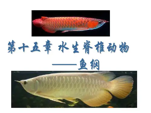
鱼类比较组织解剖AnatomyandHistology
- 格式:ppt
- 大小:1.62 MB
- 文档页数:41
1.组织学(Histology ),是研究人体细微结构和功能关系的科学。
这门科学是借助于显微镜进行观察的,又称微视解剖学或小体解剖学。
2.解剖学(Anatomy),是研究人体宏观结构和机能关系的科学。
解剖学是借助于肉眼观察的,又称巨视解剖学,或大体解剖学。
3.人体组织学与解剖学:是研究人体从宏观到微观结构与其与功能关系的科学。
4.细胞(Cell):是人体结构和功能的最小基本单位。
5.组织(tissue):是由许多在形态和功能上具有密切联系的细胞和细胞间质所组成的基本结构称为组织。
6.器官(organ):由几种不同的组织结合在一起构成具有一定形态和功能的结构称器官。
7.系统(system):许多在结构和功能上具有密切联系的器官结合在一起,共同执行某种特定生理活动。
8.人体(Human body):由许多系统、器官和组织共同组成的有机整体。
9.上皮组织:简称上皮,是由密集的细胞和少量的细胞间质组成。
大部分上皮组织分布在机体的外表面和衬贴在体内各种有腔器官的腔面。
组成上皮组织的细胞称上皮细胞。
上皮细胞排列紧密而规律,细胞间质很少。
上皮细胞具有明显的极性,可分为游离面和基底面。
10.结缔组织:是由细胞和大量的细胞间质组成。
与上皮细胞比较,结缔组织的主要结构特点是细胞数量少而种类多,细胞形态多样,无极性,分散在细胞间质内;结缔组织的细胞间质多。
11.神经纤维:是由神经元的长突起和包在其外表的神经胶质细胞所组成的纤维状结构。
根据神经纤维有无髓鞘,分为有髓神经纤维和无髓神经纤维两种。
12.小循环:全身返回心的含有二氧化碳较多的静脉血自右心室泵出,经肺动脉与其分流到肺泡毛细血管进行气体交换,使静脉血变成动脉血,再经肺静脉流回左心房。
血液沿上述路径的循环称小循环或肺循环。
13.大循环:含氧和营养物质较多的肺动脉血,自左心房泵出,经主动脉与其分支流到全身毛细血管(肺泡毛细血管除外),进行物质和气体交换,使动脉血变成静脉血,静脉血再汇入各级静脉,经上、下腔静脉与冠状窦流回右心房。
人体解剖与组织胚胎学专业英语精选英文人体解剖与组织胚胎学专业英语:The Intricate Nature of Human Anatomy and HistologyHuman anatomy and histology, two fields at the forefront of medical science, offer a profound understanding of the structure and organization of the human body. These disciplines, which delve into the very essence of our existence, are not only critical for medical professionals but also fascinating for those interested in the wonders of the human body.Anatomy: The Blueprint of the Human BodyAnatomy, often referred to as the "blueprint" of the human body, is the study of the structure and organization of the body's systems and organs. It involves the examination of bones, muscles, organs, and other tissues that make up the human frame. This field is crucial for understanding how the body functions in health and disease, guiding surgical procedures, and informing diagnostic methods.Anatomists study the gross structure of the body through dissection, which allows them to visualize and understand the relationships between different organs and tissues. They also investigate the neural, muscular, and vascular systems, revealing the intricate networks that control movement, sensation, and vital functions.Histology: The Microscopic World of Cells and TissuesHistology, on the other hand, focuses on the microscopic structure of cells and tissues. It is the study of the organization and function of cells and their interactions within tissues andorgans. Histologists use microscopy to observe and analyze the structure and arrangement of cells, their nuclei, cytoplasm, and organelles.This field is particularly important in understanding how cells specialize to perform specific functions within the body. For example, cardiomyocytes in the heart are specifically adapted to contract and pump blood, while neurons in the brain are designed to transmit electrical signals. Histology also plays a crucial role in diagnosing diseases by examining cellular changes that occur in various pathological conditions.Embryology: The Development of LifeEmbryology, which is closely related to histology, deals with the development of the human body from fertilization to birth. It studies the processes involved in the growth and differentiation of cells, tissues, and organs during embryogenesis. Embryologists investigate the complex interactions between genes, proteins, and the environment that govern embryonic development.This field is fascinating as it reveals the remarkable transformation of a single cell into a fully functional human being. It also provides insights into congenital defects and other developmental disorders, informing research into potential treatments and interventions.In conclusion, human anatomy, histology, and embryology offer profound insights into the structure, function, and development of the human body. These disciplines are not only crucial for advancing medical science but also serve to marvel at the wonders of life itself. As we continue to explore the intricacies of the human body, these fields will remain at the forefront of medical research and education.中文对照翻译:人体解剖学和组织学的复杂性人体解剖学和组织学是医学前沿的两个领域,对人体的结构和组织有着深刻的理解。
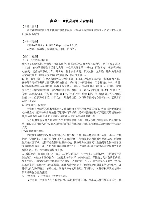
实验3 鱼的外形和内部解剖【目的与要求】通过对鲤鱼或鲫鱼外形和内部构造的观察,了解硬骨鱼类的主要特征及适应于水生生活的形态结构特征【材料与用品】活鲤鱼(或鲫鱼),以体重2.0kg、2龄以上为宜;放大镜、解剖盘、解剖器具、棉球、直尺等。
【操作与观察】(一)外形观察和测量鲤鱼体呈纺续捶形,略侧扁,背部灰黑色,腹部近白色。
身体可区分为头、躯千和尾3部分。
1、头部自吻端至鳃盖骨后缘为头部。
口位于头部前端(口端位),两侧各有2条触须(鲫鱼无触须)。
吻背面有鼻孔1对,眼1对,位于头部两侧,形大而圆,无眼睑。
眼后头部两侧为宽扁的鳃盖,鳃盖后缘有膜状的鳃盖膜,籍此覆盖鳃孔。
2、躯干部和尾部自鳃盖后缘至肛门为躯干部;自肛门至尾鳍基部最后一枚椎骨为尾部。
躯干部和尾部体表被以覆瓦状排列的圆鳞,鳞外覆有一薄层表皮,用手抚摸鱼体表,粘滑。
躯体两侧从鳃盖后缘到尾部,各有1条由鳞片上的小孔排列成的点线结构,此即侧线,被侧线孔穿过的鳞片称侧线鳞。
体背和腹侧有鳍,背鳍1个,较长,约为躯干的3/4;臀鳍1个,较短;尾鳍末端凹入分成上下相称的2叶,为正尾型;胸鳍I对,位于鳃盖后方左右两侧;腹鳍1对,位于胸鳍之后,肛门之前,属腹鳍腹位;肛门紧靠臀鳍起点基部前方,紧接肛门后有1泄殖孔。
3、硬骨鱼的一般测量:全长指自吻端至尾鳍末端的长度;体长指自吻端至尾鳍基部的长度;体高指躯干部最高处的垂直高;躯干长指由鳃盖骨后缘到肛门的长度;尾柄长指臀鳍基部后端至尾鳍基部的长度;尾柄高指尾柄最低处的垂直高;尾长指由肛门至尾鳍基部的长度。
头长指由吻端至鳃盖骨后缘(不包括鳃盖膜)的长度;吻长指由上颌前端至眼前缘的长度。
眼径指眼的最大直径;眼间距指两眼间的直线距离。
眼后头长指眼后缘至鳃盖骨后缘的长度。
(二)内部解剖与观察将活鲤鱼置解剖盘,便其腹部向上,用手术刀在肛门前与体轴垂直方向剪一小口。
使鱼侧卧,左侧向上,自肛门前的开口向背方剪到脊柱,沿侧线下方向前剪至鳃盖后缘,再沿鳃盖后缘剪至下颌,这样可将左侧体壁肌肉揭起,使心脏和内脏暴露。
实验鱼的解剖一、实验目的通过对鲤鱼的结构观察,从形态学角度认识生物体的结构组成,了解其适应水生生活的结构特点。
学习鱼内部解剖的基本操作方法。
二、实验材料与用具鲜活鲤鱼、解剖器、解剖盘等。
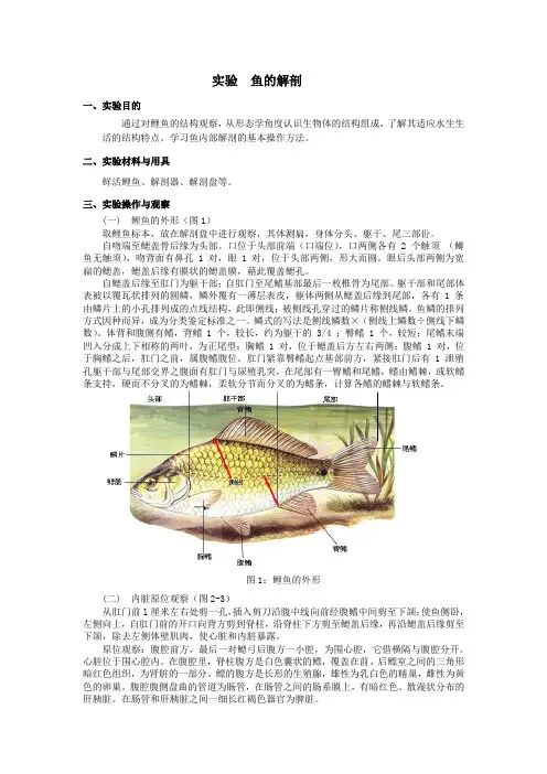
三、实验操作与观察(一) 鲤鱼的外形(图1)取鲤鱼标本,放在解剖盘中进行观察,其体测扁,身体分头、躯干、尾三部份。
自吻端至鳃盖骨后缘为头部。
口位于头部前端(口端位),口两侧各有 2 个触须(鲫鱼无触须),吻背面有鼻孔 1 对,眼 1 对,位于头部两侧,形大而圆。
眼后头部两侧为宽扁的鳃盖,鳃盖后缘有膜状的鳃盖膜,藉此覆盖鳃孔。
自鳃盖后缘至肛门为躯干部;自肛门至尾鳍基部最后一枚椎骨为尾部。
躯干部和尾部体表被以覆瓦状排列的圆鳞,鳞外覆有一薄层表皮,躯体两侧从鳃盖后缘到尾部,各有 1 条由鳞片上的小孔排列成的点线结构,此即侧线;被侧线孔穿过的鳞片称侧线鳞。
鱼鳞的排列方式因种而异,成为分类鉴定标准之一。
鳞式的写法是侧线鳞数×(侧线上鳞数÷侧线下鳞数)。
体背和腹侧有鳍,背鳍 1 个,较长,约为躯干的 3/4 ;臀鳍 1 个,较短;尾鳍末端凹入分成上下相称的两叶,为正尾型;胸鳍 1 对,位于鳃盖后方左右两侧;腹鳍 1 对,位于胸鳍之后,肛门之前,属腹鳍腹位。
肛门紧靠臀鳍起点基部前方,紧接肛门后有 1 泄殖孔躯干部与尾部交界之腹面有肛门与尿殖乳突,在尾部有一臀鳍和尾鳍,鳍由鳍棘,或软鳍条支持,硬而不分叉的为鳍棘,柔软分节而分叉的为鳍条,计算各鳍的鳍棘与软鳍条。
图1:鲤鱼的外形(二) 内脏原位观察(图2-3)从肛门前l厘米左右处剪一孔,插入剪刀沿腹中线向前经腹鳍中间剪至下颌;使鱼侧卧,左侧向上,自肛门前的开口向背方剪到脊柱,沿脊柱下方剪至鳃盖后缘,再沿鳃盖后缘剪至下颌,除去左侧体壁肌肉,使心脏和内脏暴露。
原位观察:腹腔前方,最后一对鳃弓后腹方一小腔,为围心腔,它借横隔与腹腔分开。
心脏位于围心腔内。
在腹腔里,脊柱腹方是白色囊状的鳔,覆盖在前、后鳔室之间的三角形暗红色组织,为肾脏的一部分。
经济动物学报 2004,8(3):163~166Journal of EconomicAnimal大鲵精巢的解剖及组织形态结构研究Ξ刘进辉1,江 辉1,谭理琦2,周玉林3,李丽平1 (11湖南农业大学动物科学技术学院,长沙410128;21广东省深圳市福田区兽医卫生防疫检疫所,深圳518033;31湖南怀化职业技术学院,怀化418000)摘 要:对3只成年不同时期的大鲵精巢进行大体解剖及组织形态结构观察,结果表明,大鲵精巢由被膜和实质构成。
被膜由外层的腹膜和内层的白膜构成,白膜从精巢头部向实质伸入形成隔膜,把精巢实质分成许多形状和大小不同的精小叶;大鲵精巢实质由皮质部和髓质部两部分构成,皮质部主要由精小叶和少量的间质组织构成,精小叶内分布有生殖细胞聚集而成的精小囊和壶腹。
雄性大鲵的生殖细胞的整个发育过程都是在精小囊和壶腹中进行,不同时期的精巢内的生殖细胞发育有一定的规律;髓质部主要由丰富的血窦、毛细淋巴管和结缔组织构成。
关键词:大鲵;精巢;精小叶;组织形态结构中图分类号:S86413 文献标识码:A 文章编号:100727448(2004)0320163204StudyonAnatomy,MorphologyandHistologyStructureofAndrias davidianus testisLIUJin2hui1,JIANGHui1,TANLi2qi2,ZHOUYu2lin3,LILi2ping1 (11College of Animal Science and Technology,Hu’nan Agricultural University,Changsha410128,Chi2 na;21Futian Veterinary and Hygiene Institute for Prevention and Inspection,Shenzhen518033,China;31College of Vocational Technology,Huaihua418000,China)Abstract:Anatomy,morphologyandhistologystructureof3Andriasdavidianustestiswasstudiedwithregularlymethodsanditwasfoundthattheseweresomecharacteristicsofhistologystructurewhichwasconstitutedbypeitoneumintheouterlayerandtunicaalbugmeaintheinnerlayer.Tunicaalbugineastretchedintoparenchymaandformeddissepiment,Whichdividedtestisintoseminifereuslobulesmadeof cortexandmedullar.Moreover,themajorityofcortexwascomposeofseminiferouslobulesandinterstitialtissue,themedullarypartconnetivetissue.Attheborderofseminiferouslobules,somespermatogeniccellsformedtestiscorpsulewheredevelopmentalprocessofsepermatogeniccellsofmaleMegalobatrachus wascarriedout.Keywords:Andrias davidianus;spermary;spermarylobule;histologyandmorphologystructure 大鲵(Andrias davidianus),俗名娃娃鱼,隶属于两栖纲、有尾目、大鲵科,为隐鳃鲵科动物,属国家二级保护动物,曾广泛分布于我国中南部各省,具有重要的经济价值[123]。

一、与解剖学有关的副主题词(一)anatomy&histology 解剖学和组织学2.含义:用于各器官、部位和组织的正常解剖学和组织学,也用于动植物的正常解剖学或组织结构。
5.举例胰腺的构造——胰腺/*解剖学和组织学狗肝脏的形态学——肝/*解剖学和组织学+狗/*解剖学和组织学加壳纲眼的组织学——眼/*解剖学和组织学+甲壳纲/*解剖学和组织学子宫大小的测量——子宫/*解剖学和组织学垂体的重量——垂体/*解剖学和组织学(二)/cytology 细胞学2.含义:用以表明单细胞或多细胞的正常细胞形态学。
5.举例胰腺的细胞学——胰腺/*细胞学月经周期的胰腺细胞学——胰腺/*细胞学+*月经周期非典型性结核分枝杆菌的结构——分枝杆菌属,非典型性/*细胞学肝细胞膜——肝/*细胞学+细胞膜狗胰腺细胞学——胰腺/*细胞学+狗/*解剖学和组织学不应标:狗/细胞学利用大鼠肾细胞系研究细胞分化——肾/细胞学+*细胞分化+细胞系+大鼠(CT)+动物(CT)对比研究结肠和肾的细胞分化情况(分别采用大鼠的肾细胞系和结肠细胞系)——结肠/*细胞学+肾/*细胞学+*细胞分化+细胞系+大鼠(CT)+动物(CT)+对比研究(CT)脊髓灰质炎病毒的结构——脊髓灰质炎病毒/*超微结构不应标:脊髓灰质炎病毒/*细胞学(三)/pathology 病理学2.含义:与组织、器官及疾病主题词组配,表明在疾病状态时器官、组织及细胞的结构。
5.举例正常和病理性肝细胞——肝/*细胞学+肝/*病理学肝肿瘤的细胞学——肝肿瘤/*病理学肝炎时的肝病理学——肝/*病理学+肝炎/*病理学测量心肌梗塞范围的方法——心肌梗塞/*病理学风湿性心内膜炎是心瓣膜纤维变性——心脏病,风湿性/*病理学+回肠/*病理学恶性贫血时不常见的巨母红细胞的形态异常——贫血,恶性/*血液+巨母红细胞/*病理学肌营养障碍时内亚线粒体颗粒——肌营养障碍/*病理学+线粒体,肌/*超微结构(四)/ultrastructure 超微结构2.含义:与组织及细胞(包括肿瘤)、以及微生物主题词组配,表明通常用光学显微镜观察不到的细微解剖结构。
第二章硬骨鱼的解剖和生理一前言这一章的目的是在把硬骨鱼类的解剖和生理的大纲介绍给读者,使读者能够在病理组织中所发生的同时也了解鱼组织病变的机转。
鱼类的许多基本功能与其它脊椎动物甚相似,所以有关哺乳动物的知识对于了解鱼的功能上也有所帮助,但不能将硬骨鱼类看作为哺乳动物的原始形态。
其实鱼类是进步的脊椎动物,在演化上较近代发生的动物,而且也广布到不同的生存区域。
事实上,硬骨鱼包括不同的种,比任何一科的脊椎动物所含的种都要多,因此许多原则性的结论的真正价值都不甚确切。
但主要被研究的鱼种并不甚广,因此有利用价值的结论还是可以获得的。
在这里,我们主要着重的是在于熟悉的哺乳动物的解剖和生理的差异方面。
主要考虑的对象在于水域环境以及它加于鱼身上的限制。
为了要抗拒自然水环境施加到鱼体的破坏性影响,鱼体内在的环境必须保持正常,而且与外在的环境要不断地做必要的物质交换。
在环境因子中最主要的因子就是水的比热高,一般鱼类是变温性动物,其体温需要顺应环境的温度。
因此像鱼的心跳速度、消化速度和生长等都没有一定的数据可供参考,且都因温度而改变。
因此,凡是研究硬骨鱼类的人,都要将这些因子记在心中。
同样的一种鱼在不同的温度水环境中,几乎变成难以辨认的形式,像虹鳟肾上腺的增大或心跳率的降低,均以温度而变(Randall 1970)。
二皮肤系统皮肤是面对环境的第一道障碍,保证内部生理作用的正常进行。
皮肤的实际情况对于病害的过程极为重要。
硬骨鱼的皮肤包括角质层、表皮层、基质膜、真皮层和下皮层,各层的排列情形与说明见图2.1。
(一)角质层角质层是外层,1970年,Whiter是第一位对角质层做详细报导的人,角质层的主要成分是黏多糖,约1微米的厚度,通常是由表皮细胞分泌而成,而非由杯状黏液细胞组成,它含有细胞中的原生质、脱落的细胞、加上其它由杯状细胞组成的分泌物。
角质层的物理性一致程度在种与种之间有很大的差异。
在潮间水潭和海底,鱼种特别发达,角质层中含有特殊的免疫球蛋白和分解酶(后者因种的不同,在含量上差异很大)及游离的脂肪酸,这些成分都具有抗病原的作用。
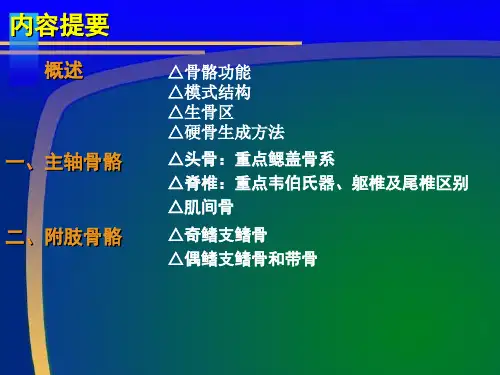
鱼类的内部形态构造(第146期)1.骨骼骨骼是支持身体和保护体内器官的组织,它和动物体的运动也有密切关系。
骨骼有内外之分,外骨骼包括鳞甲、鳍条和棘刺等;内骨骼通常是指埋在肌肉里的骨骼部分,包括头骨、脊柱和附肢骨骼。
头骨由脑颅和咽颅两部分组成、硬骨鱼类(常见的淡水养殖鱼类均为硬骨鱼类)的脑颅由许多骨片所合成,其主要作用是保护脑;咽颅由一对颌弓、一对舌弓和五对鳃弓所组成,分别具有支持颌、舌和鳃的功能。
脊柱由体椎和尾椎两种脊椎骨组成,体椎附有肋骨,尾椎无肋骨着生,两者容易区别。
每个脊椎的椎体前后两面都是凹形的,故称之为双凹椎体,这是鱼类所特有。
附肢骨骼是指支持鱼鳍的骨骼,支持背鳍、臀鳍和尾鳍的骨骼是不成对的奇鳍骨骼;支持胸鳍和腹鳍的骨骼为成对的偶鳍骨骼。
鱼类的偶鳍骨没有和脊柱联接,与其他陆生脊椎相比,显然又是一个特点。
2.肌肉鱼类的摄食、逃避敌害、繁殖等等一系列的生命活动,都要依靠肌肉的规律性收缩所起的运动来完成。
鱼类的躯干部和尾部的肌肉由许多肌节组成,肌节之间有隔膜连接而呈分节现象。
体侧肌肉被一水平走向的肌隔分为两段,上段叫轴上肌,下段叫轴下肌。
轴上肌分化出背鳍部分的肌肉。
尾部肌节分化出尾鳍肌。
轴下肌分化为腹部与胸、腹鳍等部肌肉。
3.消化系统消化系统包括消化道和消化腺。
消化道的起端为口,经口腔、食道、胃、肠而终于肛门。
口腔内有齿和鳃耙等构造。
一般鱼类具有颌齿和咽齿两种,前者多起摄取食物的作用,后者则有压碎和咀嚼食物的功能。
鳃耙着生在鳃弓内缘,它是咽部的滤食器官。
草食性和杂食性的鱼类(如草鱼、鲤、鲫等)的鳃耙较疏短,吃浮游生物的鱼类(如鲢鱼、鳙鱼等)的鳃耙则密而长。
鱼类没有明显的舌,紧接口腔的一段为食道,一般短宽而壁厚,具有较强的扩张性,以利吞食比较大型的食物。
胃在食道的后方,是消化道中最膨大的部分。
鲤科鱼类通常没有明显的胃,其外表与食道并无多大差别,但鲇科鱼类等肉食性鱼类的胃却很发达,界线也很明显。
大麻哈鱼消化器官的形态学和组织学观察作者:李培伦刘伟鲁万桥唐富江王继隆来源:《南方农业学报》2022年第05期摘要:【目的】摸清大麻哈鱼消化器官的形态结构特征,为其人工养殖过程中饵料调配和优化提供参考。
【方法】以人工养殖的2龄大麻哈鱼为研究对象,采用解剖学和常规石蜡切片技术对其消化器官的形态学和组织学进行研究。
【结果】大麻哈鱼的消化道由口咽腔、食道、胃、幽门盲囊和肠组成。
口咽腔中颌齿、咽骨齿发达;食道粗短且肌层发达;胃呈V形,由贲门部、胃体部和幽门部组成,黏膜褶皱较多;胃黏膜层表面为排列紧密的单层柱状上皮细胞,黏膜层内陷形成大量胃小凹,在胃小凹的基部具有数量丰富的胃腺组织,其中贲门部和胃体部胃腺数量较多,而幽门部较少;幽门盲囊呈螺旋式纵行排列,其黏膜层由单层柱状上皮细胞和大量杯状细胞构成;肠道较短,前肠有较薄的黏膜褶皱而黏膜层界限不明显,后肠则未见明显的黏膜层和黏膜褶皱结构。
肝脏不分叶,肝小叶界限模糊,中央静脉和胆管结构清晰。
【结论】大麻哈鱼消化器官的形态结构特征与其食性功能相适应,具有典型肉食性鱼类消化器官特征。
关键词:大麻哈鱼;消化器官;形态学;组织学中图分类号: S965.229 文献标志码: A 文章编号:2095-1191(2022)05-1457-09Morphology and histology observation of the digestive organs and liver of chum salmon (Oncorhynchus keta)LI Pei-lun, LIU Wei, LU Wan-qiao, TANG Fu-jiang, WANG Ji-long(Heilongjiang River Fisheries Research Institute,Chinese Academy of Fishery Sciences/Key Open Laboratory of Cold Water Fish Germplasm Resources and Breeding of Heilongjiang Province,Harbin, Heilongjiang 150070, China)Abstract:【Objective】To find out the morphological and structural characteristics of the chum salmon’s (Oncorhynchus keta) digestive organs,so as to provide an effective reference for the optimization of artificial breeding feed. 【Method】The morphology and histology of the digestive organs of the second-year chum salmon were taken as objectives. Morphological and histological characteristics were studied using the anatomy and conventional paraffin section techniques. 【Result】The digestive tract of chum salmon was composed of oropharynx cavity,esophagus,stomach,pyloric caecum,intestine. The jaw teeth and pharyngeal teeth were well developed in the oropharynx cavity,and the chum salmon had a very coarse and short esophagus with thick muscle layer. The stomach was in a “V” shape,consisting of the cardiac stomach,gastric body and pyloric stomach,with many folds in mucosal. The surface of the gastric mucosal layer was a densely-arranged single layer of columnar epithelial cells. The mucosal layer was inverted to form a large number of gastric pits,and there were abundant gastric gland tissues at the base of the gastric pits. Among them,the cardiac stomach,gastric body of the stomach had more gastric glands,but the pyloric stomach had less. The pyloric caecum was spirally and longitudinally arranged,and its mucosal layer was composed of a single layer of columnar epithelial cells and a large number ofgoblet cells. The intestine was short,the foregut had thin mucosal folds and the boundary of the mucosal layer was not obvious,and there was no obvious mucosal layer and mucosal fold structure in the hindgut. The liver has no lobes,the boundaries of the liver lobules were blurred,but the structures of central vein and bile ducts were clear. 【Conclusion】The morphological and structural characteristics of the chum salmon’s digestive organs are adapted to its diet function,and had the typical characteristics of the digestive organs of carnivorous fish.Key words: chum salmon (Oncorhynchus keta); digestive organ; morphology; histologyFoundation items: Ministry of Agriculture and Rural Affairs Finance Special Species Resources Protection Project (2010-2020); The Fundamental Research Funds for the Central Public Welfare Research Institutes (HSY201712Q)0 引言【研究意义】鱼类通过消化器官对摄入体内的食物进行消化、吸收,以维持鱼体的正常生长、发育和繁殖,其对食物的消化和吸收过程与高等脊椎动物相似,但也具有其自身的结构特点,主要因其对栖息水体环境及摄取的食物类型不同所产生的适应性(林浩然,2011)。