Broadly Neutralizing Alphavirus Antibodies Bind an Epitope on E2 and Inhibit Entry and Egress
- 格式:pdf
- 大小:5.07 MB
- 文档页数:14

俄研制出多用途抗病毒新药——帕纳维尔
佚名
【期刊名称】《养殖与饲料》
【年(卷),期】2003(000)002
【摘要】@@ 据俄罗斯<消息报>报道,以俄罗斯医学科学院通讯院士瓦列里·谢尔吉延科为首的10人研究小组,经过多年努力研制出一种用途广泛的抗病毒新药,并已通过临床试验.这种命名为"帕纳维尔"抗病毒新药使用了经过特殊机械清洗后提取的植物成分.研究人员波斯佩洛娃在介绍"帕纳维尔"的药用机理时说,常规使用的抗病毒药物直接作用于病毒,其作用机理是复制;而"帕纳维尔"作用机理是阻碍病毒粒子同机体细胞的联系.因此,不管是何种病毒,含有DNA分子链还是RNA分子链,对"帕纳维尔"并不重要,"帕纳维尔"能直接保护细胞.
【总页数】1页(P46)
【正文语种】中文
【相关文献】
1.抗病毒新药阿尔比多尔的合成 [J], 丁德生;李振菊
2.俄研制出新药品可除体内重金属 [J],
3.俄研制出多用途抗病毒新药 [J],
4.抗病毒新药阿尔比多尔(Arbidole)的合成 [J], 丁德生;李振菊
5.俄研制出治疗艾滋病的新药 [J], 黎文
因版权原因,仅展示原文概要,查看原文内容请购买。

加拿大将开始进行博拉病毒疫苗的临床试验
牛津大学临床研究工作者Felicity Hartnell博士正拿着一小瓶埃博拉病毒疫苗
本周一,加拿大官方将向世卫组织总部运送埃博拉病毒疫苗VSV-EBOV,以便在人类身上开展临床试验。
加拿大广播公司表示:世卫组织已挑选了来自肯迪亚、加蓬、德国、瑞士的250名志愿者来接受此次疫苗试验。
试验将会在十月底或十一月初开始,而世卫方面也希望在十二月初看到疫苗的效果。
如果疫苗真能有效地抑制埃博拉并没有任何严重的副作用,那么试验将在年内继续在几内亚、利比亚以及塞拉利昂着三个受到埃博拉病毒严重侵扰的国家实行。
此次疫苗由加拿大公共卫生局开发的。
加拿大政府的一项声明表示该疫苗已经“在动物实验中取得了非常显著的效果”。
加拿大首席公共卫生官员格雷戈里·泰勒表示“本次疫苗将成为抑制此次灾难的重要工具,我们会好好运用这个工具来与WHO开展密切合作,解决一些道德上与逻辑上的问题,共同应对灾难。
”
疫苗将会被分成三个不同的批次进行空运,以免在运输过程中出现问题。
WHO将会在零下80摄氏度的条件下保存这些疫苗。
VSV-EBOV是目前两种较为成熟的埃博拉病毒疫苗之一(另一种是由英美两国组织共同研发的)。
这两支疫苗都在今年九月份WHO召开的一次健康专家会议上引发了讨论。
两
支疫苗都还不够成熟,而且相应的制药公司都无法进行大批量的生产。
但这两支疫苗都可以再明年早些时候占据大量的市场份额。
但与此同时,健康专家们在如何管理埃博拉病毒疫苗上产生了分歧。
在病毒盛行的时候少量发放病毒疫苗很明显将会遭遇困境,更何况我们还无法知道疫苗的具体效果。

预防疟疾的英文作文英文:Malaria is a serious disease that affects millions of people worldwide. It is caused by a parasite that is spread through mosquito bites. In order to prevent malaria, there are several things that can be done.Firstly, it is important to avoid mosquito bites as much as possible. This can be done by using insect repellent, wearing long-sleeved clothing and pants, and sleeping under mosquito nets. Mosquitoes are most active at dawn and dusk, so it is important to take extra precautions during these times.Secondly, it is important to take antimalarial medication if you are traveling to an area where malaria is prevalent. These medications can help prevent the disease, but it is important to follow the instructions carefully and take the medication as directed.Lastly, it is important to be aware of the symptoms of malaria. These can include fever, headache, chills, andflu-like symptoms. If you experience any of these symptoms after traveling to a malaria-prone area, it is important to seek medical attention immediately.中文:疟疾是一种严重的疾病,全球有数百万人受到影响。

Scientists Develop Therapy That Someday Might Protect Public Against Flu PandemicsJessica BermanMay 29, 2013Researchers have developed a gene therapy against pandemic influenza in laboratory animals, one that stops infection at the point of entry - the nose. The therapy could potentially thwart the most aggressive viral pathogens, saving the lives of an estimated 500,000 people who die worldwide each year from the flu.The genetic therapy developed by researchers at the University of Pennsylvania expresses so-called broadly neutralizing antibodies, giving lab mice and ferrets almost complete protection against a number of lethal avian influenza strains, including those isolated from the deadly 1918 and 2009 pandemics.Unlike conventional vaccines whic h stimulate the body’s natural immune system to fight an infection, broadly neutralizing antibodies halt a virus’s biological activity so it cannot make people sick by infecting cells in the first place. The antibodies can become effective in two to three days.The head of the University of Pennsylvania's Gene Therapy Program, James Wilson, says scientists created a nasal spray to introduce protective genes.“And create what I call a "bioshield" around the nose and the mouth to prevent the influenza viru s from replicating,” Wilson said.The genes, which engineer the tissue to produce protective antibodies, were delivered by a harmless cold virus in the nasal spray.The therapy uses a single gene that produces antibodies against many different flu strains, hence the term, "broadly neutralizing antibodies." Wilson says this broad-based strategy protected all mice exposed to lethal amounts of three strains of H5N1 and two strains of H1N1.But the microbes replicated or reproduced rapidly in untreated rodents. The nasal spray was also successfully tested in ferrets, a good model for human flu because the tiny, furry animals cough and sneeze when sick.Conventional vaccines to protect against seasonal influenza are not 100 percent effective in preventing illness. The viral strains mutate rapidly, so there is little or no immune-system protection stimulated by the previous year’s flu shot, and the pathogens can evade experts' predictions of what virus is likely to be in circulation during the coming flu season.Wilson says a different approach to flu protection is needed. Researchers are currently in discussions with U.S. drug regulators about quickly testing the therapy in humans using a safe flu strain. According to Wilson, they are aiming to manufacture and stockpile the drug in anticipation of a serious influenza pandemic.“So then there is a pretty direct path into first, in human safety and then efficacy studies, which we have charted out. And with the right resources, we could move very quickly on t hat,” Wilson said.Wilson says the U.S. government has also expressed an interest in using the broadly neutralizing antibody approach to protect against bioweapons, such as anthrax and other toxic agents.An article on the development of a gene therapy against pandemic influenza is published in Science Translational Medicine .。

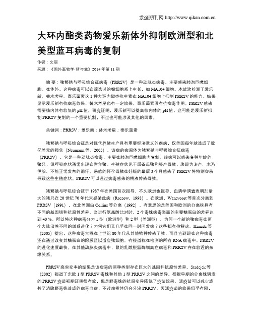
大环内酯类药物爱乐新体外抑制欧洲型和北美型蓝耳病毒的复制作者:文丽来源:《国外畜牧学·猪与禽》2014年第11期摘要:猪繁殖与呼吸综合征病毒(PRRSV)是一种动脉炎病毒,主要感染肺泡巨噬细胞。
在体外,这种病毒可以在筛选过的猴细胞系上生长,如MA104细胞。
本试验检测了爱乐新、替米考星、泰乐菌素这3种大环内酯类抗生素在MA104细胞上抑制PRRSV的能力。
结果显示爱乐新有抗病毒效果,替米考星也有一定效果。
泰乐菌素没有抗病毒作用。
PRRSV感染需要核内体有较低的pH值。
研究证明,爱乐新可以提高核内体的pH值,这可能是爱乐新抑制PRRSV复制的一个重要机制,不过也可能涉及其他的因素。
关键词:PRRSV;爱乐新;替米考星;泰乐菌素猪繁殖与呼吸综合征是对现代养猪生产具有重要经济意义的疾病,仅美国每年就造成了数亿美元的损失(Neumann等,2005)。
该病的病原体为猪繁殖与呼吸综合征病毒(PRRSV),它是一种动脉炎病毒,主要在肺泡巨噬细胞内复制。
该病可以感染各种年龄的猪只,但呼吸症状通常出现在青年猪。
生殖症状见于后备母猪和经产母猪,表现为流产、木乃伊胎、不能正常发育的弱仔。
易感的怀孕母猪在妊娠的最后3个月感染了PRRSV将特别容易导致这些生殖症状。
PRRSV可以通过病毒感染的精液传染母猪。
猪繁殖与呼吸综合征于1987年在美国首次报导,不久欧洲也报导。
血清学调查表明加拿大的猪只在20世纪70年代末感染此病(Rossow,1998)。
在欧洲,Wensvoort等首次分离到PRRSV(1991),在北美洲由Collins等分离(1992)。
有意思的是美国和欧洲的分离株具有不同的基因组和抗原性差异。
当进行氨基酸比对时,2个毒株病毒表面的主要糖蛋白的差异达到40 %。
所以将这种病毒分为1型(欧洲型)和2型(美洲型)。
为何一个新的猪病毒在两个大陆沿着不同的谱系进化?为何它们又几乎在同一时间发病?这些都有待解决。
Broadly Neutralizing AntibodiesStructural studies of broadly neutralizing antibodies are paving the way to vaccines for HIV, influenza and RSVViruses like HIV and influenza have evolved sneaky methods for evading our immune system. The immune system searches for foreign molecules, but several viruses have found ways to hide their unique parts and masquerade as normal human molecules. They do this in many ways. As viral surface glycoproteins are synthesized in infected cells, they are decorated with the same sugar chains that coat human proteins, providing an effective camouflage. The conserved functional sites of the viral protein are hidden deep in a pocket surrounded by these sugars, and thus are difficult for antibodies to reach. In addition, these viruses have error-prone replication machinery, which creates a great diversity in the viral glycoproteins. So unfortunately, once the immune system has found antibodies to recognize the infecting virus, other viruses rapidly mutate to change the site that is recognized.The Immune System Fights BackAntibodies of the immune system tend to focus on easily accessible loops on the viral surface, which often have great sequence and conformational variability. This is a problem for two reasons: the virus population can quickly evade these antibodies, and the antibodies are attacking portions of the protein that are not essential for function. Amazingly, however, after several years of battle with the infection, some people develop broadly neutralizing antibodies, termed "broadly" because they attack many strains of the virus, and "neutralizing" because they attack key functional sites in the virus and block infection. Unfortunately, however, these antibodies usually come too late and do not provide effective protection from the disease.Attacking HIVResearchers are studying these broadly neutralizing antibodies and trying to find ways to spur the immune system into creating them quickly with a vaccine. Because they recognize unusual targets, they are unusual antibodies. The one shown here, from PDB entry 4nco , uses an approach seen in many of these antibodies. The structure includes one Fab arm of the antibody (in blue) and the outer portion of the HIV envelope glycoprotein (in yellow and red, with sugars in orange). The antibody has an unusually long extension of one of the loops, which pokes like a finger through the coating of sugar chains and into a conserved site of the viral glycoprotein. The rest of the antibody also has dozens of mutations that refine the interaction with surrounding protein and sugars.The Fab portion of a broadly-neutralizing antibody (blue) bound to HIV envelope glycoprotein (yellow, orange, and red).Attacking InfluenzaFab portions of three broadly-neutralizing antibodies (blue) bound to influenza hemagglutinin (yellow, orange, and red).Broadly neutralizing antibodies for influenza also focus on conserved functional targets, attacking a vulnerable site on the viral protein hemagglutinin . The one at the top uses a similar approach as the HIV-binding antibody, with a loop that extends into the receptor binding site. The two at the bottom attack another conserved function of the hemagglutinin: the machinery involved in membrane fusion. Structures for these three antibodies are available in PDB entries 3sm5, 4fqi and 3sdy, and are shown all bound to one hemagglutinin in this illustration.Exploring the StructureThe ultimate goal of this research is to find way to make vaccines that will spur production of these broadly neutralizing antibodies, to provide protection against infection. Remarkably, this goal of structure-based vaccine design has been achieved for respiratory syncytial virus (RSV). The work started with a structure of a particularly potent antibody that neutralizes the receptor-binding site of the viral fusion glycoprotein, shown here on the left (PDB entry 4jhw). Based on this structure, researchers engineered a soluble form of the glycoprotein that adopts the same shape as the antibody-bound form, requiring a number of mutations, shown here on the right (PDB entry 4mmv). When mice and macaques are vaccinated with this engineered protein, it provides immunity from the virus. To explore these proteins in more detail, click on the image for an interactive Jmol.Topics for Further DiscussionPDB entry 1op5 includes an unusual domain-swapped antibody, which creates a third binding site in addition to the two conventional binding sites on the two Fab domains. It is a broadly neutralizing antibody that attacks the sugars on the surface of HIV envelope glycoprotein. Because they are so flexible, crystallographers typically study a fragment of antibodies which includes only one of the Fab arms. To see the structure of some intact antibodies, look at the examples in the Molecule of the Month on antibodies.References1.4nco: J. P. Julien, A. Cupo, D. Sok, R. L. Stanfield, D. Lyumkis, M. C. Deller, P. J.Klasse, D. R. Burton, R. W. Sanders, J. P. Moore, A. B. Ward & I. A. Wilson (2013) Crystal structure of soluble cleaved HIV-1 envelope trimer. Science 342,1477-1483.2.4mmv: J. S. McLellan, M. Chen, M. G. Joyce, M. Sastry, G. B. E. Stewart-Jones, Y.Yang, B. Zhang, L. Chen, S. Srivatsan, A. Zheng, T. Zhou, K. W. Graepel, A.Kumar, S. Moin, J. C. Boyington, G. Y. Chuang, C. Soto, U. Baxa, A. Q. Bakker, H.Spits, T. Beaumont, Z. Zheng, N. Xia, S. Y. Ko, J. P. Todd, S. Rao, B. S. Graham & P. D. Kwong (2013) Structure-based design of a fusion glycoprotein vaccine forrespiratory syncytial virus. Science 342, 592-598.3.4jhw: J. S. McLellan, M. Chen, S. Leung, K. W. Graepel, X. Du, Y. Yang, T. Zhou,U. Baxa, E. Yasuda, T. Beaumont, A. Kumar, K. Modjarrad, Z. Zheng, M. Zhao, N.Xia, P. D. Kwong & B. S. Graham (2013) Structure of RSV fusion glycoproteintrimer bound to a prefusion-specific neutralizing antibody. Science 340, 1113-1117.4.4fqi: C. Dreyfus, N. S. Laursen, T. Kwaks, D. Zuijdgeest, R. Khayat, D. C. Ekiert, J.H. Lee, Z. Metlagel, M. V. Bujny, M. Jongeneelen, R. van der Vlugt, M. Lamrani, H.J. W. M. Korse, E. Geelen, O. Sahin, M. Sieuwerts, J. P. Brakenhoff, R. Vogels, O.T. W. Li, L. L. Poon, M. Peiris, W. Koudstaal, A. B. Ward, I. A. Wilson, J. Goudsmit & R. H. Rriesen (2012) Highly conserved protective epitopes on influenza Bviruses. Science 337, 1343-1348.5.J. P. Julien, P. S. Lee & I. A. Wilson (2012) Structural insights into key sites ofvulnerability on HIV-1 Env and influenza HA. Immunological Reviews 250,180-198.6.3sdy: D. C. Ekiert, R. H. Friesen, G. Bhabha, T. Kwaks, M. Jongeneelen, W. Yu, C.Ophorst, F. Cox, H. J. W. M. Korse, B. Brandenberg, R. Vogels, J. P. Brakenhoff, R.Kompier, M. J. Koldijk, L. A. Cornelissen, L. L. Poon, M. Peiris, W. Koudstaal, I. A.Wilson & J. Goudsmit (2011) A highly conserved neutralizing epitope on group 2influenza A viruses. Science 333, 843-850.7.3sm5: J. R. Whittle, R. Zhang, S. Khurana, L. R. King, J. Manischewitz, H. Golding,P. R. Dormitzer, B. F. Haynes, E. B. Walter, M. A. Moody, T. B. Kepler, H. X. Liao & S. C. Harrison (2011) Broadly neutralizing human antibody that recognizes thereceptor- binding pocket of influenza virus hemagglutinin. Proceedings of theNational Academy of Science USA 108, 14216-14221.。
姜黄素减轻小鼠癫痫发作吴连连;秦滢;胡安康【期刊名称】《实验动物科学》【年(卷),期】2022(39)4【摘要】目的探讨姜黄素对小鼠癫痫发作的作用及机制。
方法C57/BL6雄性小鼠60只,随机分为3组:正常组、癫痫模型组和姜黄素治疗组,每组20只。
观察记录每组小鼠癫痫发作Racine分级评分和癫痫发作的潜伏期。
免疫荧光染色检测海马CA3区GFAP和IBA-1表达,蛋白免疫印迹(Western blot)检测海马IL-1β、NLRP3、caspase1的表达水平。
结果姜黄素治疗组小鼠癫痫发作Racine分级评分显著低于癫痫模型组(P<0.05),发作潜伏期显著延长(P<0.05)。
与正常组相比,模型组海马CA3区GFAP和IBA-1的表达显著增加(P<0.01),胶质细胞呈现明显激活状态,IL-1β、NLRP3、caspase1蛋白的相对表达量显著升高(P<0.01),与模型组相比,姜黄素组海马CA3区的GFAP和IBA-1阳性细胞数显著减少(P<0.05),海马IL-1β、NLRP3、caspase1蛋白相对表达量也显著降低(P<0.05)。
结论姜黄素可以减轻癫痫发作程度,这可能通过调节NLRP3/caspase1通路抑制海马炎性反应来实现。
【总页数】5页(P39-43)【作者】吴连连;秦滢;胡安康【作者单位】徐州医科大学实验动物中心【正文语种】中文【中图分类】Q95-3【相关文献】1.盐酸小檗碱预处理对青霉素钠诱发小鼠癫痫发作程度、发作潜伏期的影响及其机制2.TLR4/NF-κB信号通路在姜黄素减轻癫痫小鼠神经元损伤中的作用3.姜黄素减轻小鼠深部组织压力性损伤4.匹罗卡品致痫小鼠自发性癫痫发作模型的建立及癫痫后海马新生神经细胞增生的研究5.姜黄素通过TLR4/NF-кB/NLRP3信号通路减轻LPS/D-GalN诱导的小鼠急性肝损伤因版权原因,仅展示原文概要,查看原文内容请购买。
may also depend upon genetic diversification. It is instructive to consider highly antigenically diverse pathogens in the general context of pathogen variability. A number of pathogens have evolved rapid genetic diversification, including various bacteria and parasites, but the champions of diversity are viruses. RNA viruses in particular have high mutation rates, largely due to the involvement of virus-encoded error-prone RNA and DNA polymerases in their replication cycle, leading to mutation rates as high as 1.5×10⁻³mutations per nucleotide per genomic replication (1). Certain DNA viruses, in particular single-strand DNA viruses, can also have high mutation and substitution rates, sometimes as high or higher than RNA viruses (1). For many viruses, sequence variability is crucial to escape host immune cellular and humoral responses, leading to great antigenic diversity in the proteins targeted by the adaptive immune system. However, other variable viruses have evolved modes of replication and transmission that allow them to survive without the need to escape the pressure of their host adaptive immune responses, e.g. by transmitting from one host to the next prior to an effective immune response being mounted as for measles and polio viruses. As a consequence, these viruses do not display a high level of antigenic diversity despite an inherent capacity to do so. Therefore, high sequence diversity per se is not necessarily an obstacle to vaccine development and effective vaccines have been developed against relatively highly diverse viruses, such as measles virus, hepatitis B virus,polio virus and rabies virus. Conversely, it should be noted that, vaccines have been difficult to develop against pathogens with low diversity, such as Herpes Simplex Virus (HSV). In contrast, high antigenic diversity, in which there is a very high level of variability in the viral protein sites principally targeted by the immune system (immunodominant or immunoprominent epitopes), does consistently impair vaccine development. Classical vaccine approaches will tend to afford protection against a very limited fraction of circulating virus populations. Since the most important contributor to protection by the majority of anti-viral vaccines is neutralizing antibodies targeting the surface Env proteins,the highly dynamic antigenic diversity of these proteins is the major obstacle to the development of practical vaccines for viruses such as HIV, influenza virus and HCV.HIV establishes a chronic infection that, over a period of years, if left untreated, leads to AIDS. About 25 million people have died of AIDS and about 35 million are currentlyinfected (2). The epicenter of the plague is sub-Saharan Africa with an adult infection rate attaining 5%. The high relative cost of treatment has begun to consume a large proportion of development aid to this region, with diverse negative consequences. Influenza virus infection produces acute respiratory and systemic symptoms and leads to between one quarter and one half million deaths on average per year and societal costs of billions of dollars annually in healthcare and lost productivity (3). At 10-50 year intervals, the virus triggers recurring deadly pandemics; the great influenza pandemic of 1918 killed about 50million people worldwide (4), and the other pandemics of the past century in 1957, 1968 and 2009 caused millions of deaths. HCV chronically infects 120-170 million people globally(5) and is a major cause of chronic liver disease and liver cancer in the developing world.The societal costs of HCV in the US are expected to approach $85 billion by 2024 (6).HIV, influenza and HCV show great sequence heterogeneity in their envelope (Env)proteins, which are the essential targets of an effective antibody response. Thisheterogeneity is seen between different isolates or strains, particularly in their surface-exposed amino acid residues. Nevertheless, at least two regions on these viral Env proteins are expected to be both conserved and accessible, even if only transiently in some instances,to permit virus infection. All enveloped viruses are required to bind to one or more receptors on their target host cell and have a mechanism for entry into that cell. Therefore, the Env proteins contain a receptor binding site(s) and the viral fusion machinery, both of which are likely to be exposed, at least transiently, during cell attachment and viral entry. However,these conserved accessible regions may be relatively small; for example for influenza virusNIH-PA Author ManuscriptNIH-PA Author ManuscriptNIH-PA Author Manuscriptinfectivity, the hemagglutinin (HA) protein that mediates viral entry is only required to recognize a relatively small glycan moiety, containing a terminal sialic acid, as its receptor on target cells. The small size of the receptor severely limits the conserved footprint that can be specifically recognized by an antibody in order to acquire cross reactivity against different strains and subtypes. For HIV-1, that footprint is larger as the receptor is a protein,CD4, but CD4 has a molecular width of only a single immunoglobulin (Ig) domain whereas an antibody antigen-binding fragment (Fab) has twice that width with two Ig domains.Suggestions that extended conserved regions may be exposed at the Env surface came originally from observations of serum cross-neutralizing activity against diverse influenza and HIV isolates (7, 8) from individuals infected with the corresponding viruses. Serum cross-neutralizing activity could arise, in principle, from a combination of a large number of antibodies directed to variable regions. However, the isolation of individual monoclonal antibodies that were able to neutralize in vitro multiple, diverse isolates (broadly neutralizing monoclonal antibodies, bnMAbs (9)) confirmed the presence of conserved antibody footprint-sized accessible regions on Env antigens on the viral surface. BnMAbs to HIV were first isolated from infected individuals in 1992 by electrofusion (10) and by phage display (11), but only shown convincingly to be broadly neutralizing to primary as distinct from laboratory-adapted virus isolates in 1994 (12, 13). A mouse bnMAb to influenza virus was isolated and characterized in 1993 (14) and a mouse bnMAb to HCV was first generated in 2001 (15) and shown to be broadly functional in 2005 (16). Until the late 2000s, only a small number of bnMAbs against these viruses had been isolated and characterized but in the last 3 years an explosion in the rate of generation and characterization of such Abs has occurred. These exciting breakthroughs have unearthed many novel antibodies with unexpected epitopes, as well as novel modes of antibody-antigen recognition and have suggested a plethora of new vaccine and drug targets.For many years, only a handful of bnMAbs against HIV, all human, were known (17, 18).Of these antibodies, two that were directed to the Env glycoprotein gp120 (b12, 2G12) andtwo to the transmembrane Env glycoprotein gp41 (2F5, 4E10) were the most notable and were intensely studied. Importantly, these bnMAbs were all shown to protect against free virus challenge in relatively robust macaque models of HIV infection (19-22). Crystalstructures of these MAbs in complex with Env antigens were all determined and a variety of novel antibody features were identified in their hypervariable regions (23, 24). These features included long heavy chain variable complementarity determining regions 3(HCDR3s), which had scarcely been reported at that time, antibody domain exchange to recognize a glycan cluster on gp120 (2G12), and very hydrophobic HCDR3s (2F5, 4E10),which appeared to be associated with recognition of epitopes very close to the virusmembrane (i.e. membrane proximal external region or MPER) for the two anti-gp41 MAbs.Many immunogens have been designed based, to varying degrees, on molecularunderstanding of the interaction of these bnMAbs with HIV Env antigens. No immunogen tested to date has “re-elicited” antibodies with broadly neutralizing character. Several explanations have been proposed, including the limitations imposed by the availability of such a small panel of bnMAbs that makes the drawing of general conclusions forimmunogen design hazardous. Therefore, considerable effort by a number of laboratories went into the discovery of more bnMAbs. Two factors appear to have been most crucial in the success of that effort; first, the screening of large cohorts of infected donors to identify individuals with broadly neutralizing sera using reproducible high-throughput neutralization assays (25) and second, the application of novel single B cell technologies to samples from these donors to facilitate the isolation of bnMAbs from a background of many other non-neutralizing anti-Env antibodies. The application of direct neutralization screening to a large number of B cells (about 30,000) from a donor with broad and potent serum neutralizing activity led to the isolation of a pair of bnMAbs to a novel epitope in 2009 (26). The newNIH-PA Author ManuscriptNIH-PA Author ManuscriptNIH-PA Author ManuscriptMAbs were approximately an order of magnitude more potent in standard neutralization assays than the previously identified bnMAbs while generally maintaining or improving breadth. This development was quickly followed by the sorting of B cells using an engineered gp120 molecule to identify a potent bnMAb directed to the CD4 binding site of gp120 of similarly enhanced potency and even greater breadth (27). The application of direct neutralization screening to further donors with broadly neutralizing sera led to the identification of yet more bnMAbs that targeted novel mixed glycan/protein epitopes on gp120 with even greater potency (28). Larger panels of highly potent anti-CD4bs bnMAbs have also been isolated by single B cell sorting using Env baits and specially designed PCR primers to account for a high degree of antibody mutation (29). Engineering of one such MAb has generated remarkable potency and breadth (31). Deep sequencing of antibody repertoires from donors from whom bnMAbs have been isolated has revealed many additional potent related MAbs (30). BnMAb targets on HIV Env are represented in Fig 1.The structures of many of the newer HIV bnMAbs in complex with Env antigens (31-34),together with analysis of the bnMAb sequences, provide opportunities and potential lessons for vaccine and drug discovery. Several new targets are available for immunogen and drug design and the understanding of existing targets has also been greatly enhanced. For example, recognition of a favored vaccine target, the CD4 binding site has now been shown to not necessarily require a long HCDR3, but appears to be strongly dependent on certain V H gene segments (29, 32). High affinity glycan recognition on Env can also be achieved by conventional antibodies in the absence of 2G12-like domain exchange (33). Other recurring themes of HIV bnMAbs have been a relatively high degree of somatic hypermutation, a prevalence of insertions and deletions in the antibody variable regions regions and an inability of germ line versions of the bnMAbs to bind Env (26-29, 35-37). It is still unclear whether a high degree of hypermutation reflects a general property of HIV Abs that has arisen due to the chronic antigen stimulation of natural infection or is a requirement for the evolution of such Abs towards broad cross-reactivity. It is also unclear the affinity thresholdthat is required for the Env-germ line antibody to trigger a response. Nevertheless, a number of immunogens are currently being designed to bind to germ line, as well as mature,antibodies to activate naive B cells (37).Besides informing vaccine design, the discovery of conserved exposed regions, particularly associated with the glycan shield of gp120 (Fig. 1), provides a number of new potential viral entry inhibitor targets.For influenza virus, the first bnMAb was isolated in 1993 by immunization of a mouse with H2N2 virus (hemagglutinin (HA) subtype 2, neuraminidase subtype 2) and shown toneutralize group 1 viruses bearing HAs of subtypes 1, 2, 5, 6 and 9 (14). The antibody was proposed to bind to a conserved region in the stem of HA in contrast to typical strain-specific antibodies that bind to hypervariable regions on the head of HA (Fig 2). In 2008 and 2009, a number of novel human bnMAbs that cross-neutralized group 1 subtypes were isolated from phage libraries by different laboratories. The antibodies had remarkablysimilar sequences to one another-close to the corresponding human germ line sequence of the V H 1-69 family with relatively few somatic mutations, showed similar patterns of group I subtype neutralization (representing 10 of the 16 known flu subtypes) and were protective in mouse models (38-41). Structural studies showed this group of V H 1-69 bnMAbs recognized a conserved region of the stem of HA using the heavy chain alone (41, 42) (Fig 2). More recently, high-throughput screening of immortalized antibody-secreting cells generated a bnMAb that recognizes this conserved region in a different way using both heavy and light chains and is able to neutralize both group 1 and group 2 viruses (43). In addition, a different cell-based method has been used to isolate a bnMAb that binds to a second site on the stem of HA, again quite close to the virus membrane, neutralizes group 2 viruses, and protectsNIH-PA Author ManuscriptNIH-PA Author ManuscriptNIH-PA Author Manuscriptmice against virus challenge (42) (Fig. 2). Furthermore, bnMAbs have recently been described that target the head region of HA (38, 44-47) around the receptor binding site. One of these bnMAbs neutralizes both selected isolates from both group 1 and group 2 viruses (48), but most neutralize only a single subtype. The structure of a MAb that broadly neutralizes viruses from the H1 subtype does so by targeting an epitope that overlaps the HA receptor binding site (47) (Fig. 2).The holy grail of influenza virus vaccine research is a universal vaccine that protects against all strains and subtypes of the virus, including seasonal and pandemic strains, and thereby renders the annual vaccine redundant. If the vaccine is long acting, as for example for the yellow fever or smallpox vaccines, it could be given at a relatively young age and protect into old age. There is a great need for such a vaccine since current seasonal influenza vaccines tend to be less effective in elderly individuals as the capacity of the immune system wanes (49). Also, the vaccine needs to be reformulated annually to include the best predictions of which strains will circulate in any given year and the match is not often optimal. Indeed, the efficacy and effectiveness of current influenza vaccines may be notably lower than previously thought (49). Furthermore, the vaccine takes some months to develop and manufacture, which can be problematic in the face of a potential pandemic or epidemic.As a new approach to the standard live or attenuated influenza vaccines, the identification of the stem of HA as a vaccine target has already led to design of “headless” HA immunogens,which have thus far produced modest improvements in neutralization breadth (50, 51), and the engineering of scaffolds incorporating crucial structural elements recognized by the stem bnMAbs, which are currently being evaluated. A number of different immunization strategies are also being intensively investigated resulting in some success in improving the breadth of neutralizing anti-influenza virus responses (52, 53). It is also worth noting that the pandemic H1N1 vaccine does induce broadly cross-reactive antibodies to the HA stem region (54). Finally, as with HIV, the bnMAbs identify promising drug targets and, indeed, a small protein has been computationally designed to bind to the HA stem region defined bybnMAbs and experimentally shown to neutralize influenza virus by inhibiting critical HA conformational changes required for fusion (55), by a mechanism similar to that used by the stem antibodies. Presumably, a small molecule drug could also be targeted to this region and used to specifically inhibit influenza virus infection.For HCV, the first bnMAb was isolated in 2001 (15) and shown to be broadly neutralizing in 2005 (16). The mouse bnMAb and a human bnMAb identified later (56) define a continuous epitope on the E2 Env glycoprotein of the virus and inhibit interaction of E2 with thereceptor CD81. In the last few years, a range of human and murine bnMAbs, mostly directed to discontinuous epitopes on E2, have been identified (57-62) with one report of a bnMAb directed to the E1 glycoprotein (63). Of note, a single bnMAb was shown to provideprotection against HCV quasispecies challenge in an animal model (59). Unfortunately, no structure is yet available for E2 or the E1 E2 complex limiting the value of the bnMAbs for immunogen and drug design. However, the crystal structure of a human bnMAb complexed with a peptide corresponding to a continuous epitope on E2 (56) has recently beendetermined (64) providing a template for immunogen design.For the highly antigenically diverse viruses described above, an effective vaccine would seek to induce antibodies that recognize conserved regions and neutralize as broadly aspossible. Challenges will always remain due to the diverse sequences and constant antigenic variation in these viruses, which will lead to differences in neutralization sensitivity across the spectrum of circulating isolates and could lower overall efficacy of vaccine-induced antibody responses or as in the case of dengue virus, enhance disease (65).NIH-PA Author ManuscriptNIH-PA Author ManuscriptNIH-PA Author ManuscriptFinally, we have focused here on harnessing the information derived from bnMAbs to help define vaccine and drug targets on highly antigenically diverse viruses. These bnMAbs may also have direct application as prophylactic and/or therapeutic passively administered reagents. For HIV in a prophylactic setting, one might consider systemic passiveadministration of bnMAbs for high-risk individuals or topical application in a microbicide.In a therapeutic setting, combining passively administered antibody with anti-retroviral drugs could be considered. One advantage of systemic antibody is long half-life, but a serious disadvantage is high cost. This problem could be ameliorated by the expression of bnMAbs from vectors such as adeno-associated virus (AAV) (66, 67). For influenza virus,therapy with bnMAbs could be considered, especially given indications that antibody administration relatively late in disease course may be beneficial (68). For HCV, a clear application of bnMAbs is to prevent re-infection of the grafted liver that typically occurs following liver transplant (69)In summary, highly antigenically diverse pathogens are a major health concern and are particularly difficult to counter through vaccination. The generation of a whole newarmamentarium of broadly neutralizing antibodies to several highly antigenically diverse viruses in the last three years has revealed new and unexpected vulnerabilities in these previously impregnable viruses. The stage is now well and truly set for rational vaccinedesign based on exploiting these vulnerabilities. Immunogen design based on computational approaches is advancing rapidly (70, 71). Small animal models expressing human antibody repertoires suitable for immunogen evaluation are being evaluated and developed.Technologies for the detailed evaluation of antibody responses including deep sequencing and single B cell approaches will facilitate iterative improvements of immunogens.Immunization strategies based on a better understanding of the antibody requirements for broad neutralization and of the roles of innate immunity and T cell help in eliciting theappropriate antibody responses will also likely be crucial. The challenges are manifest, but the advances made against HIV-1, influenza viruses and HCV described here may haveramifications and implications that go well beyond these highly diverse viruses to the many other microbial pathogens that threaten the health and well being of mankind.AcknowledgmentsWe acknowledge the financial support of the National Institutes of Health, the International AIDS Initiative (IAVI)and the Ragon Institute. We thank C. Corbaci for help in preparation of the manuscript. We thank all our past and present laboratory members for their many contributions to our research efforts.References and Notes1. Duffy S, Shackelton LA, Holmes EC. Rates of evolutionary change in viruses: patterns and determinants. Nat. Rev. Genet. 2008; 9:267. [PubMed: 18319742]2. UNAIDS World Aids Day Report. UNAIDS; Geneva: 2011. How to get to zero: Faster. Smarter.Better..3. Graham-Rowe D. Epidemiology: Racing against the flu. Nature. 2011; 480:S2. [PubMed:22158296]4. Basler CF, Aguilar PV. Progress in identifying virulence determinants of the 1918 H1N1 and the Southeast Asian H5N1 influenza A viruses. Antiviral Res. 2008; 79:166. [PubMed: 18547656]5. Shepard CW, Finelli L, Alter MJ. Global epidemiology of hepatitis C virus infection. Lancet Infect.Dis. 2005; 5:558. [PubMed: 16122679]6. Combating the silent epidemic of viral hepatitis: Action plan for the prevention, care and treatment of viral hepatitis. United States Department of Health and Human Services. 20117. Graves PN, Schulman JL, Young JF, Palese P. Preparation of influenza virus subviral particles lacking the HA1 subunit of hemagglutinin: unmasking of cross-reactive HA2 determinants.Virology. 1983; 126:106. [PubMed: 6189287]NIH-PA Author Manuscript NIH-PA Author ManuscriptNIH-PA Author Manuscript8. Vujcic LK, Quinnan GV Jr. Preparation and characterization of human HIV type 1 neutralizing reference sera. AIDS Res. Hum. Retroviruses. 1995; 11:783. [PubMed: 7546904]9. Burton DR. Antibodies, viruses and vaccines. Nat. Rev. Immunol. 2002; 2:706. [PubMed:12209139]10. Buchacher, A., et al. Vaccines '92: Modern approaches to new vaccines including prevention of AIDS. Brown, F.; Chanock, R.; Ginsberg, HS.; Lerner, R., editors. Cold Spring Harbor Laboratory Press; Cold Spring Harbor, NY: 1992. p. 191-194.11. Barbas CF 3rd, et al. Recombinant human Fab fragments neutralize human type 1immunodeficiency virus in vitro. Proc. Natl. Acad. Sci. U. S. A. 1992; 89:9339. [PubMed:1384050]12. Conley AJ, et al. Neutralization of divergent human immunodeficiency virus type 1 variants and primary isolates by IAM-41-2F5, an anti-gp41 human monoclonal antibody. Proc. Natl. Acad. Sci.U. S. A. 1994; 91:3348. [PubMed: 7512731]13. Burton DR, et al. Efficient neutralization of primary isolates of HIV-1 by a recombinant human monoclonal antibody. Science. 1994; 266:1024. [PubMed: 7973652]14. Okuno Y, Isegawa Y, Sasao F, Ueda S. A common neutralizing epitope conserved between the hemagglutinins of influenza A virus H1 and H2 strains. J. Virol. 1993; 67:2552. [PubMed:7682624]15. Owsianka A, Clayton RF, Loomis-Price LD, McKeating JA, Patel AH. Functional analysis of hepatitis C virus E2 glycoproteins and virus-like particles reveals structural dissimilarities between different forms of E2. J. Gen. Virol. 2001; 82:1877. [PubMed: 11457993]16. Owsianka A, et al. Monoclonal antibody AP33 defines a broadly neutralizing epitope on the hepatitis C virus E2 envelope glycoprotein. J. Virol. 2005; 79:11095. [PubMed: 16103160]17. Burton DR, et al. HIV vaccine design and the neutralizing antibody problem. Nat. Immunol. 2004;5:233. [PubMed: 14985706]18. Mascola JR, Montefiori DC. The role of antibodies in HIV vaccines. Annu. Rev. Immunol. 2010;28:413. [PubMed: 20192810]19. Mascola JR. Defining the protective antibody response for HIV-1. Curr. Mol. Med. 2003; 3:209.[PubMed: 12699358]20. Hessell AJ, et al. Effective, low-titer antibody protection against low-dose repeated mucosal SHIVchallenge in macaques. Nat. Med. 2009; 15:951. [PubMed: 19525965]21. Hessell AJ, et al. Broadly neutralizing human anti-HIV antibody 2G12 is effective in protectionagainst mucosal SHIV challenge even at low serum neutralizing titers. PLoS Path. 2009;5:e1000433.22. Hessell AJ, et al. Broadly neutralizing monoclonal antibodies 2F5 and 4E10 directed against thehuman immunodeficiency virus type 1 gp41 membrane-proximal external region protect against mucosal challenge by simian-human immunodeficiency virus SHIVBa-L. J. Virol. 2010; 84:1302.[PubMed: 19906907]23. Burton DR, Stanfield RL, Wilson IA. Antibody vs. HIV in a clash of evolutionary titans. Proc.Natl. Acad. Sci. U S A. 2005; 102:14943. [PubMed: 16219699]24. Douek DC, Kwong PD, Nabel GJ. The rational design of an AIDS vaccine. Cell. 2006; 124:677.[PubMed: 16497577]25. Stamatatos L, Morris L, Burton DR, Mascola JR. Neutralizing antibodies generated during naturalHIV-1 infection: good news for an HIV-1 vaccine? Nat. Med. 2009; 15:866. [PubMed: 19525964]26. Walker LM, et al. Broad and potent neutralizing antibodies from an African donor reveal a newHIV-1 vaccine target. Science. 2009; 326:285. [PubMed: 19729618]27. Wu X, et al. Rational design of envelope surface identifies broadly neutralizing human monoclonalantibodies to HIV-1. Science. 2010; 329:856. [PubMed: 20616233]28. Walker LM, et al. Broad neutralization coverage of HIV by multiple highly potent antibodies.Nature. 2011; 477:466. [PubMed: 21849977]29. Scheid JF, et al. Sequence and structural convergence of broad and potent HIV antibodies thatmimic CD4 binding. Science. 2011; 333:1633. [PubMed: 21764753]NIH-PA Author ManuscriptNIH-PA Author ManuscriptNIH-PA Author Manuscript30. Wu X, et al. Focused evolution of HIV-1 neutralizing antibodies revealed by crystal structures and deep sequencing. Science. 2011; 333:1593. [PubMed: 21835983]31. Diskin R, et al. Increasing the potency and breadth of an HIV antibody by using structure-based rational design. Science. 2011; 334:1289. [PubMed: 22033520]32. Zhou T, et al. Structural basis for broad and potent neutralization of HIV-1 by antibody VRC01.Science. 2010; 329:811. [PubMed: 20616231]33. Pejchal R, et al. A potent and broad neutralizing antibody recognizes and penetrates the HIV glycan shield. Science. 2011; 334:1097. [PubMed: 21998254]34. McLellan JS, et al. Structure of HIV-1 gp120 V1/V2 domain with broadly neutralizing antibody PG9. Nature. 2011; 480:336. [PubMed: 22113616]35. Corti D, et al. Analysis of memory B cell responses and isolation of novel monoclonal antibodies with neutralizing breadth from HIV-1-infected individuals. Plos One. 2010; 5:e8805. [PubMed:20098712]36. Huber M, et al. Very Few Substitutions in a Germline Antibody Are Required to Initiate Significant Domain Exchange. J. Virol. 2010; 84:10700. [PubMed: 20702640]37. Xiao X, et al. Germline-like predecessors of broadly neutralizing antibodies lack measurable binding to HIV-1 envelope glycoproteins: implications for evasion of immune responses and design of vaccine immunogens. Biochem. Biophys. Res. Commun. 2009; 390:404. [PubMed:19748484]38. Kashyap AK, et al. Combinatorial antibody libraries from survivors of the Turkish H5N1 avian influenza outbreak reveal virus neutralization strategies. Proc. Natl. Acad. Sci. U. S. A. 2008;105:5986. [PubMed: 18413603]39. Throsby M, et al. Heterosubtypic neutralizing monoclonal antibodies cross-protective against H5N1 and H1N1 recovered from human IgM+ memory B cells. Plos One. 2008; 3:e3942.[PubMed: 19079604]40. Ekiert DC, et al. Antibody recognition of a highly conserved influenza virus epitope. Science.2009; 324:246. [PubMed: 19251591]41. Sui J, et al. Structural and functional bases for broad-spectrum neutralization of avian and humaninfluenza A viruses. Nat. Struct. Mol. Biol. 2009; 16:265. [PubMed: 19234466]42. Ekiert DC, et al. A highly conserved neutralizing epitope on group 2 influenza A viruses. Science.2011; 333:843. [PubMed: 21737702]43. Corti D, et al. A neutralizing antibody selected from plasma cells that binds to group 1 and group 2influenza A hemagglutinins. Science. 2011; 333:850. [PubMed: 21798894]44. Ohshima N, et al. Naturally occurring antibodies in humans can neutralize a variety of influenzavirus strains, including H3, H1, H2, and H5. J. Virol. 2011; 85:11048. [PubMed: 21865387]45. Krause JC, et al. Epitope-specific human influenza antibody repertoires diversify by B cellintraclonal sequence divergence and interclonal convergence. J. Immunol. 2011; 187:3704.[PubMed: 21880983]46. Krause JC, et al. A broadly neutralizing human monoclonal antibody that recognizes a conserved,novel epitope on the globular head of the influenza H1N1 virus hemagglutinin. J. Virol. 2011;85:10905. [PubMed: 21849447]47. Whittle JR, et al. Broadly neutralizing human antibody that recognizes the receptor-binding pocketof influenza virus hemagglutinin. Proc. Natl. Acad. Sci. U. S. A. 2011; 108:14216. [PubMed:21825125]48. Yoshida R, et al. Cross-protective potential of a novel monoclonal antibody directed againstantigenic site B of the hemagglutinin of influenza A viruses. PLoS Path. 2009; 5:e1000350.49. Osterholm MT, Kelley NS, Sommer A, Belongia EA. Efficacy and effectiveness of influenzavaccines: a systematic review and meta-analysis. Lancet Infect. Dis. 2012; 12:36. [PubMed:22032844]50. Bommakanti G, et al. Design of an HA2-based Escherichia coli expressed influenza immunogenthat protects mice from pathogenic challenge. Proc. Natl. Acad. Sci. U. S. A. 2010; 107:13701.[PubMed: 20615991]51. Steel J, et al. Influenza virus vaccine based on the conserved hemagglutinin stalk domain. mBio.2010; 1NIH-PA Author ManuscriptNIH-PA Author ManuscriptNIH-PA Author Manuscript。
Article Broadly Neutralizing Alphavirus Antibodies Bind an Epitope on E2and Inhibit Entry and EgressGraphical AbstractHighlightsd Broadly neutralizing MAbs bind an epitope in the B domain ofalphavirus E2proteind Broadly neutralizing MAbs protect in vivo against infectionby multiple alphavirusesd B domain MAb binding re-positions the A domain and cross-links adjacent E2spikesd MAbs that cross-neutralize alphavirus infection block viralentry and egress steps AuthorsJulie M.Fox,Feng Long,Melissa A.Edeling,...,Daved H.Fremont, Michael G.Rossmann,Michael S.DiamondCorrespondencediamond@In BriefA class of broadly neutralizing monoclonal antibodies identified here protects against infection and diseasein vivo against multiple alphaviruses, including chikungunya.These antibodies bind a discrete epitope on the alphavirus E2glycoprotein,block viral entry and egress,and allow cross-linking of adjacent E2protein spikes,suggesting avenues for possible vaccine-or antibody-based therapeutic development against multiple alphaviruses.Accession Numbers5ANY,EMD-3144 Fox et al.,2015,Cell163,1095–1107November19,2015ª2015Elsevier Inc./10.1016/j.cell.2015.10.050ArticleBroadly Neutralizing Alphavirus AntibodiesBind an Epitope on E2and Inhibit Entry and EgressJulie M.Fox,1Feng Long,5Melissa A.Edeling,2Hueylie Lin,1Mareike K.S.van Duijl-Richter,6Rachel H.Fong,7Kristen M.Kahle,7Jolanda M.Smit,6Jing Jin,8Graham Simmons,8Benjamin J.Doranz,7James E.Crowe,Jr.,9Daved H.Fremont,2Michael G.Rossmann,5and Michael S.Diamond1,2,3,4,*1Department of Medicine2Department of Pathology and Immunology3Department of Molecular Microbiology4Center for Human Immunology and Immunotherapy ProgramsWashington University School of Medicine,St.Louis,MO63110,USA5Department of Biological Sciences,Purdue University,West Lafayette,IN47907,USA6University of Groningen and University Medical Center Groningen,9713GZ Groningen,the Netherlands7Integral Molecular,Inc.,Philadelphia,PA19104,USA8Blood Systems Research Institute,San Francisco,CA94118,USA9Departments of Pediatrics,Pathology,Microbiology,and Immunology and the Vanderbilt Vaccine Center,Vanderbilt University,Nashville, TN37235,USA*Correspondence:diamond@/10.1016/j.cell.2015.10.050SUMMARYWe screened a panel of mouse and human mono-clonal antibodies(MAbs)against chikungunya virus and identified several with inhibitory activity against multiple alphaviruses.Passive transfer of broadly neutralizing MAbs protected mice against infection by chikungunya,Mayaro,and O’nyong’nyong ing alanine-scanning mutagenesis,loss-of-function recombinant proteins and viruses,and multiple functional assays,we determined that broadly neutralizing MAbs block multiple steps in the viral lifecycle,including entry and egress,and bind to a conserved epitope on the B domain of the E2glycoprotein.A16A˚resolution cryo-electron mi-croscopy structure of a Fab fragment bound to CHIKV E2B domain provided an explanation for its neutralizing activity.Binding to the B domain was associated with repositioning of the A domain of E2 that enabled cross-linking of neighboring spikes. Our results suggest that B domain antigenic determi-nants could be targeted for vaccine or antibody ther-apeutic development against multiple alphaviruses of global concern.INTRODUCTIONAlphaviruses are arthropod-transmitted single-stranded posi-tive-sense-enveloped viruses of the Togaviridae family and cause disease worldwide.The two surface glycoproteins on the mature virion,E2and E1,facilitate binding and entry through receptor-mediated endocytosis and low-pH-mediated fusion within endosomes(Lescar et al.,2001;Smith et al.,1995).Alpha-virus virions have T=4quasi-icosahedral symmetry,with240copies of the E2-E1heterodimer assembling into80trimeric spikes on the viral surface(Cheng et al.,1995).Twenty of these spikes(‘‘i3’’)are coincident with the icosahedral3-fold axes,and 60are in general positions at quasi3-fold axes(‘‘q3’’).X-ray crys-tallographic structures have been determined of the E1glyco-protein,the p62-E1precursor,the E2-E1heterodimer,and the (E1-E2)3trimer(Lescar et al.,2001;Li et al.,2010;Roussel et al.,2006;Voss et al.,2010).The mature E2protein contains three domains:an A domain,which is located centrally on the surface of the spike and possesses the putative receptor binding site;the B domain,located on the distal end of the spike, covering the fusion loop on E1;and the C domain,at the proximal end of the spike.The E1protein is a type II membrane fusion pro-tein containing three b-barrel domains.Domain I is located spatially between domains II and III,with the fusion peptide lying at the distal end of domain II(Lescar et al.,2001;Voss et al., 2010).The E1protein lies at the base of the trimeric spike with E2positioned on top of it.Chikungunya virus(CHIKV)is transmitted to humans by Aedes species of mosquitoes and causes a debilitating infection char-acterized by fever,rash,myositis,and arthritis,with joint disease lasting in some individuals for several years(Schilte et al.,2013). CHIKV historically caused outbreaks in Africa and Asia.In2013, transmission of CHIKV occurred in the Western Hemisphere,and in just18months,CHIKV has caused more than1.4million cases in the Americas in more than40countries,including locally ac-quired infections in Florida(Kendrick et al.,2014).In comparison, other arthritogenic alphaviruses(e.g.,Ross River[RRV],Semliki Forest[SFV],Mayaro[MAYV],and Sindbis[SINV]viruses)circu-late with more limited global distribution,with outbreaks in Oce-ania,Africa,and South America.Although currently there are no available licensed vaccines or therapies for CHIKV or any other alphavirus,studies have demonstrated the importance of antibody-mediated protection (Kam et al.,2012;Lum et al.,2013).Passive transfer of g-globulin purified from the plasma of CHIKV-immune patients tomice Cell163,1095–1107,November19,2015ª2015Elsevier Inc.1095prevented mortality following a lethal CHIKV infection (Couderc et al.,2009).Analogously,monoclonal antibodies (MAbs)neutralize CHIKV infection in vitro and protect against disease in mice and non-human primates (Fong et al.,2014;Fric et al.,2013;Goh et al.,2013;Pal et al.,2013,2014;Smith et al.,2015).One goal of vaccine and therapeutic efforts against viruses is the development of broadly neutralizing antibodies that inhibit most strains within a genetically diverse virus family.Broadly neutralizing MAbs have been described for human immunodefi-ciency (HIV),influenza A (IAV),dengue (DENV),and hepatitis C (HCV)viruses (reviewed in Corti and Lanzavecchia,2013).Although broadly neutralizing MAbs against alphaviruses have not been described,polyclonal antibodies (induced by a CHIKV vaccine candidate)protected against O’nyong’nyong virus (ONNV)infection (Partidos et al.,2012)and convalescent serum from RRV-infected mice protected against CHIKV pathogenesis (Gardner et al.,2010).Earlier reports described cross-protection between different alphaviruses using hyperimmune serum (Wust et al.,1987).These studies suggest that conserved epitopes exist across different alphaviruses that are recognized by pro-tective antibodies.We screened a panel of murine and human MAbs against CHIKV (Pal et al.,2013;Smith et al.,2015)for neutralization of different alphaviruses.We identified ten MAbs that neutralized at least two different alphaviruses and showed that these MAbs blocked multiple steps in the viral lifecycle,including entry and egress.Two broadly neutralizing MAbs,CHK-187and CHK-265,protected in vivo against CHIKV,ONNV,and MAYV.Ge-netic analyses established that broadly neutralizing anti-alphavi-rus MAbs recognized an epitope centered on the B domain of the E2protein.Cryo-electron microscopic studies showed that binding of CHK-265to the B domain on CHIKV was associ-ated with repositioning of the A domain away from its native position in the E2-E1heterodimer,which facilitated interaction with an edge of the A domain and cross-linking of adjacent E2protein spikes.Overall,these studies describe a class of broadly neutralizing antibodies with protective activity that inhibit entry and egress of distantly related viruses within the alphavirus genus.RESULTSAnti-CHIKV MAbs Cross-Neutralize Related Arthritogenic AlphavirusesPreviously,we identified a panel of neutralizing mouse and hu-man MAbs that inhibited infection of multiple CHIKV strains (Pal et al.,2013;Smith et al.,2015).As a first step toward evalu-ating whether MAbs against CHIKV had inhibitory activity against distinct alphaviruses with envelope protein amino acid identities ranging from 42.2%to 86.3%(Figure S1),we assessed immuno-reactivity by flow cytometry (Figure S2).From the panel of 60neutralizing anti-CHIKV MAbs,ten mouse MAbs and eight human MAbs bound to three or more different viruses (Table 1).However,these cross-reactive MAbs did not bind to cells in-fected with Venezuelan equine encephalitis virus (data not shown),which is more divergent (45.3%amino acid identity withCHIKV).Table 1.Cross-Reactivity of Mouse and Human MAbs against Different Alphaviruses a GETV,Getah virus;BEBV,Bebaru virus;MIDV,Middelburg virus;and BFV,Barmah Forest virus.See also Figures S1and S2.a‘‘++’’denotes positive staining,and an absence of a symbol denotes negative staining by flow cytometry on infected cells.1096Cell 163,1095–1107,November 19,2015ª2015Elsevier Inc.We evaluated the neutralization potential of the cross-reactive MAbs against alphaviruses that are closely (ONNV)or distantly (MAYV,RRV,and SFV)related to CHIKV.As anticipated,each of the MAbs neutralized CHIKV infection efficiently (Figures 1A and 1H),as reported previously (Pal et al.,2013;Smith et al.,2015).Of the ten cross-reactive mouse MAbs tested,eight neutralized MAYV,seven neutralized SFV,six neutralized ONNV,and three neutralized RRV (Figures 1B-1E and 1H).Unex-pectedly,cross-neutralization of MAYV was greater than ONNV even though the latter virus is more closely related to CHIKV (Fig-ure S1).Three MAbs (CHK-48,CHK-187,and CHK-265)neutral-ized all alphaviruses tested,with CHK-187and CHK-265showing the greatest potency (Figure 1H).Of the eight cross-reactive human MAbs tested,only two (2H1and 8I4)cross-neutralized MAYV,RRV,and/or SFV (Figure 1F and 1G).The sequences of human MAb 8I4antibody variable genes were con-ventional;it used the most commonly expressed V H gene (V H 3-23),had a high level of identity with germline sequences (99%[278of 282nucleotides]with V H 3-23*04and 90%[45of 50nu-cleotides]with JH5*02),and had an HCDR3length of 18.Broadly Neutralizing MAbs Protect In Vivo against Multiple AlphavirusesWe assessed the efficacy of CHK-187and CHK-265in vivo against CHIKV using an arthritis model in wild-type (WT)mice (Morrison et al.,2011).A single 100m g dose ofCHK-187,Figure 1.Murine and Human Anti-CHIKV MAbs Neutralize Infection of MAYV,RRV,ONNV,and SFV(A–G)MAbs were incubated with 102FFU of (A,F,G)CHIKV,(B,F,G)Mayaro,(C,F,G)Ross River,(D)O’nyong’nyong,or (E,F,G)Semliki Forest viruses for 1hr at 37 C followed by addition of MAb-virus mixture to Vero cells for 18hr.Virally infected foci were stained and counted.Wells containing MAb were compared to wells containing no MAb to determine the relative infection.DENV1-E98was included as an isotype control MAb.(H).EC 50values were determined by non-linear regression and are shown as ng/ml (95%CI).Each graph represents the mean and standard deviation (SD)from at least two independent experiments.See also Figure S1.Cell 163,1095–1107,November 19,2015ª2015Elsevier Inc.1097CHK-265,or an isotype control MAb was administered1day prior to infection with103FFU of CHIKV in the footpad.Treat-ment with CHK-187or CHK-265reduced ankle joint swelling to nearly baseline at3days after infection when compared to the isotype control MAb(Figure2A).CHK-187diminished the CHIKV burden in the ipsilateral ankle and prevented virus dissemination,whereas CHK-265reduced spread to the contra-lateral ankle joint(Figure2B).Since the greatest cross-neutralization by CHK-265or CHK-187was against MAYV,we assessed the protective efficacy of these two MAbs in vivo against MAYV infection.To do this,we developed a new arthritis model of MAYV in WT mice.After inoc-ulation with103FFU of MAYV,mice developed joint swelling, similar to that observed after CHIKV ing this model, 100m g of CHK-265,CHK-187,or an isotype control MAb was administered1day prior to infection,and ankle size was measured.Additionally,serum,spleen,quadriceps muscle, and ankles were collected on day3after infection.Treatment with CHK-265or CHK-187reduced joint swelling compared to isotype control MAb-treated animals(Figure2C).The reduced disease correlated with decreased viral burden,as CHK-187 diminished viral load in the spleen,muscle,and contralateral ankle(Figure2D).Remarkably,CHK-265completely protected against MAYV infection,with no detectable virus at the site of inoculation or in any other tissue analyzed.As an additional test,we evaluated the efficacy of CHK-265 and CHK-187against ONNV infection.Since ONNV does not replicate extensively in WT mice(Seymour et al.,2013),we developed an arthritis model in IfnarÀ/Àimmunodeficient mice. After infection with ONNV,IfnarÀ/Àmice developed ankle swelling and hind limb weakness,with variable rates of recovery. All mice receiving the isotype control MAb developed joint swelling and limb weakness.In contrast,mice receiving CHK-187or CHK-265showed minimal clinical disease(Figure2E), reduced joint swelling from day5through day14after infection (Figure2F),and greater weight gain(Figure2G).To confirm that reduced disease was linked to decreased ONNV infection, we measured viral burden in the spleen,quadriceps muscles, and ankles5days after infection.CHK-187reduced ONNV infection in the ipsilateral foot as well as at distant sites compared to isotype control MAb-treated animals(Figure2H). CHK-265limited ONNV spread to the contralateral joint and muscle.Thus,at least two broadly neutralizing MAbs can protect against infection and disease caused by multiple arthritogenic alphaviruses.Cross-Protective MAbs Map to the B Domain of the E2 GlycoproteinThe binding sites of broadly neutralizing mouse and human MAbs were mapped by alanine-scanning mutagenesis and mammalian cell display(Davidson and Doranz,2014)of the E2, 6K,and E1proteins(Figure S3).All cross-neutralizing MAbs bound primarily to sites within the B domain of the E2protein (Figures3A and3B).Eight amino acids(Q184,S185,I190, V197,Y199,G209,L210,I217)emerged as critical for binding (Figure3A and3B).These residues are highly conserved across CHIKV E2proteins,as determined by alignment of415genome sequences()(Figure3C).Variation was detected only at amino acid position210on E2,with leucine in 386of415sequences,glutamine in28of415sequences,and threonine in1of415sequences.Alignment of other arthritogenic alphaviruses with CHIKV showed that I190,Y199,G209,and I217are conserved,whereas Q184,S185,V197,and L210are divergent,particularly in RRV(Figure3A).To corroborate the alanine-scanning mapping results,we introduced amino acid substitutions into CHIKV E2ectodomain and generated recombinant proteins in E.coli(Pal et al.,2013) for binding studies(Figures4A–4J).Amino acids in CHIKV E2B domain were changed to the corresponding amino acids in RRV(Q184T,S185A,V192A,N193G)to previously defined escape mutations(G209E,L210P,K215E,K233E)against other neutralizing mouse MAbs or to residues(R68A and D250A)in the E2A domain that showed loss of binding to other human MAbs (Pal et al.,2013;Smith et al.,2015).Binding of CHK-84,which maps to the A domain(data not shown),was not altered by any of the mutations,suggesting that the recombinant proteins folded correctly(Figure4D).The majority of the E2protein resi-dues identified by alanine-scanning mutagenesis as part of the epitope(Figure3A)were confirmed,and additional substitutions (e.g.,Q184T,G209E,and L210P)that disrupted binding were de-tected.Binding of all cross-neutralizing MAbs tested was affected to varying levels by mutations at Q184,G209,and L210(Figures4A–4J).Mutation at S185also was associated with loss of binding of several broadly neutralizing MAbs(Figures 4B,4C,4E,4G,and4H).These residues all are located within or immediately adjacent to the cryo-EM-determined footprint of CHK-265(see structural analysis in Figure5D)and thus comprise an epitope for broadly neutralizing MAbs.When CHK-48,CHK-65,CHK-77,CHK-88,CHK-124,CHK-265,and8I4were tested for inhibition of RRV infection,they were poorly(CHK-65,CHK-77,CHK-88,and CHK-124),weakly (CHK-48and8I4),or only moderately(CHK-265)neutralizing (Figures1C and1H).To explore whether virus-specific amino acid differences in the epitope explained the reduced neutraliza-tion of RRV,we changed two residues in the E2protein of the RRV cDNA clone to the corresponding CHIKV residues(184 [T/Q]and185[A/S]).The introduction of the two CHIKV amino acids into RRV resulted in improved binding and neutral-ization of RRV by CHK-48,CHK-65,CHK-77,CHK-88,CHK-124, CHK-187,and CHK-265(Figures S4and4K–4Q).Engineering of two other CHIKV residues into RRV(192[A/V]and193[G/N]) also improved binding(Figure S4)and neutralization(Figure4R) of MAb CHK-98,which mapped to residues189,191,192,and 193in the B domain(Figure3A).Binding of B Domain MAbs Is Coincident with Structural Rearrangement of CHIKV E2We determined the structure of CHIKV virions in complex with Fab fragments of CHK-265by cryo-electron microscopy(cryo-EM)at$16A˚resolution(Figure5A).Three Fab molecules were bound to each of the q3and i3trimeric spikes within the60 icosahedral asymmetric units.Unexpectedly,the virus density remaining after subtraction of thefitted CHK-265Fab density could not be interpreted byfitting of the crystal structure of the E1-E2heterodimer(Voss et al.,2010)(Figures S5A and S5B). Visual inspection suggested that the A and B domains in the1098Cell163,1095–1107,November19,2015ª2015Elsevier Inc.Figure2.CHK-187or CHK-265Protect against Alphavirus Disease and Dissemination In Vivo(A–D)Four-week-old WT mice were pretreated with100m g of CHK-187,CHK-265,or WNV E60(isotype control)MAb1day prior to inoculation with103FFU of (A,B)CHIKV or(C,D)MAYV in the footpad.(A and C)Footpad swelling(width3height)in the ipsilateral and/or contralateral joint was measured prior to and 3days following inoculation(n=10to12).(B and D)Viral load was determined in indicated tissues3days following inoculation(n=6–7).(E–H)Six-to seven-week-old IfnarÀ/Àmice were administered MAbs as described above1day prior to inoculation with10FFU of ONNV in the footpad.(E)Mice (n=8)were monitored for18days,and disease was scored as described in the Supplemental Experimental Procedures section.(F)Footpad swelling in the ipsilateral foot was followed during the course of infection(n=8).(G)Weight was monitored each day and normalized to starting weight(n=8).(H)The indicated tissues were collected5days after infection,and viral load was determined(n=6).For clinical measurements(panels A,C,F,and G),the mean and SD are shown,with the dashed line indicating the baseline prior to infection.For(A)and(C), statistical significance was determined by a one-way ANOVA with a Bonferroni post hoc test.For(F)and(G),statistical significance was determined by a two-way ANOVA with a Bonferroni post hoc test adjusting for repeated measures.For viral titers(B,D,and H),the median value is shown with the limit of sensitivity of the assay displayed as a dashed line.Statistical significance was determined by a Kruskal-Wallis with a Dunn’s post hoc test.Each graph represents data obtained from at least two independent experiments(*p<0.05,**p<0.01,***p<0.001,****p<0.0001).Cell163,1095–1107,November19,2015ª2015Elsevier Inc.1099heterodimer had undergone substantial conformational change.To define this change,the A and B domains were removed from the crystal structure before fitting the remainder of the modified E1-E2dimer,and then the A and B domains were fitted manually into the density using EMfit to maximize the average densities for each domain (Rossmann et al.,2001)(Table S3).This process showed that the position and orientation of the B domain had moved further over the fusion loop in domain II of E1protein.In addition,the A domain had undergone a large repositioning (atranslation of 21A˚and rotation of 71 )around the domain II of E1(Figures 5B and S5C–S5E and Movies S1and S2).In thisnew position,CHK-265Fab binds the B domain on one spike and contacts the A domain on a neighboring spike,effectively cross-linking the spikes on the virion surface (Figure 5C and Movie S3);each q3spike is linked to two neighboring q3spikes and one i3spike,and each i3spike is linked to three neighboring q3spikes.The C termini of the constant domains of Fab mole-cules that are bound to neighboring spikes make contacts with each other across quasi 2-fold axes in a manner consistent with the T4quasi symmetry surface lattice.This result suggests that the intact CHK-265IgG might be able to bind and cross-link many of the spikes together.The interface betweenCHK-265Figure 3.Broadly Neutralizing MAbs Map to Domain B of the E2Protein(A)CHIKV,ONNV,SFV,MAYV,and RRV were aligned using MegaAlign (DNA Star)with strain names following the virus.The B domain of CHIKV is highlighted in yellow.Residues mapped by alanine-scanning mutagenesis (see Figure S3)are in solid colored circles (mouse MAbs)or triangles (human MAbs).Additional residues identified as critical for MAb binding to the recombinant CHIKV E2protein (Figure 4)are shown as an ‘‘X’’in the MAb color.(B)Mapped residues are shown as spheres on the CHIKV p62-E1structure using PyMOL (PDB 3N42).Residues identified for a single MAb are indicated with a colored sphere corresponding to the MAb color in (A).Residues important for binding of multiple MAbs are colored in increasing shades of gray (light gray,2–3MAbs;medium gray,4MAbs;dark gray,5MAbs;and black,7–8MAbs).Residues identified for R 4MAbs are indicated on the E2structure by an arrow.E1is shown in yellow and E3in tan.E2-A is in cyan,E2-B in dark green,and E2-C in purple.(C)Blow-up of E2B domain with key residues (R 3MAbs with loss of binding)shown as sticks using Pymol (PDB:3N42).The percent variation of amino acids in CHIKV strains is indicated to the right of the residue and was determined by aligning 415different CHIKV E2protein sequences.See also Figure S3.1100Cell 163,1095–1107,November 19,2015ª2015Elsevier Inc.and the virus consists of19residues in the B domain and4in the A domain(Figure5D and Table S4).The cryo-EM-determined footprint of CHK-265on the B domain(amino acids180–220) is consistent with the identified loss-or gain-of-binding residues (e.g.,Q184,S185,V192,N193,G209,and L210)from the muta-genesis-based strategies described above.Broadly Neutralizing MAbs Inhibit Both Viral Entry and EgressWe evaluated the mechanism of inhibition for two broadly neutralizing MAbs,CHK-187and CHK-265.Inhibition of viral attachment was assessed by pre-incubating CHK-187,CHK-265,or an isotype control MAb with CHIKV and then adding the mixture to cells at4 C.CHK-187and CHK-265did not block viral attachment any more strongly than did the isotype control MAb(Figure6A).Entry blockade was tested by pre-incubating CHIKV with CHK-187,CHK-265,or with the isotype control MAb and then allowing it to bind to cells at37 C.One hour later, unbound virus and MAb were removed by extensive washing, and infectivity was assessed18hr later.Exposing CHIKV to CHK-187and CHK-265only at the time of entry resulted in neutralization that was comparable to when MAbs were main-tained throughout infection,suggesting that entry blockade is a dominant mode of inhibition(Figure6B).To determine whether MAb valency affected entry blockade,studies were repeated with Fab fragments.The Fab fragments were somewhat less potent(5-to10-fold)than their IgG counterparts(Figure6B). Saturating amounts of either CHK-187or CHK-265Fab frag-ments could not inhibit infection completely and resulted in a substantial neutralization-resistant fraction.This result suggests that,while monovalent binding of B domain MAbs can inhibit the entry step of infection,bivalent binding is required for complete neutralization.Since CHK-187and CHK-265blocked at a post-attachment entry step,we tested whether they inhibited fusogenic activity using a liposomal fusion assay(Smit et al.,1999).Pyrene-labeled CHIKV was incubated with MAbs and mixed with liposomes,and a low-pH(5.1)buffer was added to trigger fusion.In contrast to results with potently neutralizing type-specific MAbs that bind preferentially to the A domain and completely block fusion(Pal et al.,2013),the cross-neutralizing B domain MAbs showed var-iable inhibition:fusion was blocked weakly($20%)by CHK-187, moderately($60%)by CHK-265,and more strongly($80%)by CHK-88,although none inhibited completely(Figure6C).We next evaluated whether B domain MAbs also could block viral egress,presumably by inhibiting assembly or budding from the plasma membrane.Cells were inoculated with CHIKV and then washed extensively to remove free virus.Subsequently, CHK-187,CHK-265,or isotype control MAb was added,and viral RNA was analyzed from supernatants harvested at1or 6hr;6hr corresponds to the initial round of virion secretion.Addi-tion of CHK-187or CHK-265reduced the amount of CHIKV RNA in the supernatant compared to cells treated with the isotype control MAb(Figure6D).Fab fragments of CHK-187or CHK-265were less potent than intact IgG,suggesting that cross-link-ing of E2proteins on the cell or virion surface might contribute to blockade of egress(Figure6E).To confirm these results,we transfected CHIKV RNA directly into cells,then added CHK-187,CHK-265,or isotype control MAb,and monitored accumu-lation of RNase-A-resistant encapsidated CHIKV RNA in the cells and supernatant.CHK-187and CHK-265had equivalent levels of intracellular viral RNA but had reduced accumulation of viral RNA in the supernatant compared to the isotype control MAb at24hr(Figure6F).Finally,we determined the relative contribution of entry and egress blockade to cross-neutralization of MAYV.CHK-187or CHK-265inhibited MAYV infection at the entry(Figure6G)and egress(Figure6H)steps,although the ef-fects on egress were less than that observed with CHIKV.Taken together,these results indicate that,while broadly neutralizing MAbs can inhibit multiple steps(including fusion and egress) in the alphavirus lifecycle,they preferentially cross-neutralize infection by blocking entry at a post-attachment pre-fusion step.DISCUSSIONThis study describes a panel of broadly neutralizing MAbs against multiple and distantly related arthritogenic alphaviruses. We identified ten mouse and human cross-neutralizing MAbs and showed that two of these MAbs protected in vivo against infection with homologous and heterologous alphaviruses.A conserved epitope in the B domain of the E2protein contributed to the recognition of these broadly neutralizing MAbs.Structural analysis of CHIKV complexed with CHK-265showed a large conformational change in the A domain of E2.Mechanistically, the B domain MAbs blocked CHIKV infection at both viral entry and egress steps,although the cross-neutralizing activity was due primarily to inhibition of entry.Collectively,these results describe a class of broadly neutralizing MAbs with substantive inhibitory activity against different members of the alphavirus genus.We detected a larger number of broadly neutralizing mouse compared to human MAbs.While this could reflect a sampling bias of a small number of mice and a single human(Smith et al.,2015),it could suggest that the epitope repertoire is different between the species,as has been observed with anti-bodies against other viruses.In contrast to individuals who develop broadly neutralizing antibodies to HIV through constant exposure to the escaping viral envelope protein and extensive somatic hypermutation over time(Doria-Rose et al.,2014), sequencing of8I4,the broadly neutralizing anti-alphavirus hu-man MAb,revealed no evidence of such selection.Thus,selec-tion for clones with specific and extensive somatic mutations,as required for HIV envelope protein antigens(Dosenovic et al., 2015),may not be required to elicit broadly neutralizing anti-bodies against alphaviruses.Although CHK-187and CHK-265neutralized infection of CHIKV and MAYV equivalently in cell culture,greater protection in mice was observed against MAYV compared to CHIKV.The phenomenon in which an antibody raised against one virus pro-tects to greater levels against a related virus was observed pre-viously withflaviviruses.MAbs recognizing the conserved fusion loop of WNV E protein provided greater protection against DENV than WNV(Oliphant et al.,2006;Williams et al.,2013).In contrast to CHK-187and CHK-265,theflavivirus-specific fusion loop-specific MAbs showed greater neutralizing activity in cell culture against DENV than WNV.The differences in protection in vivo Cell163,1095–1107,November19,2015ª2015Elsevier Inc.1101。