神经内科英文课件-脑囊虫病
- 格式:ppt
- 大小:339.50 KB
- 文档页数:12


概述脑囊虫病(cerebral cysticercosis)是猪带绦虫的幼虫(囊尾蚴)寄生脑组织形成包囊所致,50%-70%的囊虫病累及脑。
也可寄生于身体的其他部位以皮下、肌肉、眼、口腔多见,肺、心脏、骨骼少见。
流行病学本病在墨西哥、中南美、非洲西部和南部、印度、中国和东南亚常见。
在我国以东北、华北地区多见,西北地区及云南省次之,长江以南少见。
好发于青壮年,14-50岁发病占80%,男性多于女性,比例为5:1. 病因和发病机制人是猪带绦虫的中间和终末宿主。
其感染方式有:①内在自身感染:患有绦虫的病人,由于呕吐或肠道逆蠕动,使绦虫妊娠节片回流至胃内,虫卵在十二指肠内孵化逸出六钩蚴,钻过肠壁进入肠系膜小静脉与淋巴循环而输送至全身和脑,发育成囊虫蚴。
②外在自身感染:绦虫病人的手部沾染虫卵,污染食物,经口而感染。
③外来感染:病人自身并无绦虫寄生,因摄入附有虫卵的蔬菜或瓜果后而感染。
病理改变典型包囊有薄壁包膜或多个囊腔,为5-10mm.儿童常见数百个囊尾蚴组成的粟粒样包囊,脑膜包囊使CSF淋巴细胞增多,脑实质包囊内蚴虫很少引起炎症,通常在感染数年内蚴虫死亡时出现明显炎症反应,表现相应的临床症状。
临床表现由于囊虫侵入颅内的数目、部位不同,以及囊虫的发育过程和死亡不一,因此临床症状复杂多变,病情波动。
少数病例由于大量囊虫进入脑内,发病急骤,出现明显的精神和神经障碍,甚至迅速死亡。
一般而言本病神经损害取决于囊虫数目和位置所致的机械效应及囊虫引起的炎性和中毒反应。
表现为颅内压增高、局灶神经体征、癫痫、精神障碍等。
按临床特点可分下列类型:①脑实质型:由一次大量感染后引起弥漫性脑水肿,反应性炎症变化等。
临床表现有精神异常、全身性癫痫、瘫痪、失语、感觉障碍、脑膜刺激征、共济失调和昏迷等症状,不能以脑的局灶损害解释。
②蛛网膜或脑膜型:脑膜包囊破裂或死亡引起头痛,交通性脑积水和脑膜炎等;基底池内包囊转化为葡萄状后不断扩大,引起梗阻性脑积水;累及蛛网膜下腔出现蛛网膜下腔梗阻和炎症。

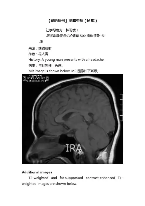
【双语病例】脑囊虫病(MRI)让学习成为一种习惯!医学影像服务中心拥有500病例征象+讲座来源:熊猫放射作者:花人青History: A young man presents with a headache.病史:年轻男性,头痛。
MR image is shown below. MR图像如下所示。
Additional imagesT2-weighted and fat-suppressed contrast-enhanced T1-weighted images are shown below.T2WI及T1WI压脂增强图像如下所示。
FindingsA cystic structure is seen in the splenium of the corpus callosum with a small internal nodule and smooth rim enhancement. There is an additional small cyst with internal signal consistent with fluid in the anterior horn of the left lateral ventricle.影像表现:胼胝体压部见一囊性结构,内见一小壁结节,增强扫描壁环形强化,表面光滑。
左侧侧脑室前角另见一小液性囊性灶。
Differential diagnosis•Neurocysticercosis•Abscesses•Metastatic disease鉴别诊断:•脑囊虫病•脑脓肿•转移瘤Diagnosis: Neurocysticercosis (NCC), colloidal vesicular stage诊断:脑囊虫病(NCC),胶样囊泡期Key pointsNeurocysticercosis (NCC)Neurocysticercosis is a parasitic infection of the central nervous system is caused by the pork tapeworm Taenia solium.Highest incidence is in Latin America.Depending on location of the infection, patients may present with headache, seizures, hydrocephalus, etc.Symptoms are caused by the immune reaction to NCC, not the lesions themselves.Typically occur in subarachnoid spaces, ventricles/cisterns, and at the gray/white matter interface.Variable-size cysts may be seen with or without rim enhancement and a small central nodule (scolex).Four stages of infection:•Vesicular: Viable larva; smooth-walled cyst with central scolex•Colloidal vesicular: Degenerating larva; rim-enhancing cyst and marked surrounding edema•Granular nodular: Healing stage; mild edema and rim enhancement•Nodular calcified: Healed stage; small, involuted cyst with or without calcificationsMetastatic disease is far more common and must always be considered in the differential.Large, complex cysts with edema may mimic a neoplasm.知识点脑囊虫病是猪肉绦虫所致的一种中枢神经系统寄生虫感染疾病。