
16层螺旋CTA在颅内血管性病变的诊断应用
- 格式:pdf
- 大小:333.10 KB
- 文档页数:2
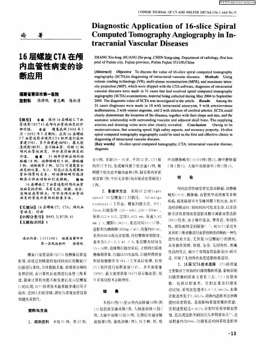

显堡趁堑皇佥△趑越堂呈QQ垒生筮!曼鲞箜3塑・147-图l左大脑后动脉发自左大脑中动脉图2右大脑中动脉水平段(M1)狭窄图3前交通支动脉瘤图4AVM:清楚显示供血动脉及引流静脉3);AVM1例(图4)供血血管及引流血管显示清楚明确。
本组病例显示前交通支29例(100%),后交通支26例34支(58.62%),小脑上动脉29例54支血管(93.1%),小脑下前动脉20例36支血管(62.07%),小脑下动脉26例46支血管(79.31%)。
讨论当临床要了解脑血管的情况,传统方法是插管脑血管造影,其检查结果也作为其他检查的金标准,介入放射学的发展及先进仪器设备的作用,脑血管造影进一步简便化,DSA对血管显示能力突显其在该领域的优越性;但是其创伤性的检查以及该检查诱发再出血的可能性,均限制了其在急性脑血管意外的临床应用[II。
此外,由于不能早期获得脑出血的量和病因,往往给I阻床选择治疗方案带来困难,延误治疗,增加了死亡率和后遗症发生率。
CTA检查前一般先作CT平扫,可早期鉴别脑血管意外的类型(梗死或出血),比较准确i贝4定脑出血的量,很大程度改善了传统DSA的不足。
据研究121与DSA相比,CTA有以下优点:①仅做静脉穿刺,创伤小,避免了DSA麻醉和插管所引起的并发症。
②可任意角度动态观察颅内血管,更加客观和细致地显示血管的解剖结构,而常规DSA只能l一2个角度进行投照。
③可同时显示颈内动脉系、椎动脉系和Willis环血管全貌,而DSA每次只能显示一条大血管及其属支。
④时间短费用少。
⑤受患者病情因素限制少,急性脑出血或蛛网膜出血患者,属危重症,当临床怀疑动脉瘤或AVM可能为出血诱因时,DSA检查受限,CTA可作为早期检查可靠方法。
1.与普通螺旋CT相比,16排螺旋CT有明显优越性:(1)扫描时间明显缩短,普通螺旋CTA扫描时间30—40S,16排旋CT扫描时问9~10S。
(2)成功率高,由于扫描时间短,在造影剂血管充盈期间可进行重复扫描,本组2例,由于年龄较大(8l岁和85岁),血液循环较慢,第1次扫描动脉充盈欠佳,接着作第2次扫描,血管充盈满意,总时间24S。
16层螺旋CT脑血管成像的临床应用价值[摘要] 目的探讨16层螺旋ct脑血管成像的临床应用价值。
方法选择我院2009年10月~2012年10月收治的脑血管病变患者70例,采用16层螺旋ct进行诊断,就临床资料进行回顾性分析。
结果肝血管主干及其主要分支采用mscta均可显示,大脑前、中动脉及其5级分支可清晰显示。
29例动脉狭窄的患者,动脉粥样硬化斑块及狭窄部位cta可清晰显示。
4例动脉闭塞患者中断部位、范围、形态cta或清楚显示。
脑动脉瘤18例患者其形态、大小、位置、载瘤动脉及瘤颈关系cta均可清晰显示。
23例动静脉畸形的患者引流静脉、供血动脉、畸形血管团cta可清晰显示。
静脉畸形cta显示静脉呈海蛇头样畸形。
颈动脉海绵窦患者cta显示患侧眼上静脉扩张,海绵窦扩大。
动静脉畸形及动脉瘤后均经手术证实,无其他异常情况发生。
结论临床颅内血管性病变检查中,多层螺旋ct脑血管造影成像技术具有无创、定位明确、定性、快速、方便的特点,明显提高了确诊率,为临床治疗提供了强有力的保障。
[关键词] 16层螺旋ct;脑血管成像;临床应用价值[中图分类号] r445 [文献标识码] b [文章编号] 2095-0616(2013)14-101-0216-slice spiral ct of cerebral vascular imaging and its clinical application valuetan yingjiect room, lufeng people’s hospital in guangdong province,lufeng 516500,china[abstract] objective to investigate the effect of 16 slice spiral ct cerebral imaging and its clinical application value. methods 70 cases of patients with cerebral vascular disease in our hospital from 2009 october to 2012 october were,by 16 slice spiral ct diagnosis,clinical data were retrospectively analyzed. results the liver vascular trunk and main branches of the mscta can be displayed,the anterior cerebral artery,the 5 branches can clearly display.29 cases of artery stenosis in patients with atherosclerotic plaque and stenosis,cta can clearly display.4cases of patients with artery occlusion interrupt site and range,morphology of cta or clear.cerebral aneurysm in 18 cases of patients with its shape,size,position, the parent artery and aneurysm neck can clearly show the relationship between cta.23 cases of patient with arteriovenous malformation of the draining vein,artery, nidus cta can clearly display. venous cta display the left parietal vein in caput deformity.cta patients with carotid cavernous sinus showed dilated vein ipsilateral eye,enlargement of cavernous sinus.avm and aneurysm were confirmed by operation,no other abnormal situations. conclusion the clinical intracranial vascular lesions,multislice spiral ct angiography imaging is non-invasive,clear positioning,qualitative,fast,convenient, can significantly improve the diagnosis rate,has provided the powerful safeguard for the clinical treatment of.[key words] 16 slice spiral ct;angiography;clinical application近年来,社会经济的飞速发展带动了医疗科技的进步,颅内血管性病变诊治中多层螺旋ct血管造影(mscta)的应用使效果得到了明显改善,特别是16层螺旋ct,具有高时间和空间图像分辨力、扫描层厚薄、扫描速度快的特点,可合理设计扫描参数,对扫描延迟时间可个性化选择,具有较为重要的临床价值[1]。

16排螺旋CT血管造影在脑血管病变诊治中的临床应用邓晓彬【期刊名称】《河北医学》【年(卷),期】2012(018)001【摘要】Objective: To explore the clinical value of 16-slice spiral CT angiography for cerebral vascular disease. Method: Retrospectively analyzed the clinical datas of 52 patients with cerebral vascular disease who were examined by 16-slice spiral CT in our hospital, and evaluated the image of cerebral vascular by volume rendering (VR) , maximum intensity projection (MIP) reconstruction and multi-planar reconstruction (MPR) technique. Result: 49 cases with intracranial aneurysms, 3 cases with brain arteriovenous malformation (AVM) in 52 cases cerebral vascular disease.52 cases of 16-slice CTA of VR, MIP and MPR reconstruction imaged to the trunk and main branches of cerebral blood vessels show 100% , and clearly showed the aneurysm's location, shape, size, and with the surrounding blood vessels and the relationship between the skull, also clearly showed the AVM's abnormal vascular group, feeding arteries and draining veins. Conclusion: 16 slice spiral CT angiography is a non-invasive imaging technique of intracranial vascular disease screening method. It has non-invasive, fast, safe, high accuracy characteristics, and can be used as the preferred and effectived screening method for the diagnosis of cerebral vascular disease.%目的:探讨16螺旋CT脑血管造影在脑血管病变诊治中的临床应用价值.方法:对我院收治的52例脑血管病变患者的16排CTA检查资料进行回顾性分析,并以容积重建(VR)、最大密度投影(MIP)重建和多平面重建(MPR)技术对脑血管影像表现进行评价分析.结果:52例脑血管病变中诊断出颅内动脉瘤49例,脑内动静脉血管畸形(AVM)3例.52例16排CTA的VR、MIP和MPR重建图像对脑血管主干及其主要分支显示100%,并且清晰显示动脉瘤的位置、形态、大小、以及与周围血管和颅骨的关系,也清晰显示了AVM的畸形血管团、供血动脉和引流静脉.结论:16排螺旋CT脑血管造影成像技术是一种无创伤的颅内血管性病变检查方法.它具有无创、快捷、安全、准确性高的特点,可作为脑血管病变首选而有效的检查方法.【总页数】4页(P61-64)【作者】邓晓彬【作者单位】重庆市渝北区中医院放射科, 重庆 401120【正文语种】中文【相关文献】1.16排螺旋CT血管造影在脑血管病变诊断中的临床应用价值 [J], 李鹏2.16排螺旋CT血管造影在脑血管病变诊断中的临床应用价值 [J], 李鹏;3.16层螺旋CT血管造影在诊断脑血管病变中的临床应用价值 [J], 刘永辉;蔡云;夏广明;梁家宁4.16层螺旋CT血管造影在诊断脑血管病变中的临床应用价值 [J], 刘永辉;蔡云;夏广明;梁家宁5.16排螺旋CT血管造影对脑血管病变的临床应用价值 [J], 李雯;钟毅;邓晓峰因版权原因,仅展示原文概要,查看原文内容请购买。