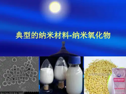
二氧化钛ppt课件
- 格式:ppt
- 大小:1.11 MB
- 文档页数:28

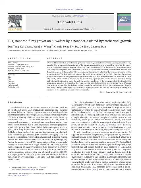
TiO 2nanorod films grown on Si wafers by a nanodot-assisted hydrothermal growthDan Tang,Kui Cheng,Wenjian Weng ⁎,Chenlu Song,Piyi Du,Ge Shen,Gaorong HanDepartment of Materials Science and Engineering,State Key Laboratory of Si Materials,Zhejiang University,Hangzhou 31027,Chinaa b s t r a c ta r t i c l e i n f o Article history:Received 12September 2010Received in revised form 6May 2011Accepted 10May 2011Available online 18May 2011Keywords:TiO 2Film Nanorod NanodotsHydrothermal Assisted growthWe report the controlled hydrothermal growth of rutile TiO 2nanorods on Si wafers by using an anatase TiO 2nanodot film as an assisted growth layer.The anatase nanodot film was prepared on the wafer by phase-separation-induced self-assembly and subsequent heat-treatment at 500°C.The nanodots on the wafer were then subjected to hydrothermal treatment to induce the growth of rutile TiO 2nanorod films.The size and dispersion density of the resulting TiO 2nanorods could be varied by adjusting the Ti ion concentration in the growth solution.The TiO 2nanorods were of the rutile phase and grew in the [001]direction.The growth mechanism reveals that the growth of the rutile nanorods was wholly dependent on the existence of rutile TiO 2seeds,which could be formed by the dissolution –reprecipitation of the anatase nanodots during hydrothermal treatment or under the high-temperature conditions of the subsequent heat-treatment of the as-prepared nanodots.In controlling the rutile nanorod growth,the anatase nanodots show more ef ficiency than a dense anatase film.Preliminary evaluations of the rutile nanorod films have demonstrated that the wettability changed from highly hydrophobic to superhydrophilic and that the photocatalytic activity was enhanced with increasing nanorod dispersion density.©2011Elsevier B.V.All rights reserved.1.IntroductionTitania (TiO 2)is attractive for use in various applications by virtue of its photochemical and photovoltaic properties and chemical inertness.TiO 2exists in three crystal structures,of which rutile has advantages over the other two phases (anatase and brookite)in terms of chemical stability,dielectric constant,and ultraviolet (UV)ray absorption rate [1–3].Nanostructured forms of TiO 2,such as nanoparticles,nanospheres,nanorods,and nanosheets,have received considerable attention due to their physical and chemical properties,which differ greatly from those of their bulk counterparts.In recent years,increasing applications of nanostructured TiO 2in different fields have been explored,for example in photocatalysts,photovol-taics,cosmetics,gas sensors,gate insulator antifogging and self-cleaning coatings for devices,biological areas such as protein folding,micelle and membrane formation,and molecular recognition [4–10].Among the various types of TiO 2nanostructures,there is a particular interest in one-dimensional single-crystalline TiO 2nanostructures such as nanorod or nanowire arrays or films because of their lower recombination rate for excited electron –hole pairs,large interface for charge separation,unique optical and electrical properties [11–13],and larger spatial area for the grafting and assembling of functional molecules in dye-sensitized solar cells and biological surface modi fications.Since the applications of one-dimensional single-crystalline TiO 2nanostructures are strongly dependent on their shapes,size,density,and crystallinity,it is of great signi ficance to explore synthetic techniques by which the nanostructural characteristics can be well de fined and controlled [14].To date,many groups have investigated different paths for the preparation of rutile TiO 2nanorod arrays,for example through the sol –gel template method,hydrothermal treatment,liquid-phase deposition template routes,gas-phase methods,combustion synthesis,metal-organic chemical vapor depo-sition,or anodic oxidation [1,2,15,16].Among these methods,considerable attention has been paid to the hydrothermal method because of its convenience,versatility,high productivity,and low cost.In order to achieve growth of nanorods on substrates such as Si,sapphire,polyethylene terephthalate,and polystyrene by hydrother-mal synthesis,an assisted growth layer is essential.Such a layer on a substrate serves not only to facilitate nucleation but also to strengthen bonding between the substrate and the nanorods [17].Vertical ZnO nanowire arrays on substrates have been grown through the use of textured ZnO and indium-doped tin oxide seeds [18–21],and rutile TiO 2nanorod and nanowire arrays have been grown on transparent conductive film coated glass substrates such as fluorine-doped tin oxide and indium tin oxide films [22–25].Since the assisted growth layers have been proven to play an indispensable role in the formation of nanorods,the in fluence of the nanostructures of these layers on the growth should be of great value to explore in order to achieve better control of the process.Si wafers are widely used in various optical-electronic devices,and TiO 2nanorods have been applied in photo-sensors/photocatalysts [6–10].Hence,TiO 2nanorods on Si wafersThin Solid Films 519(2011)7644–7649⁎Corresponding author.Tel./fax:+8657187953787.E-mail address:wengwj@ (W.Weng).0040-6090/$–see front matter ©2011Elsevier B.V.All rights reserved.doi:10.1016/j.tsf.2011.05.011Contents lists available at ScienceDirectThin Solid Filmsj o u r n a l h o m e p a g e :w w w.e l s ev i e r.c o m /l o c a t e /ts fcould provide a basis for creating integrated optical-electronic devices.In this work,phase-separation-induced self-assembly[26]was adopted to pre-prepare an anatase TiO2nanodotfilm as an assisted growth layer on a Si wafer.A rutile nanorodfilm was grown on the wafer by an induced function of the nanodot layer under hydrother-mal treatment.The role of the anatase TiO2nanodotfilm as an assisted growth layer was investigated,and the wettabilities and photocata-lytic performances of the grown TiO2nanorods with different densities were measured.A growth mechanism for the TiO2nanorods is proposed.2.Experimental2.1.MaterialsAll chemicals were of analytical reagent grade and were used without further purification.All aqueous solutions were prepared using deionized water.2.2.Preparation of the assisted growth layer on Si wafersFor the phase-separation-induced self-assembly to prepare an anatase TiO2nanodotfilm on a Si wafer,an ethanol solution with a molar ratio of acetylacetone(AcAc)/tetrabutyl titanate(TBOT)/H2O of 0.3:1:1,containing40mg/L polyvinyl pyrrolidone(PVP),was applied to the wafer by spin-coating at8000rpm for40s[26].The spin-coated wafer was then heated at500°C for2h in a muffle furnace.The resulting nanodotfilms were used as assisted growth layers.Through the above procedure,thicker TiO2nanodotfilms for X-ray diffraction(XRD)measurement were prepared by repeating the spin-coating process six times.For comparison,a dense anatase TiO2film was prepared by using a spinning solution without PVP.Also,to verify the growth mechanism,a TiO2nanodotfilm with rutile phase was prepared by heating at800°C.2.3.Hydrothermal growth of TiO2nanorodsTypically,a certain amount of concentrated hydrochloric acid (36.5–38%by weight)was mixed with30mL of deionized water and the requisite amount of TBOT to obtain a total volume of60mL.In this study,the amount of hydrochloric acid was varied by tuning the TBOT concentration from0.02mol/L to0.05mol/L.The mixture of water and hydrochloric acid was stirred under ambient conditions for5min before the addition of the TBOT.To obtain a clear solution,the whole mixture had to be stirred under ambient conditions for a further period of time.Then,the solution was transferred to a50mL Teflon-lined stainless steel autoclave,and a piece of TiO2-coated wafer was placed at an angle against the wall of the Teflon liner with the coated side facing down.The hydrothermal growth was conducted at160°C for2h in an electric oven.After the growth hadfinished,the autoclave was cooled to room temperature in ambient air,and then the wafer was taken out,rinsed extensively with deionized water and ethanol, and allowed to dry in ambient air.An ultrasonically cleaned,uncoated Si wafer was used as a control to study the effect of the assisted growth layer.2.4.Characterizations and measurementsThe crystal structures of the nanodotfilm,the densefilm and the nanorodfilms were examined by XRD.The XRD patterns were recorded using an X'Pert PRO PANalytical diffractometer using Cu-K R radiation(λ=1.5406Å),scanning from15°to80°at a rate of 2°min−1.The X-ray tube voltage and current were set at40kV and 40mA,respectively.Morphological and lattice structural information was obtained byfield-emission scanning electron microscopy (FESEM,HITACHI,S-4800)with operating voltage of5.0kV,trans-mission electron microscopy(TEM/HRTEM,TECNAI,F-30)with accelerating voltage of300kV,and selected-area electron diffraction (SAED)analysis.To prepare specimens for TEM observation,a piece of wafer with grown nanorods was added to5mL of ethanol in a small glass vial and sonicated for30min.A few drops of the sonicated suspension were then applied to a carbon-coated200mesh copper grid and dried under ambient conditions before imaging.The dispersion density of the nanorods was obtained by counting the amount of nanorods in a given area(25μm2).To ensure the accuracy of the dispersion density,the average value was taken after five different areas on the substrate had been counted.Wettability was measured by means of an optical contact angle meter(DATAPHYSISE,OCA20).The photocatalytic performance of the TiO2nanorods was determined by the degradation of Rhodamine dye solution.Three wafers(1cm×1cm)bearing nanorods with different densities were immersed in5mL aliquots of aqueous Rhodamine B dye solution with a concentration of10−5mol/L in glass vessels.Prior to irradiation,the vessels were placed in the dark for60min to obtain saturation adsorption of Rhodamine B onto the catalysts.The vessels were then placed in a Solar Box ready for UV-irradiation to induce the photochemical reaction[27,28].The nanorod-bearing wafer/dye solution samples were irradiated in the horizontal direction for 60min,and the distance between the UV lamp(150W xenon arc-lamp with a420nm cut-offfilter)and the sample was kept within 15cm.The change of Rhodamine B concentration was measured by means of a double-beam UV/Vis spectrophotometer(TU-1901).3.Results and discussion3.1.The role of the nanodot layerDuring hydrothermal treatment,the TiO2nanorod growth completely depends on whether an assisted growth layer is present on the wafer.Fig.1shows typical FESEM images of hydrothermally treated wafers without any layer(Fig.1a),with a TiO2nanodotfilm (Fig.1b),and with a TiO2densefilm,respectively.The images reveal that no growth occurred on the bare wafer,dense tetragonal nanorods with a dispersion density of about83μm−2grew on the nanodotfilm, and that the same nanorods grew on the densefilm but with a lower dispersion density of9μm−2.The nanodotfilm thus displayed a stronger assisting role for nanorod growth.3.2.Effect of initial Ti concentrationFig.2shows that the density and size of the nanorods could be varied by changing the TBOT concentration in the growth solution. The density of the nanorods increased significantly from almost0to 83μm−2for the wafers with the nanodotfilm(Fig.2a,b,c,d)and to 10μm−2for the wafers with the densefilm(Fig.2a1,b1,c1,d1)when the TBOT concentration in the growth solution was increased from 0.02to0.05mol/L.Accordingly,the diameter of the nanorods increased from~35nm to~120nm.In addition,the surface of the nanodots(~100nm)became rough(inset in Fig.2a)andfine particles of average diameter~27nm from the deposition process appeared on the wafer(inset in Fig.2b).3.3.Microstructural characteristics of the grown TiO2nanorodsFig.3displays the XRD patterns of the coated wafers before and after hydrothermal growth.The nanodots were originally of anatase phase (Fig.3a);however,the diffraction peaks of the grown nanorods changed to match those of the rutile phase(SG,P42/mnm;JCPDS No.65–0191, a=b=0.4517nm and c=0.2940nm)(Fig.3b).Compared to the standard powder diffraction pattern,the remarkably enhanced(101)7645D.Tang et al./Thin Solid Films519(2011)7644–7649and (002)peaks in Fig.3b indicate that the grown nanorods are oriented against the wafer surface.The intensi fication of the (002)diffraction peak means that the TiO 2nanorods grew in the [001]direction,resulting in an increase in the (101)diffraction peak due to a contact angle between the (101)and (002)planes of approximately 33°[29].The microstructure of the TiO 2nanorods was further characterized by TEM and HRTEM.In some previous studies,TiO 2nanorods have been grown by hydrothermal treatment in the [110],[101],and other directions through the use of seed layers [30–33].In this work,the HRTEM image (Fig.4a)shows that the nanorod was single-crystalline and that the lattice fringes had interplanar spacings of 0.327nm and0.294nm,corresponding to the interplanar distances of the (110)and (001)planes of rutile TiO 2.The [110]axis is perpendicular to the nanorod side walls,indicating that the nanorod grew along the [001]direction,consistent with the XRD data.The sharp SAED pattern of a nanorod examined along the [110]zone axis (Fig.4b)is also indicative of good crystallization.3.4.Growth mechanism of the rutile nanorodsIn this work,anatase TiO 2nanodots were deployed to play an assisting role to grow TiO 2nanorods.However,the resulting TiO2Fig.1.The in fluence of different wafers on the nanorod growth under hydrothermal treatment in solutions containing 0.05mol/L TBOT:(a)uncoated Si wafer;(b)a wafer coated with a TiO 2nanodot film;(c)a wafer coated with a dense TiO 2film;(a1)hydrothermally treated (a);(b1)hydrothermally treated (b);(c1)hydrothermally treated(c).Fig.2.The in fluence of TBOT concentration in the solution on the nanorod growth:(a,a ′)0.02mol/L;(b,b ′)0.03mol/L;(c,c ′)0.04mol/L;(d,d ′)0.05mol/L;(a –d)wafers coated with TiO 2nanodot film;(a1–d1)wafers coated with dense TiO 2film.7646 D.Tang et al./Thin Solid Films 519(2011)7644–7649nanorods after hydrothermal treatment were of the rutile phase.The growth mechanism of the TiO 2nanorods is suggested to proceed as follows.Considering the structures,both anatase and rutile phases employ [TiO 6]octahedra as fundamental structural units,but they have different connections.Edge-shared bonding of this unit leads to anatase,while corner-shared bonding results in rutile.In solutions highly acidi fied with HCl,rutile preferentially nucleates due to the growing unit of Ti(IV)oxo species with a small number of OH ligands,which suppresses edge-shared bonding and enhances vertex-shared bonding [26].In addition,anatase TiO 2in highly acidic solution tends to be unstable because its surface is easily protonated and begins to dissolve [34].In this work,when the anatase nanodots on the wafer were immersed in a hydrothermal solution with high acidity,they were partially dissolved.Consequently,the Ti concentration around the nanodots increased,leading to enhanced supersaturation to yield rutile in the hydrothermal solution.Finally,rutile nanoparticles were deposited around the nanodots when a certain amount of the nanodots had been dissolved.This dissolution –reprecipitation process was con firmed by SEM and TEM images,as shown in Fig.5.TheFig.3.XRD patterns of wafers coated with the assisted growth layer:(a)before hydrothermal treatment;(b)after hydrothermal treatment in the growth solution with 0.05mol/LTBOT.Fig.4.HRTEM image (a)and SAED pattern (b)of a TiO 2nanorod grown in a solution containing 0.04mol/LTBOT.Fig.5.Microstructures of the deposited nanoparticles and rough nanodots in a nanorod film grown in a solution containing 0.04mol/L TBOT:(a)cross-sectional view of the film;(b)TEM image of nanoparticles,nanodots,and nanorods from the film;(c)TEM image and SAED pattern of the rough nanodots;(d)HRTEM image of thenanoparticle.Fig.6.Schematic illustration of the hydrothermal growth process of the rutile nanorods originated from the anatase nanodot film.7647D.Tang et al./Thin Solid Films 519(2011)7644–7649smaller nanoparticle of size ~35nm showed lattice fringes (Fig.5d)that matched those of the rutile phase.This particle is believed to be a deposited nanoparticle because its size is as small as the fine ones in the SEM micrograph (Fig.2b).The SAED pattern of the larger particle of diameter ~150nm shows that it consists of rutile and anatase phases.This particle is considered to originate from a nanodot.The former phase comes from the deposition of rutile nanoparticles on the surface of the nanodot,while the latter phase results from the undissolved part.All of the deposited rutile nanoparticles on the surface of the nanodots and wafer can serve as seeds for rutile growth.The orientation attachment mechanism [35]indicates that whether or not a particle will play a seeding role strongly depends on the spontaneous self-organization of the fine grains in the particle to expose a common crystallographic orientation.Here,it is suggested that only the deposited rutile nanoparticles with [001]crystallo-graphic orientation organization are capable of seeding the growth of nanorods.Therefore,higher Ti concentration in the hydrothermal solution results in the deposition of more and larger nanoparticles.As a result,both the number and size of the nanoparticles increase,and the resulting nanorods have high density and large diameter,as shown in Fig.2.Fig.6schematically illustrates the growth mechanism of the rutile nanorods on the wafer with an anatase nanodot film as an assisted growth layer.When a dense anatase film was used as the assisted growth layer,the hydrothermal growth of TiO 2nanorods was due to the sameformation process.Since the dense film has a much smaller speci fic surface area than the nanodot film,the assisting activity should be much weaker than that with the latter,resulting in the appearance of only a few nanorods (Figs.1and 2).An additional experiment was carried out in order to verify the above-proposed growth mechanism.As-prepared nanodots on the wafer were heated at 800°C rather than 500°C,and the resulting nanodots were shown to consist of rutile besides anatase (Fig.7a).Under hydrothermal treatment (with 0.02mol/L TBOT),the nanodots containing rutile clearly induced nanorod growth (Fig.7-b2)in comparison with the anatase nanodots (Fig.7-b1).Hence,the growth of rutile nanorods completely depends on the existence of rutile seeds rather than anatase nanodots on the wafers because of the large lattice mismatch between anatase (a =b =0.3785nm)and rutile (a =b =0.4594nm)of 17.6%.This conclusion is in good agreement with the fact that rutile TiO 2nanorods could be grown on fluorine-doped tin oxide (a =b =0.4687nm)with 2%lattice mismatch [23].3.5.Wettability of the TiO 2nanorodsThe wettability of a solid surface depends on both the chemical composition and the geometrical structure [19].The wettability of the nanorods grown on the wafers depends on their dispersion density.Fig.8indicates the wettabilities of nanorods with densities between 9μm −2and 83μm −2.The water contact angle decreases from 119±2.0°to about 0°with increasing density of the nanorods.AtransitionFig.7.The in fluence of heat-treatment temperature of wafers with a TiO 2nanodot film on nanorod growth under hydrothermal conditions in a solution containing 0.02mol/L TBOT:(a)XRD patterns of two nanodot-coated wafers heated at 500°C and 800°C;(b)hydrothermally treated wafer heated at 500°C;(c)hydrothermally treated wafer heated at 800°C.Fig.8.The in fluence of different densities of TiO 2nanorod films on the water contact angle.7648 D.Tang et al./Thin Solid Films 519(2011)7644–7649from high hydrophobicity to superhydrophilicity could be achieved by controlling the density of the TiO 2nanorods.3.6.Photocatalytic decomposition of Rhodamine B on TiO 2nanorods The photocatalytic activity of TiO 2thin films is often coupled with their switchable wettability.In the present work,it was indeed found that an increase in nanorod density led to more extensive degradation of Rhodamine B dye solution (Fig.9).This could be attributed to the larger speci fic surface area for a higher nanorod density.4.ConclusionsAn anatase TiO 2nanodot film on a Si wafer has been demonstrated to show a good assisting ability in inducing the hydrothermal growth of nanorods,and represents a good alternative for tailoring the nanorod density and diameter.The resulting TiO 2nanorods are of the rutile phase and grow in the [001]direction.An anatase nanodot film showed greater growth-assisting ef ficiency than a dense anatase film.Here,the growth of rutile nanorods originates from rutile TiO 2seeds formed by the dissolution –reprecipitation of the anatase nanodots during hydro-thermal treatment.The rutile nanorod films displayed tunable wetta-bility from highly hydrophobic to superhydrophilic,and enhanced photocatalytic activity with increasing nanorod dispersion density.AcknowledgmentThis work has been financially supported by the National Natural Science Foundation (51072178,30870627and 50872122),the Research Fund of the Doctoral Program of Higher Education of China (200703351015),and the Research Fund of Shenzhen Key Laboratory of Functional Polymers.References[1] E.Bae,N.Murakami,T.Ohno,J.Mol.Catal.A:Chem.300(2009)72.[2] E.Bae,T.Ohno,Appl.Catal.,B:Environ.91(2009)634.[3]X.P.Huang,C.X.Pan,J.Cryst.Growth 306(2007)117.[4]W.X.Hou,Q.H.Wang,Langmuir 25(2009)6875.[5]T.X.T.Sayle,D.C.Sayle,ACS Nano 4(2010)879.[6]L.Jiang,Y.J.Zhong,G.C.Li,Mater.Res.Bull.44(2009)999.[7]K.L.Ding,Z.J.Miao,B.J.Hu,G.M.An,Z.Y.Sun,B.X.Han,Z.M.Liu,Langmuir 26(2010)10294.[8] F.Dong,W.R.Zhao,Z.B.Wu,Nanotechnology 19(2008).[9]S.Kim,S.H.Ehrman,Langmuir 23(2007)2497.[10]J.P.Wei,J.F.Yao,X.Y.Zhang,W.Zhu,H.Wang,M.J.Rhodes,Mater.Lett.61(2007)4610.[11]W.Y.Wu,Y.M.Chang,J.M.Ting,Cryst.Growth Des.10(2010)1646.[12]Y.X.Li,M.Guo,M.Zhang,X.D.Wang,Mater.Res.Bull.44(2009)1232.[13]Q.S.Wei,K.Hirota,K.Tajima,K.Hashimoto,Chem.Mater.18(2006)5080.[14]S.W.Yang,L.Gao,Mater.Chem.Phys.99(2006)437.[15]M.N.Tahir,P.Theato,P.Oberle,G.Melnyk,S.Faiss,U.Kolb,A.Janshoff,M.Stepputat,W.Tremel,Langmuir 22(2006)5209.[16]H.B.Li,X.C.Duan,G.C.Liu,X.B.Jia,X.Q.Liu,Mater.Lett.62(2008)4035.[17]J.Song,S.Lim,J.Phys.Chem.C 111(2007)596.[18]L.E.Greene,w,D.H.Tan,M.Montano,J.Goldberger,G.Somorjai,P.D.Yang,Nano Lett.5(2005)1231.[19]H.K.Sun,M.Luo,W.J.Weng,K.Cheng,P.Y.Du,G.Shen,G.R.Han,Nanotechnology 19(2008)125603.[20]H.K.Sun,M.Luo,W.J.Weng,K.Cheng,P.Du,G.Shen,G.R.Han,Nanotechnology 19(2008)395602.[21] C.K.Xu,P.H.Shin,L.L.Cao,J.M.Wu,D.Gao,Chem.Mater.22(2010)143.[22] E.Vigil,J.A.Ayllon,A.M.Peiro,R.Rodriguez-Clemente,X.Domenech,J.Peral,Langmuir 17(2001)891.[23] B.Liu,E.S.Aydil,J.Am.Chem.Soc.131(2009)3985.[24]X.J.Feng,K.Shankar,O.K.Varghese,M.Paulose,tempa,C.A.Grimes,Nano Lett.8(2008)3781.[25]J.C.Lee,T.G.Kim,W.Lee,S.H.Han,Y.M.Sung,Cryst.Growth Des.9(2009)4519.[26]M.Luo,K.Cheng,W.Weng,C.Song,P.Du,G.Shen,G.Xu,G.Han,Nanotechnology 20(2009)215605.[27] F.Sayilkan,M.Asiltürk,Mater.Res.Bull.43(2008)134.[28]K.Xu,G.Zhu,Appl.Surf.Sci.13(2009)255.[29] E.Hosono,S.Fujihara,K.Kakiuchi,H.Imai,J.Am.Chem.Soc.126(2004)7790.[30] C.W.Zou,X.D.Yan,Chem.Phys.Lett.84(2009)476.[31]Thuy-Duong Nguyen Phan,J.Cryst.Growth 79(2009)312.[32] B.Xue,R.Liu,Mater.Lett.63(2009)4723.[33]P.A.Morris,A.Roshko,J.Cryst.Growth 174(1997)433.[34]K.Yanagisawa,J.Ovenstone,J.Phys.Chem.B 103(1999)7781.[35]R.L.Penn,J.F.Ban field,Science 281(5379)(1998)969.Fig.9.The in fluence of different densities of TiO 2nanorod films on the photodegrada-tion of Rhodamine B under UV light irradiation.7649D.Tang et al./Thin Solid Films 519(2011)7644–7649。

