蛋白翻译后修饰组学实验方法
- 格式:pdf
- 大小:2.23 MB
- 文档页数:4
百泰派克生物科技
蛋白质药物的翻译后修饰
随着生物医药技术的发展,越来越多的蛋白质类药物被开发出来用于各种疾病的治疗,常见的蛋白质类药物如蛋白质、多肽、单克隆抗体、疫苗和抗体偶联药物等。
这些蛋白质类药物有时需要进行一些修饰才能发挥预想的生物学功能,达到治疗效果。
常见的蛋白质药物翻译后修饰包括糖基化、二硫键、乙酰化和磷酸化等,不同的修饰类型、氨基酸修饰位点以及修饰的含量都会严重影响药物终产品的安全性和疗效性。
研究表明,糖基化程度的高低与促红细胞生成素(一种蛋白类药物)的活性和半衰期密切相关,单克隆抗体的糖链类型影响其与受体的亲和力。
因此,对蛋白质药物的翻译后修饰的分析与鉴定是必不可少的。
目前主要依靠液相色谱-串联质谱技术对蛋白质药物进行翻译后修饰鉴定,其原理和分析流程与常规的蛋白质翻译后修饰鉴定类似。
百泰派克生物科技采用Thermo Fisher的Q ExactiveHF质谱平台结合Nano-LC色谱,提供快速高效的蛋白质药物翻译后修饰鉴定服务技术包裹,您只需要将您的实验目的告诉我们并将您的样品寄给我们,我们会负责项目后续所有事宜,包括蛋白提取、蛋白酶切、修饰肽段富集、肽段分离、质谱分析、质谱原始数据分析、生物信息学分析,欢迎免费咨询。


蛋白质修饰的研究方法人类基因组包含23,000个基因,然而,这些基因产生了显著更大的蛋白质组。
这是由于在转录后和翻译后水平的多样性。
单个基因在转录后,可以通过可变剪接,偶尔通过RNA编辑,产生多个mRNA分子。
许多蛋白质在翻译后,通过蛋白质修饰和/或蛋白质剪切,产生成熟的和功能型的形态。
通过广泛的翻译后修饰,蛋白质的结构和功能就多样性了。
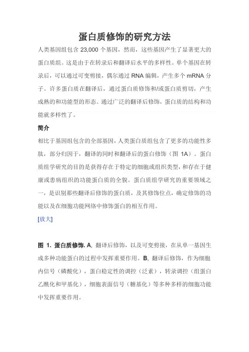
简介相比于基因组包含的全部基因,人类蛋白质组包含了更多的功能性多肽,部分归因于,翻译的同时和翻译后的蛋白修饰(图1A)。
蛋白质组学研究的目的是获得存在于特定的细胞或组织类型,和存在于健康或患病组织的功能蛋白质的全貌。
蛋白质组学研究的重要领域之一,是识别那些翻译后修饰的蛋白质,及其修饰位点,确定修饰的功能以及在细胞功能网络中修饰蛋白的相互作用。
[放大]图 1.蛋白质修饰.A, 翻译后修饰,以及可变剪接,在从单一基因生成多种功能蛋白的过程中发挥重要作用。
B, 翻译后修饰,作为细胞内信号(磷酸化),蛋白稳定性的调控(泛素),转录调控(组蛋白乙酰化和甲基化),细胞表面信号(糖基化)等多种多样的细胞功能中发挥重要作用。
在过去的几十年,开发了多种方法测定蛋白质修饰。
在这里,我们重点介绍识别蛋白质修饰的一般方法,质谱;识别磷酸化的特异性方法,磷酸盐标记;识别泛素化的方法;在染色质重塑过程中识别组蛋白乙酰化和甲基化的方法;识别蛋白质糖基化的方法(表1和图1B )。
详细的实验步骤以后将会整理归纳。
修饰 功能检测泛素化 蛋白质降解 •质谱• 泛素化检测糖基化 细胞外信号转导 • 质谱•高效液相色谱法 表格 1. 蛋白质修饰的类型,其主要的细胞功能,和主要的研究方法。
初步识别新的修饰位点的一个主要方法是计算机分析。
已经广泛应用计算机程序根据蛋白质的氨基酸序列识别假定修饰位点,这不是本文的讨论范围。
计算机程序的概述见Liu and Li, 2011 [1] 。
质谱在过去20年来,质谱分析已成为确定蛋白质修饰类型和位点的必不可少的工具。
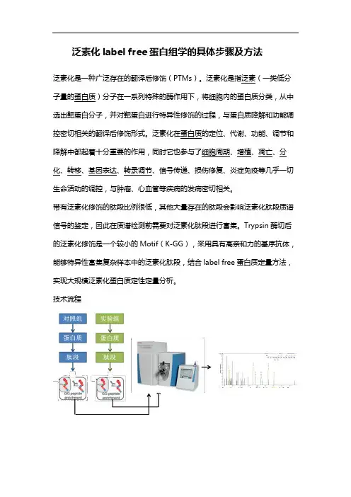
泛素化label free蛋白组学的具体步骤及方法
泛素化是一种广泛存在的翻译后修饰(PTMs)。
泛素化是指泛素(一类低分子量的蛋白质)分子在一系列特殊的酶作用下,将细胞内的蛋白质分类,从中选出靶蛋白分子,并对靶蛋白进行特异性修饰的过程,与蛋白质降解和功能调控密切相关的翻译后修饰形式。
泛素化在蛋白质的定位、代谢、功能、调节和降解中都起着十分重要的作用,同时它也参与了细胞周期、增殖、凋亡、分化、转移、基因表达、转录调节、信号传递、损伤修复、炎症免疫等几乎一切生命活动的调控,与肿瘤、心血管等疾病的发病密切相关。
带有泛素化修饰的肽段比例很低,其他大量存在的肽段会影响泛素化肽段质谱信号的鉴定,因此在质谱检测前需要对泛素化肽段进行富集。
Trypsin酶切后的泛素化修饰是一个较小的Motif(K-GG),采用具有高亲和力的基序抗体,能够特异性富集复杂样本中的泛素化肽段,结合label free蛋白质定量方法,实现大规模泛素化蛋白质定性定量分析。
技术流程。
蛋白质精氨酸三甲基化修饰的发现、组学及功能探索
蛋白质精氨酸三甲基化修饰是一种新型的蛋白质翻译后修饰方式,在这种修饰方式中,精氨酸残基上的氨基基团被三个甲基基团取代,形成了精氨酸的甲基化形式。
精氨酸三甲基化修饰最初被发现于植物RNA结合蛋白FCA中,后来又在脊椎动物中被发现。
近年来的研究表明,这种修饰在多种生物中普遍存在,包括哺乳动物、细菌、真菌和植物等。
这种修饰具有重要的生物学功能,如在调节基因转录、细胞周期、细胞分化等方面发挥了重要作用。
随着技术的发展,包括质谱、抗体、酵母双杂交等在内的多种技术已被开发用于研究精氨酸三甲基化修饰,并取得了重要的组学信息。
这些研究揭示了精氨酸三甲基化修饰的靶点蛋白、结构域和生物过程等信息。
由于精氨酸三甲基化修饰的重要作用,研究人员已经开始探索将其作为药物靶点的可能性。
此外,该修饰还具有作为癌症治疗的潜在价值,因为它与某些癌症的发生和发展有关。
总之,蛋白质精氨酸三甲基化修饰是一个新兴的研究领域,在这个领域中的进展为我们理解蛋白质修饰的多样性和生物学功能提供了新的视角。
项目名称:肝病发生发展中的蛋白质翻译后修饰及其调控的定量蛋白质组学研究首席科学家:徐平中国人民解放军军事医学科学院放射与辐射医学研究所起止年限:2011.1至2015.8依托部门:总后勤部卫生部二、预期目标1.总体目标本课题研究的总体目标是充分发挥我国在肝病模型、标本和肝脏蛋白质组研究方面的优势,利用定量蛋白质组学的原理和方法,对肝病发生发展过程中磷酸化、泛素化、糖基化蛋白质本身及其异质体的修饰位点、修饰类型、计量关系和动态变化规律以及控制机理进行系统研究。
考察这些翻译后修饰在肝病病因、预后和治疗中的价值。
通过本研究,我们将建立高效率、低成本的蛋白质磷酸化、泛素化、糖基化修饰和糖链结构解析的定性、定量研究技术;揭示肝病发生和癌变过程中磷酸化蛋白质信号通路的开关机制、泛素化调控的蛋白质量变引起质变的规律以及糖基化在肝癌发生发展和转移中的变化趋势,阐明这些翻译后修饰在肝病发生发展中的生理病理机制;筛选、鉴定一批具有诊断和药靶意义的肝病相关翻译后修饰蛋白或翻译后修饰调控蛋白。
本项目的开展将加速我国定量疾病蛋白质组学研究体系的形成,培养一批从事蛋白质翻译后修饰研究的专门人才,产生一批具有国际影响力的论文和成果,继续保持我国肝病蛋白质组学研究基地在国际上的竞争优势。
2.五年目标1)发展一套高通量、高准确度和高精度的磷酸化、泛素化和糖基化蛋白质组的定性、定量研究技术及平台。
2)系统比较在肝细胞癌发生发展过程中,HBx侵染宿主细胞后引起宿主细胞蛋白质磷酸化修饰进而导致HCC的分子机制,和肝细胞极性蛋白磷酸化修饰及其调控的下游蛋白磷酸化在肝细胞癌发生发展中的作用机制。
阐明该过程信号途径和调控网络;发现肝病发展过程中磷酸化控制的关键蛋白;探索这些验证的关键蛋白作为药靶和肝病标志物的可能性。
3)建立特定泛素链形成和脱泛素化的酶学决定机制和关系网络;筛选以Smurf1、NEDL1/2和SCF为代表的特异性泛素化蛋白质;构建2个E3基因敲除小鼠模型,鉴定8-10个新的E3调控蛋白,揭示2-3类新的E3活性调控机制;获得2-3个在肝癌发生发展过程中发生显著变化的泛素连接酶,提供1个候选肝癌标志物。

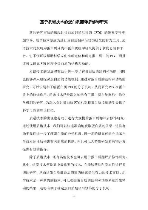
翻译后修饰蛋白质组与代谢组整合分析蛋白质的翻译后修饰(Post Translational Modifications, PTMs)是蛋白质在翻译中或翻译后经历的一个共价加工过程。
翻译后修饰蛋白质组是指细胞或组织等整体水平上的翻译后修饰蛋白质。
目前,已知的蛋白质翻译后修饰主要包括糖基化、磷酸化、酰化、泛素化、二硫键配对、甲基化和亚硝基化等等。
代谢组是细胞、组织或生物体内的小分子(通常称为代谢物)的整体水平。
翻译后修饰蛋白质可以调节细胞生物过程、影响机体的代谢变化。
影响代谢的翻译后修饰蛋白质不仅包括翻译后修饰转录因子,还包括翻译后修饰代谢酶。
因此,整合分析翻译后修饰蛋白质组和代谢组,比较它们的表达异同,有利于从不同层面解析生物的代谢机制,挖掘差异修饰蛋白质、代谢物、及它们参与的重要通路和相关基因,以进行后续深入研究。
百泰派克生物科技采用Thermo Fisher的Orbitrap Fusion Lumos质谱平台结合nanoLC-MS/MS纳升色谱,将磷酸化/糖基化/泛素化/乙酰化/甲基化/二硫键/亚硝基化等翻译后修饰鉴定服务,多种样品靶向和非靶向代谢组学分析服务,结合可定制化的生物信息学分析方法进行整合,为广大科研工作者提供基于质谱的翻译后修饰蛋白质组与代谢组整合分析服务。
翻译后修饰蛋白质组与代谢组整合分析流程翻译后修饰蛋白质组与代谢组整合分析流程。
应用领域农林领域:抗逆胁迫机制,物种保护研究等;畜牧业:致病机理研究,肉类及乳制品品质研究等;海洋水产:渔业环境与水产品安全等;微生物:致病机理,耐药机制,病原体-宿主相互作用研究等;生物医药:生物标志物,疾病机理机制,疾病分型,药物开发,个性化治疗等;环境科学:发酵过程优化,生物燃料生产,环境危害风险评估研究等;食品科学:食品储藏及加工条件优化,食品组分及品质鉴定,食品安全监检测等。
中/英文项目报告在技术报告中,百泰派克会为您提供详细的中/英文双语版技术报告,报告包括:1. 实验步骤(中英文)。
基于质谱技术的蛋白质翻译后修饰研究新的研究方法的出现让蛋白质翻译后修饰(PTM)的研究变得更加容易,质谱技术便成为进行蛋白质翻译后修饰研究的有力工具。
质谱技术的发展为蛋白质分离和蛋白质组学研究提供了新的思路和平台。
它不仅可以帮助科学家们准确定位和确定蛋白质中的PTM,而且还可以研究PTM过程中蛋白质的结构和功能。
质谱技术的发展将有助于进一步了解蛋白质的结构和功能,同时也能够深入地探讨蛋白质的功能机制。
通过对蛋白质的结构和功能的研究,可以识别和了解蛋白质PTM的分子机制,从而研究PTM在蛋白质上的修饰作用。
质谱技术已经深入地结合了蛋白质与细胞和生物化学机制的研究,为深入探讨蛋白质PTM机制和蛋白质能量谱学提供了科学可靠的理论框架。
质谱技术的出现也有助于进行大规模的蛋白质翻译后修饰研究。
通过使用质谱技术,我们可以快速准确地获取蛋白质的信息,这将有助于我们进一步了解蛋白质的分子机理。
进一步的研究可能会揭示与蛋白质翻译后修饰有关的疾病机制,并且可以为药物研发和药物开发提供有效的指导。
除了质谱技术,还有其他技术也可以用于蛋白质翻译后修饰研究。
其中,组学技术便是其中最重要的技术,它能够帮助科学家们进行系统的研究,从而给蛋白质翻译后修饰的研究提供有力的技术支持。
组学技术是一种新兴的技术,可以根据蛋白质的结构和功能系统给出精确的结果,这将有助于确定蛋白质翻译后修饰的分子机制。
质谱技术和组学技术在蛋白质翻译后修饰研究中发挥着重要作用。
质谱技术可用于定位和确定蛋白质中的PTM,同时组学技术也可以帮助科学家们系统地了解蛋白质的结构和功能,从而研究PTM过程中蛋白质的结构和功能。
新的技术的应用将有助于更好的了解蛋白质的结构和功能,并为探究蛋白质翻译后修饰的分子机制提供可靠的理论框架。
这些新的技术的应用也将有助于更好的了解蛋白质的功能机制,并有助于药物研发和药物开发。
总之,质谱技术和组学技术的出现使蛋白质翻译后修饰研究变得更加容易,同时也大大提高了研究蛋白质PTM的效率。
翻译后修饰蛋白组分析蛋白质翻译后修饰(PTMs)是指蛋白质在翻译中或翻译后的化学修饰过程。
蛋白质翻译后修饰(PTMs)通过给蛋白质添加磷酸酯,乙酸酯,酰胺基或甲基等官能团增加蛋白质组的功能多样性,并影响正常细胞生物学和发病机理的几乎所有方面。
蛋白质翻译后修饰在许多细胞过程中起着关键作用,如细胞分化、蛋白质降解、信号传导和调节过程、基因表达调节以及蛋白质相互作用。
蛋白质翻译后修饰PTMs通常包括磷酸化,糖基化,泛素化,亚硝基化,甲基化,乙酰化,脂质化和蛋白水解。
因此,PTM的特征(包括修饰类别和修饰位点)在细胞生物学以及疾病诊断和预防研究中至关重要。
蛋白质翻译后修饰(PTMs)受许多因素影响,鉴定过程比较繁琐。
例如:大多数翻译后修饰水平很低。
因此,在鉴定之前必须对修饰蛋白进行富集。
此外,修饰的稳定性以及质谱的检测效率也是PTMs分析过程中的关键因素。
百泰派克生物科技搭建有高级的分析平台,可用于表征各种翻译后修饰(PTM)。
BTP-蛋白质翻译后修饰鉴定能够解决的生物学问题百泰派克公司采用Thermo Fisher的Q ExactiveHF质谱平台,Orbitrap Fusion质谱平台,Orbitrap Fusion Lumos质谱平台结合Nano-LC,为广大科研工作者提供磷酸化/糖基化/泛素化/乙酰化/甲基化/二硫键/亚硝基化等翻译后修饰鉴定。
蛋白质氨基酸序列的特定位置可以与化学基团或者小分子量的蛋白共价结合从而发生蛋白质翻译后修饰(post-translational modifications,PTMs),相较于没有发生修饰的蛋白,PTMs会导致特定序列分子量的增加。
在蛋白翻译后修饰方式的鉴定过程中,蛋白会首先被酶切成肽段,然后进入质谱进行分析;通过质谱分析,得到的是一系列肽段的分子质量信息。
对于某一个特定肽段而言,在没有发生任何翻译后修饰的情况下,其序列信息和分子量是确定的;蛋白质翻译后修饰方式鉴定示意图当它发生了某种翻译后修饰之后,例如磷酸化修饰,由于序列信息和分子量是确定的,磷酸根的分子量也是确定的;在质谱检测过程中发现其中的部分肽段的分子量刚好增加了一个磷酸根的分子量,假设这个肽段就发生了磷酸化修饰,再通过二级质谱图进行二次确认。
Chapter VChapter VPost‐translational ModificationOf ProteinsOne gene more proteinsOne gene, more proteins•蛋白质翻译后修饰(PTM)是指蛋白质在翻译中或翻译后经历的个共价加工过程,即通过1个或几个氨基酸残基加上修饰的一个基团或通过蛋白质水解剪去基团而改变蛋白质的性质。
•从定义的角度,可以如下理解蛋白质翻译后修饰:1. 对某氨基酸的修饰包括共价连接简单的官能团(如乙酰基或磷酸基)1对某一氨基酸的修饰包括和引入一些复杂结构,如脂类和糖类。
2. 将已经结束翻译的转录本产物切割成成熟的形式,如信号肽或活性肽的加工等。
3. 氨基酸的交联,如丝氨酸和酪氨酸。
•可以说,蛋白质组中任一蛋白质都能在翻译时或翻译后进行修饰。
不同类型的修饰都会影响蛋白质的电荷状态、疏水性、构饰不同类型的修饰都会影响蛋白质的象和(或)稳定性,最终影响其功能。
•诸多实例表明蛋白质的修饰都采取一种可逆模式‐“开”或“关”的状态行或者调节蛋白质的功能或者作为个真实的分的状态进行,或者调节蛋白质的功能,或者作为一个真实的分子开关。
•目前已发现300多种不同的翻译后修饰,主要形式包括磷酸化、糖基化、乙酰化、泛素化、羧基化、核糖基化以及二硫键的配对等。
等•加入官能团乙酰化—通常于蛋白质的N末端加入乙酰。
磷酸化—加入磷酸根至Ser、Tyr、Thr或His。
糖化—将糖基加入Asn、羟离氨酸、Ser或Thr,形成糖蛋白。
烷基化加入如甲基或乙基等烷基。
—甲基化—烷基化中常见的一种,在Lys、Arg等的侧链氨基上加入甲基。
生物素化—主要有组蛋白的生物素酰化修饰,由羧化全酶合成酶与组蛋白直接相互作用完成,以及生物素附加物令赖氨酸残基酰化。
以及生物素附加物令赖氨酸残基酰化谷氨酸化—谷氨酸与导管素及其他蛋白质之间建立共价键。
甘氨酸化—一个至超过40种甘氨酸与导管素的C末端建立共价键。
蛋⽩翻译后修饰(研究⽣⾼级⽣化)蛋⽩翻译后修饰(齐以涛⽼师)上课⽼师没说重点1.蛋⽩的概念:由许多氨基酸通过肽键相连形成的⾼分⼦含氮化合物。
2.蛋⽩后修饰概念和意义(PPT4-5)3.蛋⽩后修饰种类1. 切除加⼯2. 糖基化3. 羟基化4. 甲基化5. 磷酸化6. ⼄酰化7. 泛素化8. 类泛素化9. …200. …磷酸化修饰1.概念:磷酸化是通过蛋⽩质磷酸化激酶将ATP的磷酸基转移到蛋⽩的特定位点上的过程。
⼤部分细胞过程实际上是被可逆的蛋⽩磷酸化所调控的,⾄少有30%的蛋⽩被磷酸化修饰2.作⽤位点:丝氨酸、苏氨酸和酪氨酸是主要的磷酸化氨基酸,⼤多数磷酸化蛋⽩质都有多个磷酸化位点,并且其磷酸化位点是可变的。
3.实例(MAPK途径):分裂原活化的蛋⽩激酶(MAPK)、分裂原活化的蛋⽩激酶的激酶(MAPKK)、分裂原活化的蛋⽩激酶的激酶之激酶(MAPKKK)。
在真核细胞中,这3种类型的激酶构成⼀个MAPK级联系统(MAPK cascade),通过MAPKKK-MAPKK-MAPK逐级磷酸化,将外来信号级联放⼤并传递下去。
具体过程如下:MAPKKK位于级联系统的最上游,能够通过胁迫信号感受器或者信号分⼦的受体,或者其本⾝就直接感受胞外信号刺激⽽发⽣磷酸化?MAPKKK磷酸化后变为活化状态,可以使MAPKK磷酸化?MAPKK始终存在于细胞质中,MAPKK磷酸化以后通过双重磷酸化作⽤将MAPK激活MAPK被磷酸化后有3种可能的去向:(1)停留在细胞质中,激活⼀系列其它的蛋⽩激酶(2)在细胞质中使细胞⾻架成分磷酸化(3)进⼊细胞核,通过磷酸化转录因⼦,调控基因的表达4.功能和意义:⼀:调节酶蛋⽩及⽣理代谢①糖分解代谢中糖原磷酸化酶活性的调节,被磷酸化的酶具有活性,去磷酸化的酶⽆活性②磷酸化或去磷酸化使胞内已存在酶的活性被激活或失活,调节胞内活性酶的含量⼆:调节转录因⼦活性转录因⼦通常包含DNA结合结构域和转录激活结构域.转录因⼦在转录激活结构域或调控结构域发⽣磷酸化,直接影响其转录活性. c-Jun转录激活结构域的两个丝氨酸残基磷酸化,正调控c-Jun的转录活性.三:调节转录因⼦核转位TGF-b与其I型、II型受体结合,结合后的TGF-b I型受体识别R-Smad包括Smad2和Smad3,作⽤于C末端的丝氨酸使其磷酸化⽽被激活,激活后的R-Smad与Smad4结合转⼊细胞核内,发挥转录调节活性NF-kB与其抑制因⼦IkB形成复合体时存在于胞质。
蛋白质翻译后修饰和蛋白质相互作用的研究蛋白质是细胞内重要的生物大分子之一,担负着各种功能。
蛋白质的生物功能与其结构密不可分,蛋白质分子的结构和功能是通过蛋白质翻译后的修饰和相互作用进行调控的。
蛋白质翻译后修饰是指蛋白质在合成后的修饰。
这些修饰包括蛋白质的磷酸化、甲基化、乙酰化、泛素化等,这些化学修饰可以改变蛋白质分子的三维构象,从而影响蛋白质的生物活性、局部稳定性和相互作用。
磷酸化修饰是最常见的一种蛋白质修饰方式之一,例如细胞周期蛋白激酶(CDK)磷酸化可以导致细胞周期进程的调节。
除了生物大分子内部的蛋白质修饰,蛋白质与其他生物大分子的相互作用也十分重要。
蛋白质相互作用可以改变蛋白质分子的构象和功能,从而影响细胞的生物学过程。
蛋白质与蛋白质的相互作用,例如酶的催化、信号转导等,这些相互作用往往具有特异性和多样性。
此外,蛋白质与各种配体的相互作用也是十分重要的,例如受体与荷尔蒙、免疫球蛋白与抗原、血红蛋白与氧气等。
近年来,蛋白质翻译后修饰与蛋白质相互作用已成为研究热点。
随着各种现代生物技术的迅速发展,例如蛋白质组学、蛋白质质谱学、蛋白质晶体学等,研究人员可以更准确地解析蛋白质的结构和功能。
通过对蛋白质修饰的分析和生物大分子之间的相互作用,可以进一步揭示生物体内复杂的生物学事件,例如蛋白质降解、信号转导、基因表达调节等。
同时,这些研究也为发现肿瘤、病原体、自身免疫等疾病的新靶标提供了借鉴和思路。
除此之外,同时关注蛋白质翻译后修饰和蛋白质相互作用的研究也扩大了对药物结构和药效的理解,从而有利于新药研发。
例如,因特殊修饰而激活或抑制一些特定的蛋白质,研究人员可以针对这些修饰点开发有针对性的新药物。
同时,发现特定分子间相互作用失调引起的病理状态,对这些互作分子间相互作用进行调节,也可以成为治疗某些疾病的途径。
总之,蛋白质翻译后修饰和蛋白质相互作用是研究蛋白质结构和功能的重要方向之一。
这种对蛋白质的深入研究在生物学、医学和药学等多个领域具有重要意义。
百泰派克生物科技
蛋白翻译后修饰
很多蛋白质在加工合成过程中都要经历一个共价修饰的过程,即在相应酶作用条件下,通过在氨基酸残基处加上官能基团而改变蛋白质的性质,这种过程称为蛋白质翻译后修饰(PTMs, post-translation modifications)。
目前,已发现超过400多种不同的翻译后修饰,主要形式包括糖基化、磷酸化、甲基化、乙酰化、泛素化等。
蛋白质翻译后修饰具有重要的生理意义,参与调节多项生命活动,如蛋白质的物理化学性质、活性状态、细胞定位、信号传导及蛋白之间的相互作用等。
蛋白质翻译后修饰组学主要研究蛋白质翻译后修饰的类型及发生该种修饰的水平。
目前,常用的蛋白质翻译后修饰鉴定方法有电泳法、色谱法、生物质谱等。
由于蛋白质翻译后修饰存在水平较低,形成的共价键不稳定,修饰前后差异不显著,种类多样且可能同时存在等问题,故对其进行鉴定有一定的难度,进而在研究中对修饰蛋白质进行富集分离至关重要。
百泰派克生物科技基于Thermo公司最新推出的Obitrap Fusion Lumos质谱仪结合Nano-LC,提供蛋白质翻译后修饰组学服务,包括磷酸化/糖基化/泛素化/乙酰化/甲基化/二硫键等翻译后修饰鉴定,欢迎免费咨询。
PTMScan®Proteomics Kit Protocol UNPARALLELED PRODUCT QUALITY,VALIDATION,AND TECHNICAL SUPPORTPharma Services Department ■ptmscan@ Web ■ /proteomicsS i g n a l i n g T e c h n o l o g y , I n c .o l o g y a n d P T M S c a n a r e t r a d e m a r k s o f C e l l S i g n a l i n g T e c h n o l o g y , I n c . R e v . 08/14/14v e r s i o n 3PTMScan ® Kit ProtocolCell Lysis and Protein DigestionA. Solutions and ReagentsNOTE: Prepare solutions with RODI or equivalent grade water.1.Urea Lysis Buffer: 20 mM HEPES pH 8.0, 9 M urea, 1 mM sodium orthovanadate,2.5 mM sodiumpyrophosphate, 1 mM β-glycerophosphate.NOTE: The Urea Lysis Buffer should be prepared prior to each experiment. Aliquots of the Urea Lysis Buffer can be stored in the -80°C freezer for up to 6 months.NOTE: Dissolving urea is an endothermic reaction. Urea Lysis Buffer preparation can be facilitated by placing a stir bar in the beaker and by using a warm (not hot) water bath on a stir plate. 9 M urea is used so that upon lysis, the final concentration is approximately 8 M. The urea lysis buffer should be used at room temperature. Placing the urea lysis buffer on ice will cause the urea to precipitate out of solution.2.DTT solution, 1.25 M (see stock solutions for preparation)3.Iodoacetamide solution: Dissolve 95 mg of iodoacetamide (formula weight = 184.96 mg/mmol) inwater to a final volume of 5 ml. After weighing the powder, store in the dark and add water only immedi-ately before use. The iodoacetamide solution should be prepared fresh prior to each experiment.B. Preparation of Cell Lysate, Suspension Cells1.Grow approximately 1-2 x 10⁸ cells for each experimental condition (enough cells to produce approxi-mately 10-20 mg of soluble protein).NOTE: Cells should be washed with 1X PBS before lysis to remove any media containing protein contaminants. Elevated levels of media-related proteins will interfere with the total protein determination.2.Harvest cells by centrifugation at 130 rcf (g), for 5 min at room temperature. Carefully remove supernatant,wash cells with 20 ml of cold PBS, centrifuge and remove PBS wash and add add 10 ml Urea Lysis Buffer (room temperature) to the cell pellet. Pipet the slurry up and down a few times (do not cool lysate on ice as this may cause precipitation of the urea).NOTE: If desired, the PTMScan ® protocol may be interrupted at this stage. The harvested cells can be frozen and stored at -80°C for several weeks.ing a microtip, sonicate at 15 W output with 3 bursts of 15 sec each. Cool on ice for 1 minC. Peparation of Cell Lysate, Adherent Cells1.Grow 1-2 x 10⁸ cells for each experimental condition (enough cells to produce approximately 10-20mg of soluble protein). The cell number corresponds to approximately TEN 150 mm culture dishes (depending on the cell type), grown to between 70-80% confluence.NOTE: Cells should be washed with 1X PBS before lysis to remove any media containing protein contaminants. Elevated levels of media-related proteins will interfere with the total protein determination.2.Take all 150 mm culture dishes for one sample, remove media from the first dish by decanting, andlet stand in a tilted position for 30 seconds so the remaining medium flows to the bottom edge. Remove the remainder of the medium at the bottom edge with a P-1000 micropipettor. Wash each dish with 5 ml of cold PBS. Remove PBS as described above.3.Add 10 ml of Urea Lysis Buffer (at room temperature) to the first dish, scrape the cells into the buffer,let the dish stand in tilted position after scraping the buffer to the bottom edge of the tilted dish. Remove the medium from the second dish as above. Transfer the lysis buffer from the first dish to the second dish using a 10 ml pipette, then tilt the first dish with the lid on for 30 sec and remove remaining buffer from the dish and collect. Scrape cells from the second dish and repeat the process until the cells from all the dishes have been scraped into the lysis buffer. Collect all lysate in a 50 ml conical tube.NOTE: DO NOT place Urea Lysis Buffer or culture dishes on ice during harvesting. Harvest cells using Urea Lysis Buffer at room temperature. During lysis, the buffer becomes viscous due to DNA released from the cells.4.The yield will be approximately 9-12 ml lysate after harvesting all the culture plates.NOTE: If desired, the PTMScan ® protocol may be interrupted at this stage. The cell lysate can be frozen and stored at -80°C for several weeks.ing a microtip, sonicate at 15 W output with 3 bursts of 15 sec each. Cool on ice for 1 minbetween each burst. Clear the lysate by centrifugation at 20,000 rcf (g) for 15 min at 15ºC or room temperature and transfer the protein extract (supernatant) into a new tube.NOTE: Lysate sonication fragments DNA and reduces sample viscosity. Ensure that the sonicator tip is submerged in the lysate. If the sonicator tip is not submerged properly, it may induce foaming and degradation of your sample (refer to the manufacturer’s instruction manual for the sonication apparatus).D. Reduction and Alkylation of Proteins1.Add 1/278 volume of 1.25 M DTT to the cleared cell supernatant (e.g. 36 µl of 1.25 M DTT for 10 mlof protein extract), mix well and place the tube into a 55ºC incubator for 30 min.2.Cool the solution on ice briefly until it has reached room temperature (tube should feel neither warm nor ice-cold by hand).3.Add 1/10 volume of iodoacetamide solution to the cleared cell supernatant, mix well, and incubate for15 min at room temperature in the dark.E. Protease DigestionProtease Digestion Reference Table:Recommended Motif [pSQ] Kit12235PTMScan Mono-Methyl Arginine [mme-RG] KitTrypsin*5565PTMScan Phospho-PKA Substrate Motif (RRXS*/T*)LysC*Recommended * For LysC-digested material, there is a second digestion performed after the StageTip purification of enriched peptides (see the protocol after StageTip Purification). A secondary trypsin digest is also recommended for enriched methylated tryptic peptides.Please visit /services/ptmscan_kits.html for an updated version of the table.NOTE: Alternative proteases such as GluC, chymotrypsin, and others can be used in addition to theprotease treatments outlined above to expand the coverage of modified peptides from each Motif Antibody. When considering the use of additional protease treatments it should be compatible with the respective Motif Antibody by not cleaving residues within the designated sequence motif. Protease treatments that generate larger proteolytic peptides may not be ideal if the resulting peptides do not ionize well in the mass spectrometer.1.Dilute 3-fold with 20 mM HEPES pH 8.0 to a final concentration of 2 M urea, 20 mM HEPES, pH 8.0.For example, for 10 ml of lysate add 30 ml 20 mM HEPES pH 8.0.F. Trypsin Digestion1.Add 1/100 volume of 1 mg/ml trypsin-TPCK (Worthington) stock in 1 mM HCl and digest overnight atroom temperature with mixing.2.Analyze the lysate before and after digest by SDS-PAGE to check for complete digestion.3.Continue through the Sep-Pak, IAP , and StageTip protocols prior to LC-MS analysis of enriched peptides.G. LysC DigestionA. Solutions and ReagentsReagents Not Included:1.HEPES (Sigma, H-4034)2.Sodium pyrophosphate (Sigma, S-6422)3.β-glycerophosphate (Sigma, G-9891)4.Urea, Sequanal Grade (Thermo Scientific, 29700)5.Sodium orthovanadate (Sigma, S-6508)6.Iodoacetamide (Sigma, I-6125)7.Dithiothreitol (American Bioanalytical, AB-00490)8.Trypsin-TPCK (Worthington, LS-003744)9.Trypsin (Promega, V5113)10.Lysyl Endopeptidase, LysC (Wako, 129-02541)11.Trifluoroacetic acid, Sequanal Grade(Thermo Scientific, 28903)12.Acetonitrile (Thermo Scientific, 51101)13.Sep-Pak Classic C18 columns, 0.7 ml(Waters, WAT051910)14.Burdick and Jackson Water (Honeywell, AH365-4)NOTE: Prepare solutions for cell lysis, Sep-Pak purification, and IAP enrichment with RODI or equivalent grade water. Prepare solutions for subsequent steps with HPLC grade water (Burdick and Jackson water).Stock Solutions:1.200 mM HEPES/NaOH, pH 8.0: Dissolve 23.8 g HEPES in approximately 450 ml water, adjust pH with5 M NaOH to 8.0, and bring to a final volume of 500 ml. Filter through a 0.22 µM filter (as used for cell culture), use for up to6 months.2.Sodium pyrophosphate: Make 50X stock (125 mM, MW = 446): 1.1 g/20 ml. Store at 4°C, use for up to one month.3.β-glycerophosphate: Make 1000X stock (1 M, MW = 216): 2.2 g/10 ml. Divide into 100 µl aliquots and store at -20ºC.4.Sodium orthovanadate (Na 3VO 4): Make 100X stock (100 mM, MW = 184): 1.84 g/100 ml. Sodiumorthovanadate must be depolymerized (activated) according to the following protocol:a. F or a 100 ml solution, fill up with water to approximately 90 ml. Adjust the pH to 10.0 using 1 M NaOH with stirring. At this pH, the solution will be yellow.b. B oil the solution until it turns colorless and cool to room temperature (put on ice for cooling).c. R eadjust the pH to 10.0 and repeat step 2 until the solution remains colorless and the pH stabilizes at 10.0 (usually it takes two rounds). Adjust the final volume with water.d. S tore the activated sodium orthovanadate in 1 ml aliquots at -20°C. Thaw one aliquot for each experiment; do not refreeze thawed vial.5.Dithiothreitol (DTT): Make 1.25 M stock (MW = 154): 19.25 g/100 ml. Divide into 200 µl aliquots,store at -20ºC for up to one year. Thaw one aliquot for each experiment.6.Trypsin-TPCK: Store dry powder for up to 2 years at -80ºC. Parafilm cap of trypsin container(Worthington) to avoid collecting moisture, which can lead to degradation of the reagent. Prepare 1 mg/ml stock in 1 mM HCl. Divide into 1 ml aliquots, store at -80ºC for up to one year.7.Lysyl Endopeptidase (LysC): Store dry powder up to 2 years at -80ºC. Parafilm cap of LysC con-tainer to avoid collecting moisture, which can lead to degradation of the reagent. Prepare 5 mg/ml stock in 20 mM HEPES pH 8.0. Divide into single use aliquots, store at -80ºC for up to one year.S i g n a l i n g T e c h n o l o g y , I n c .o l o g y a n d P T M S c a n a r e t r a d e m a r k s o f C e l l S i g n a l i n g T e c h n o l o g y , I n c . R e v . 08/14/14v e r s i o n 34.Continue through the Sep-Pak, IAP , and StageTip protocols before conducting the SECONDARYDIGESTION with trypsin (see end of protocol, Trypsin Digestion of Enriched LysC or Methylated Peptides).Sep-Pak ® C 18 Purification of Lysate PeptidesNOTE: Purification of peptides is performed at room temperature on 0.7 ml Sep-Pak columns from Waters Corporation, WAT051910.NOTE: Sep-Pak ® C18 purification utilizes reversed-phase (hydrophobic) solid-phase extraction. Pep-tides and lipids bind to the chromatographic material. Large molecules such as DNA, RNA, and most protein, as well as hydrophilic molecules such as many small metabolites are separated from peptides using this technique. Peptides are eluted from the column with 40% acetonitrile (MeCN) and separated from lipids and proteins, which elute at approximately 60% MeCN and above.NOTE: About 20 mg of protease-digested peptides can be purified from one Sep-Pak column. Purify peptides immediately after proteolytic digestion.A. Solutions and ReagentsNOTE: Prepare solutions with RODI or equivalent grade water. Organic solvents (trifluoroacetic acid, acetonitrile) should be of the highest grade. All percentage specifications for solutions are vol/vol.1.20% trifluoroacetic acid (TFA): add 10 ml TFA to water to a total volume of 50 ml.2.Solvent A (0.1% TFA): add 5 ml of 20% TFA to 995 ml water.3.Solvent B (0.1% TFA, 40% acetonitrile): add 400 ml of acetonitrile (MeCN) and 5 ml of 20% TFA to500 ml of water, adjust final volume to 1 L with water.B. Acidification of Digested Cell LysateNOTE: Before loading the peptides from the protein digest on the column, the digest must be acidi-fied with TFA for efficient peptide binding. The acidification step helps remove fatty acids from the digested peptide mixture.1.Add 1/20 volume of 20% TFA to the digest for a final concentration of 1% TFA. Check the pH byspotting a small amount of peptide sample on a pH strip (the pH should be under 3). After acidification, allow precipitate to form by letting stand for 15 min on ice.2.Centrifuge the acidified peptide solution for 15 min at 1,780 rcf (g) at room temperature to remove theprecipitate. Transfer peptide-containing supernatant into a new 50 ml conical tube without dislodging the precipitated material.C. Peptide PurificationNOTE: Application of all solutions should be performed by gravity flow.1.Connect a 10 cc reservoir (remove 10 cc plunger) to the SHORT END of the Sep-Pak column.2.Pre-wet the column with 5 ml 100% MeCN.NOTE: Each time solution is applied to the column air bubbles form in the junction where the 10 cc reservoir meets the narrow inlet of the column. These must be removed with a gel-loader tip placed on a P-200 micropipettor, otherwise the solution will not flow through the column efficiently. Always check for appropriate flow.3.Wash sequentially with 1 ml, 3 ml, and 6 ml of Solvent A (0.1% TFA).4.Load acidified and cleared digest (from Section B ).NOTE: In rare cases, if the flow rates decrease dramatically upon (or after) loading of sample, the purifi-cation procedure can be accelerated by gently applying pressure to the column using the 10 cc plunger after cleaning it with organic solvent. Again make sure to remove air bubbles from the narrow inlet of the column before doing so. Do not apply vacuum (as advised against by the manufacturer).5.Wash sequentially with 1 ml, 5 ml, and 6 ml of Solvent A (0.1% TFA).6.Wash with 2 ml of 5% MeCN, 0.1% TFA.7.Place columns above new 15 or 50 ml polypropylene tubes to collect eluate. Elute peptides with asequential wash of 3 x 2 ml of Solvent B (0.1% TFA, 40% acetonitrile).8.Freeze the eluate on dry ice (or -80°C freezer) for 2 hr to overnight and lyophilize frozen peptide solu-tion for a minimum of 2 days to assure TFA has been removed from the peptide sample.NOTE: The lyophilization should be performed in a standard lyophilization apparatus. DO NOT USE a SPEED-VAC apparatus at this stage of the protocol.NOTE: The lysate digest may have a much higher volume than the 10 cc reservoir will hold (up to 50-60 ml from adherent cells) and therefore the peptides must be applied in several fractions. If available a 60 cc syringe may be used in place of a 10 cc syringe to allow all sample to be loaded into the syringe at once.4.NOTE: Lyophilization, the digested peptides are stable at -80°C for several months (seal theclosed tube with parafilm for storage). The PTMScan ® procedure can be interrupted before or after lyophilization. Once the lyophilized peptide is dissolved in IAP buffer (see next step), continue to the end of the procedure.Immunoaffinity Purification (IAP)A. Solutions and ReagentsNOTE: Prepare solutions with RODI or equivalent grade water. Trifluoroacetic acid should be of the highest grade. All percentage specifications for solutions are vol/vol.1.Materials Provided in the PTMScan Kit: 10X IAP buffer; dilute with water to 1X buffer before use.Store 1X buffer up to one month at 4°C.B. Procedure1.Centrifuge the tube containing lyophilized peptide for 5 minutes at 2,000 rcf (g) at room temperatureto collect all material for dilution in IAP buffer. Add 1.4 ml IAP buffer. Resuspend pellets mechani-cally by pipetting repeatedly with a P-1000 micropipettor taking care not to introduce excessive bubbles into the solution. Transfer solution to a 1.7 ml Eppendorf tube.NOTE: After dissolving the peptide, check the pH of the peptide solution by spotting a small volume on pH indicator paper (The pH should be close to neutral, or no lower than 6.0. In the rare case that the pH is more acidic (due to insufficient removal of TFA from the peptide under suboptimal conditions of lyophilization), titrate the peptide solution with 1 M Tris base solution that has not been adjusted for pH. 5-10 µl is usually sufficient to neutralize the solution.2.Clear solution by centrifugation for 5 min at 10,000 rcf (g) at 4°C in a microcentrifuge.The insoluable pellet may appear considerable. This will not pose a problem since most of the peptide will be soluable). Cool on ice.3.Wash motif antibody-bead slurry sequentially, four times with 1mL of 1X PBS and resuspend in 40uL PBS.4.Transfer the peptide solution into the microfuge tube containing motif antibody beads. Pipet sampledirectly on top of the beads at the bottom of the tube to ensure immediate mixing. Avoid creating bubbles upon pipetting.5.Incubate for 2 hr on a rotator at 4°C. Before incubation, seal the microfuge tube with parafilm in order to avoid leakage.6.Centrifuge at 2,000 rcf (g) for 30 sec and transfer the supernatant with a P-1000 micropipettor to alabeled Eppendorf tube to save for future use. Flow-through material can be used for subsequent IAPs.NOTE: In order to recover the beads quantitatively, do not spin the beads at lower g-forces than what is specified in this procedure. Avoid substantially higher g-forces as well, since this may cause the bead matrix to collapse. All centrifugation steps should be performed at the recommended speeds throughout the protocol.NOTE: If the cells were directly harvested from culture medium without PBS washing, some Phenol Red pH indicator will remain (it co-elutes during the Sep-Pak ® C18 purification of peptides) and color the peptide solution yellow. This coloration has no effect on the immunoaffinity purification step.NOTE: All subsequent wash steps are at 0-4°C.NOTE: In all wash steps, the supernatant should be removed reasonably well. Avoid removing the last few microliters, except in the last step, since this may cause inadvertent carry-over of the beads.7.Add 1 ml of IAP buffer to the beads, mix by inverting tube 5 times, centrifuge for 30 sec, and removesupernatant with a P-1000 micropipettor.8.Repeat step 7 once for a total of TWO IAP buffer washes.NOTE: All steps from this point forward should be performed with solutions prepared with Burdick and Jackson or other HPLC grade water.9.Add 1 ml chilled Burdick and Jackson water to the beads, mix by inverting tube 5 times, centrifuge for30 sec, and remove supernatant with a P-1000 micropipettor.10.Repeat step 9 two times for a total of THREE water washes. During the last water wash, the tube mayneed to be shaken while inverting in order to ensure efficient mixing.NOTE: After the last wash step, remove supernatant with a P-1000 micropipettor as before, thencentrifuge for 5 sec to remove fluid from the tube walls, and carefully remove all remaining supernatant with a gel loading tip attached to a P-200 micropipettor.11.Add 55 µl of 0.15% TFA to the beads, tap the bottom of the tube several times (do not vortex), and letstand at room temperature for 10 min, mixing gently every 2-3 min.NOTE: In this step, the post-translationally modified peptides of interest will be in the eluent.12.Centrifuge 30 sec at 2,000 rcf (g) in a microcentrifuge and transfer supernatant to a new 1.7 mlEppendorf tube.13.Add 50 µl of 0.15% TFA to the beads, and repeat the elution/centrifugation steps. Combine both eluentsin the same 1.7 ml tube. Briefly centrifuge the eluent to pellet any remaining beads and carefully transfer eluent to a new 1.7 ml tube taking care not to transfer any beads.S i g n a l i n g T e c h n o l o g y , I n c .o l o g y a n d P T M S c a n a r e t r a d e m a r k s o f C e l l S i g n a l i n g T e c h n o l o g y , I n c . R e v . 08/14/14v e r s i o n 3Concentration and Purification of Peptides for LC-MS AnalysisNOTE: We recommend concentrating peptides using the following protocol by Rappsilber et. al. (Rappsilber, J., Ishihama, Y. & Mann, M. (2003) Stop and go extraction tips for matrix–assisted laser desorption/ionization, nanoelectrospray, and LC/MS sample pretreatment in proteomics. Anal. Chem. 75, 663–670).NOTE: We recognize there are many other routine methods for concentrating peptides using com-mercial products such as ZipTip ® (see link provided below) and StageTips (see link provided below) that have been optimized for peptide desalting/concentration. Regardless of the particular method, we recommend that the method of choice be optimized for recovery and be amenable for peptide loading capacities of at least 10 µg.StageTips: /productrange/sample_preparation_and_purification/stage_tips/index.htmlZipTip ®: /catalogue/item/ZTC18S096Concentration and Purification of Peptides for LC-MS on StageTipA. Solutions and ReagentsNOTE: Prepare solutions with HPLC grade water. Organic solvents (trifluoroacetic acid, acetonitrile) should be of the highest grade.1.Solvent C (0.1% trifluoroacetic acid, 50% acetonitrile): add 0.1 ml trifluoroacetic acid to 40ml water, then add 50 ml acetonitrile, adjust the final volume to 100 ml with water.2.Solvent D (0.1% trifluoroacetic acid): add 0.1 ml trifluoroacetic acid to 50 ml water, adjust the final volume to 100 ml with water.3.Solvent E (0.1% trifluoroacetic acid, 40% acetonitrile): add 0.1 ml trifluoroacetic acid to 30ml water, then add 40 ml acetonitrile, adjust the final volume to 100 ml with water.NOTE: Organic solvents are volatile. Tubes containing small volumes of these solutions should be prepared immediately before use and should be kept capped as much as possible, because the organic components evaporate quickly.B. Procedure1.Equilibrate the StageTip by passing 50 µl of Solvent C through (once) followed by 50 µl of Solvent D 2times by microfuge at 1,500 x g.2.Load sample by passing IP eluent through the StageTip. Load IAP eluent in 2 steps using 50 µl ineach step by microfuge at 1,500 x g.3.Wash the StageTip by passing 55 µl of Solvent D through 2 times by microfuge at 1,500 x g.4.Elute peptides off the StageTip by passing 10 µl of Solvent E through 2 times, pooling the resultingeluent by microfuge at 750 x g.NOTE: For enriched LysC or methylated peptides, a second digest with trypsin will be performed. Therefore, we recommend eluting the enriched peptides into a 0.5 ml Eppendorf tube in preparation for the trypsin digestion protocol, described below.5.Dry down the StageTip eluent in a vacuum concentrator (Speed-Vac) and redissolve the peptides inan appropriate solvent for LC-MS analysis such as 5% acetonitrile, 0.1% TFA.Trypsin Digestion of Enriched LysC or Methylated PeptidesNOTE: Continued from Section G of Cell Lysis and Protein DigestionNOTE: Trypsin digestion of enriched LysC or methylated tryptic peptides is recommended for all basophilic and methylation-specific motif antibodies.1.Prepare 1 M ammonium bicarbonate stock solution.2.Prepare digestion buffer, 50 mM ammonium bicarbonate with 5% acetonitrile.3.Dilute a stock solution of sequencing grade trypsin (Promega) with digestion buffer from 0.4 µg/µl to afinal concentration of 25 ng/µl.4.Resuspend the dried, LysC digested or tryptic methylated peptides generated from the StageTipconcentration protocol above with 10 µl of trypsin solution (25 ng/µl, 250 ng total). Vortex 3 times to redissolve the peptides and microfuge the sample to collect peptides/trypsin solution at the bottom of the microfuge tube as the final step.5.Incubate the solution at 37°C for 2 hr.6.After trypsin digestion, add 1 µl of 5% TFA to the digest solution. Vortex to mix and microfuge to collectpeptide solution at the bottom of the microfuge tube.7.Transfer the acidified peptide solution to a newly conditioned StageTip, rinse the 0.5 ml Eppendorf tubewith 40 µl of 0.1% TFA once and apply the rinse solution to the StageTip.8.Perform the StageTip desalting of the peptide digest and elute the peptides into an HPLC insert. Drypurified peptides under vacuum prior to LC-MS analysis (as described above).。