Running can suppresses tumor(细胞杂志论文:跑步明显抑制肿瘤的发展)
- 格式:pdf
- 大小:2.63 MB
- 文档页数:10

tumor-infiltrating lymphocytes review -回复"Tumor-Infiltrating Lymphocytes: Unlocking the Potential in Cancer Immunotherapy"IntroductionIn recent years, immunotherapy has emerged as a promising approach for cancer treatment. Among the various immune cells, tumor-infiltrating lymphocytes (TILs) have garnered significant attention due to their crucial role in the anti-tumor immune response. This review aims to explore the characteristics and therapeutic potential of TILs, discussing their origin, mechanisms of action, clinical applications, and ongoing research in the field.Origins and Characteristics of Tumor-Infiltrating LymphocytesTILs are a group of immune cells that infiltrate the tumor microenvironment, comprising mainly of T cells, including both CD4+ and CD8+ T cells. These lymphocytes are derived from circulating lymphocytes or are locally expanded clones that recognize tumor antigens. The ability of TILs to recognize and target tumor cells is a result of their antigen-specific T cellreceptors (TCRs) that enable them to bind to major histocompatibility complex (MHC) molecules displayingtumor-specific peptides.Activation and Mechanisms of ActionUpon recognition of tumor-specific antigens, TILs become activated and initiate various mechanisms to mount an anti-tumor immune response. One such mechanism is the release of cytotoxic molecules, including perforin and granzymes, which trigger apoptosis in tumor cells. Additionally, TILs can produce cytokines, such as interferon-gamma (IFN-γ) and tumor necrosis factor-alpha (TNF-α) that directly inhibit tumor growth and promote inflammation. Moreover, TILs can engage in antibody-dependent cell-mediated cytotoxicity (ADCC) by binding to the Fc region of tumor-specific antibodies, thereby enhancing tumor cell destruction.Clinical Applications and Current AdvancesThe therapeutic potential of TILs has sparked interest in developing clinical applications for their use in cancer treatment. Adoptive celltransfer (ACT) is a technique in which TILs are isolated, expanded ex vivo, and infused back into the patient to enhance the anti-tumor immune response. This approach has shown promising results in various cancers, including melanoma and cervical cancer, leading to durable responses and prolonged survival in some patients. However, challenges such as limited availability and the heterogeneity of TILs within tumors need to be addressed to optimize its efficacy.Combination Therapies and Ongoing ResearchTo further enhance the effectiveness of TIL-based immunotherapy, combination approaches are being explored. Immune checkpoint inhibitors, such as anti-PD-1/PD-L1 antibodies, have been combined with TIL therapy to overcome immune evasion by tumors. Preclinical studies have demonstrated synergistic effects when combining these two strategies, leading to improved response rates and prolonged survival. Additionally, advancements in techniques to identify and isolate tumor-reactive TILs and to increase their ex vivo expansion potential are ongoing research areas.ConclusionTumor-infiltrating lymphocytes hold immense promise in the field of cancer immunotherapy. Their ability to directly target tumor cells, release cytotoxic molecules, and modulate the immune response makes them an attractive therapeutic option. Further research is needed to overcome challenges associated with TIL-based therapies and optimize their efficacy. With ongoing advancements and combination approaches, TIL-based immunotherapy may become a powerful tool in the fight against cancer, offering new hope for patients worldwide.。
免疫细胞癌症英语作文范文Title: The Role of Immune Cells in Cancer。
Cancer, a complex disease characterized by uncontrolled cell growth, poses a significant challenge to human health worldwide. While traditional cancer treatments like chemotherapy and radiation therapy have been effective to some extent, the emergence of immunotherapy has revolutionized cancer treatment by harnessing the power of the immune system to target and eliminate cancer cells. In this essay, we will explore the pivotal role of immunecells in combating cancer.The immune system plays a crucial role in recognizing and eliminating foreign invaders, including cancer cells. One of the key players in this process is the T cell, a type of white blood cell that can directly recognize andkill cancer cells. T cells achieve this through the recognition of specific molecules, called antigens, present on the surface of cancer cells. Once activated, T cellsunleash a variety of mechanisms to destroy cancer cells, such as releasing cytotoxic molecules or recruiting other immune cells to the site of the tumor.Another important component of the immune system involved in cancer surveillance is the natural killer (NK) cell. NK cells are specialized immune cells that can recognize and eliminate abnormal cells, including cancer cells, without the need for prior activation. NK cells are particularly effective against cancer cells that have downregulated the expression of molecules that normally signal "self" to the immune system. By targeting such cells, NK cells contribute to the body's defense against cancer.In addition to T cells and NK cells, other immune cells also play crucial roles in the anti-cancer immune response. Dendritic cells, for example, act as sentinels of the immune system by capturing and presenting cancer cell-derived antigens to T cells, thereby initiating an immune response against the tumor. Macrophages, another type of immune cell, exhibit dual roles in cancer, depending ontheir activation state. While some macrophages promotetumor growth and progression, others have anti-tumor properties and can help eliminate cancer cells.Despite the inherent ability of the immune system to recognize and eliminate cancer cells, tumors often develop mechanisms to evade immune surveillance and promote their own growth. One such mechanism involves the expression of immune checkpoint molecules on the surface of cancer cells. These molecules serve as "brakes" on the immune response, preventing T cells from attacking the tumor. Immunotherapies targeting immune checkpoint molecules, such as programmed cell death protein 1 (PD-1) and cytotoxic T-lymphocyte-associated protein 4 (CTLA-4), have shown remarkable success in restoring anti-tumor immune responses and inducing durable remissions in a subset of cancer patients.In conclusion, immune cells play a central role in the body's defense against cancer. By recognizing and eliminating cancer cells, immune cells help prevent the development and progression of tumors. Immunotherapies that harness the power of the immune system have emerged aspromising treatments for cancer, offering new hope for patients with advanced disease. Further research into the complex interactions between cancer cells and the immune system is needed to optimize existing immunotherapies and develop new approaches to effectively target and eradicate cancer.。

肿瘤干细胞与耐受性——国自然基金模板资料文档引言肿瘤耐受性是肿瘤治疗中面临的一大挑战。
随着研究的深入,科学家们越来越意识到肿瘤干细胞在耐受性中的重要作用。
本文旨在探讨肿瘤干细胞与耐受性之间的关系,并为国家自然科学基金申请提供模板资料。
肿瘤干细胞的特点1. 肿瘤干细胞具有自我更新和不分化的能力。
2. 它们能够抵抗常规癌症治疗,如化疗和放疗。
3. 肿瘤干细胞具有高度异质性,这使得对其进行针对性干预具有一定的挑战性。
肿瘤干细胞与耐受性的关系1. 肿瘤干细胞在肿瘤耐受性中起着关键作用。
2. 它们能够通过多种机制保护自身免受治疗的影响。
3. 肿瘤干细胞能够激活耐药基因,并通过这些基因调节细胞周期、细胞凋亡等途径,提高耐药能力。
4. 通过针对肿瘤干细胞的干预,可能能够有效克服耐受性的问题。
国家自然科学基金模板资料- 项目名称:肿瘤干细胞与耐受性的相互作用及干预策略研究- 项目编号:参考国家自然科学基金的申请要求填写- 主要研究内容:1. 分析肿瘤干细胞在不同癌症类型中的存在及功能特点。
2. 研究肿瘤干细胞对不同治疗方法的耐受性机制。
3. 探索针对肿瘤干细胞的干预策略,以提高治疗效果并克服耐受性。
- 预期成果:1. 对肿瘤干细胞与耐受性之间的关系进行深入理解。
2. 提出有效的针对肿瘤干细胞的干预策略,为肿瘤治疗提供新思路。
3. 为解决耐受性问题提供科学依据和理论支持。
该模板资料可作为国家自然科学基金申请的参考,但具体填写时需根据申请要求进行调整和修改。
结论肿瘤干细胞在肿瘤耐受性中起到重要作用,对其进行研究有助于找到有效的治疗策略。
希望该文档对申请肿瘤相关国家自然科学基金的研究人员有所帮助。
(字数:XXX)。

胞内寄生菌对巨噬细胞免疫逃逸的研究进展①田丽吴显伟周伟张惠勇张少言鹿振辉(上海中医药大学附属龙华医院,上海 200030)中图分类号R392.12 文献标志码 A 文章编号1000-484X(2023)10-2086-06[摘要]病原微生物在感染宿主后,巨噬细胞作为主要的免疫哨兵细胞首先识别并吞噬入侵的病原体,而入侵的病原体可发展多种策略逃避巨噬细胞杀伤,并适应宿主细胞内环境,在胞内复制和存活。
现对胞内感染常见细菌和真菌对巨噬细胞免疫逃逸的策略进行概述。
[关键词]胞内寄生菌;固有免疫;巨噬细胞;免疫逃逸Advances in study of immune escape of macrophages by intracellular parasitic bacteriaTIAN Li, WU Xianwei, ZHOU Wei, ZHANG Huiyong, ZHANG Shaoyan, LU Zhenhui. Longhua Hospital, Shanghai University of Traditional Chinese Medicine, Shanghai 200030, China[Abstract]Following infection of host by pathogenic microorganisms, macrophages serve as primary immune sentinel cells to first recognize and engulf invading pathogens, while invading pathogens can develop multiple strategies to escape macrophage killing and adapt to intracellular environment of host to replicate and survive intracellularly. Here is a brief overview of immune escape strate‑gies of bacteria and fungi common to intracellular infections against macrophages.[Key words]Intracellular parasitic bacteria;Intrinsic immunity;Macrophages;Immune escape病原微生物侵袭机体后,机体内固有免疫系统首先启动。
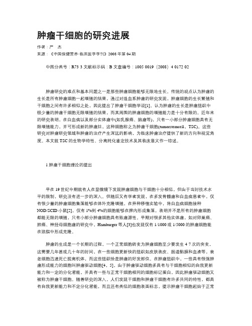
肿瘤干细胞的研究进展作者:严杰来源:《中国保健营养·临床医学学刊》2008年第04期中图分类号:R73-3 文献标示码:B 文章编号:1005-0019(2008)4-0172-02肿瘤研究的难点和基本问题之一是那些肿瘤细胞能够无限地生长。
传统的观点认为肿瘤的生长是所有肿瘤细胞一起增殖的结果,通过对造血系肿瘤的研究发现,肿瘤细胞的生长繁殖和干细胞之间有许多相似之处,因此提出了肿瘤干细胞学说[1]。
认为肿瘤的生长是肿瘤组织中极少量的肿瘤干细胞无限增殖的结果,而其周围的肿瘤细胞的增殖能力是十分有限的。
近年来的研究表明,在白血病以及部分实体瘤中(如乳腺癌、脑瘤等),只有一小部分肿瘤细胞具有无限增殖能力,并可形成新的肿瘤灶,这种细胞称之为肿瘤干细胞(tumorstemcell,TSC)。
这些研究对肿瘤研究领域和肿瘤的治疗产生深远的影响,为临床肿瘤治疗提供了新的方向和视觉角度。
本文就TSC的生物学特性、分离纯化鉴定技术及其临床意义作一综述。
1 肿瘤干细胞理论的提出早在19世纪中期就有人在显微镜下发现肿瘤细胞与干细胞十分相似,但由于当时技术水平的限制,研究没有进一步的深入。
但随后又有学者发现,在多发骨髓瘤和白血病患者中,仅有很少量的肿瘤细胞集落能够在体外克隆增殖,在异种移植实验中,将白血病细胞接种NOD/SCID小鼠[2],仅有1%到4%的细胞能够在脾内形成集落,表明并不是所有的肿瘤细胞都能无限的增殖,只有小部分肿瘤细胞具有致瘤源性。
早期对很多其他实体瘤,如对卵巢癌、肺癌、神经母细胞瘤的研究中,Hamburger等人[3]也发现仅有1/1000或1/5000的肿瘤细胞能在琼脂中形成克隆。
肿瘤的生成是一个长期的过程,一个正常细胞转变为肿瘤细胞至少要发生4-7次的突变,这需要几年甚或几十年的时间,在一些细胞更新快的组织如皮肤表皮、肠道黏膜和血液等,衰老细胞迅速死亡脱离机体,而这些组织恰是肿瘤的好发部位。

研究发现跑步有助缩小癌症肿瘤——科林·费尔南德斯(COLIN FERNANDEZ)为《每日邮报》撰稿原文发表于2016年2月16日2016年02月17日16:03 来源:Mail Online跑步也许是正遭受癌痛折磨的病人最不想做的事情,但科学家研究发现,长期而言,跑步对癌症治疗有着巨大助益。
科学家发现,高强度运动产生的肾上腺素可以让肿瘤缩小一半。
哥本哈根大学的研究人员将一组患有肺癌的老鼠放在跑步轮上,另一组作为参照的患癌老鼠则不做任何运动。
结果与运动较少的一组老鼠相比,跑步轮上的老鼠体内的肿瘤缩小了50%。
研究论文中称,在剧烈运动过程中,具有清除癌细胞功能的“自然杀伤细胞”将被导向肿瘤。
研究人员特别指出,运动中人体分泌的肾上腺素可促使免疫细胞转向生长在肝脏、肺部以及皮肤的肿瘤。
科学家将一组患有肺癌的老鼠放在跑步轮上,另一组患癌老鼠则不做运动。
跟不运动的老鼠体内的肿瘤(左)相比,患癌老鼠身上的肿瘤缩小了50%(右)。
尽管实验是在动物身上进行的,但研究人员称剧烈运动对人类癌症患者可能具有同样的效果。
哥本哈根大学的霍曼博士(Pernille Hojman)说:“我们知道自然杀伤细胞的渗透可以控制肿瘤的大小,但是运动在其中产生的作用却没人研究过。
”“在实验中,我们尝试向老鼠体内注射肾上腺素,来模拟运动过程中的肾上腺素增长,这时我们看到自然杀伤细胞在血流中被动员起来,如果有肿瘤出现,自然杀伤细胞就会发现并前往消灭。
”但如果受测老鼠体内自然杀伤细胞数量较少,即使进行运动,它们体内的肿瘤也不会缩小。
同时如果限制肾上腺素的作用,会造成相似的结果。
因此研究人员得出结论,自然杀伤细胞和肾上腺素之间相互关联。
研究人员还发现,运动时肌肉产生的一种化学讯号——白介素6(IL-6)也有助于将自然杀伤细胞导向肿瘤,霍曼博士说:“这对我们而言是一个巨大的惊喜。
”如图所示,当肾上腺素(Epinephrine)和白介素6(IL-6)水平上升时,自然杀伤细胞(NK cell)就会被激活,从而顺着血流发现肿瘤,达到抑制其增长的效果。
doi:10.3971/j.issn.1000-8578.2024.23.1094线粒体在肺癌发生中的作用机制及治疗 研究进展吴发胜1,张晖2,谢家童2,李建福2,陈慧2,鲁世金1Research Progress in Mitochondrial Treatment and Mechanism in Occurrence of Lung Cancer WU Fasheng 1, ZHANG Hui 2, XIE Jiatong 2, LI Jianfu 2, CHEN Hui 2, LU Shijin 11. Department of Radiotherapy, Ruikang Hospital, Guangxi University of Chinese Medicine, Nanning 530011, China;2. Ruikang Clinical Medical College, Guangxi University of Chinese Medicine, Nanning 530001, ChinaCorrespondingAuthor:LUShijin,E-mail:**************Abstract: Lung cancer is characterized by high incidence and mortality rates and invasiveness, and its occurrence and development are influenced by various factors. Mitochondria, as ubiquitous organelles in the human body, regulate cellular processes, such as metabolism, signal transduction, oxidative stress, and genomic instability, thereby affecting the initiation and progression of lung cancer. This article summarizes the recent research progress on mitochondrial-targeted drugs, mitochondrial transfer, and mitochondrial gene therapy for lung cancer treatment. This work also discusses the principles and prospects of mitochondrial therapy to provide new insights for lung cancer treatment.Key words: Mitochondria; Lung cancer; Mechanism; TreatmentFunding: National Natural Science Foundation of China (No. 82260944); Doctoral Research Project (No. 2018BS056)Competing interests: The authors declare that they have no competing interests.摘 要:肺癌具有高发生率、高侵袭性和高致亡率的特点,其发生发展受多方面因素影响。
《旋毛虫重组蛋白原肌球蛋白对小细胞肺癌H446细胞的影响及分子机制研究》篇一一、引言随着分子生物学技术的发展,利用蛋白质工程改造疾病治疗领域的技术已成为一种新趋势。
近年来,有学者在生物研究过程中发现,旋毛虫(Trichinella spiralis)的重组蛋白原肌球蛋白(Recombinant Proto-myosin of Trichinella spiralis)对多种癌症细胞具有潜在的抗癌效果。
本篇论文主要研究旋毛虫重组蛋白原肌球蛋白对小细胞肺癌H446细胞的影响及其分子机制。
二、材料与方法1. 材料本实验所需旋毛虫重组蛋白原肌球蛋白购自生物技术公司,小细胞肺癌H446细胞来自中国科学院生物医学研究中心。
此外,本实验还需要相应的实验耗材、化学试剂及工具软件等。
2. 方法实验过程分为多个阶段:小细胞肺癌H446细胞的培养;旋毛虫重组蛋白原肌球蛋白的制备与处理;处理后的H446细胞与重组蛋白的共培养;以及后续的细胞生长检测、分子机制分析等。
三、旋毛虫重组蛋白原肌球蛋白对小细胞肺癌H446细胞的影响实验结果显示,旋毛虫重组蛋白原肌球蛋白处理后的小细胞肺癌H446细胞的生长受到显著抑制。
具体表现为细胞增殖减缓,凋亡率增加。
此外,通过免疫荧光染色等技术,我们还发现重组蛋白能有效地促进细胞内的钙离子积累,对肿瘤细胞的正常功能造成干扰。
四、分子机制研究经过深入的分子机制分析,我们初步认为,旋毛虫重组蛋白原肌球蛋白在小细胞肺癌H446细胞中发挥作用的机制可能与激活细胞内的凋亡信号通路有关。
通过检测凋亡相关基因的表达变化,我们发现一些关键的凋亡因子如Caspase-3、Bcl-2等在处理后的H446细胞中表达水平发生了显著变化。
此外,我们还发现重组蛋白能够激活细胞内的某些信号通路,如MAPK等,从而进一步促进肿瘤细胞的凋亡。
五、讨论本实验的结果表明,旋毛虫重组蛋白原肌球蛋白在小细胞肺癌H446细胞中具有显著的抗癌效果。
免疫细胞癌症英语作文高中标题,Immunotherapy in Cancer Treatment: A Breakthrough in Medical Science。
In recent years, immunotherapy has emerged as a revolutionary approach in the treatment of cancer, marking a significant milestone in medical science. This groundbreaking therapy harnesses the body's own immune system to combat cancer cells, offering new hope to patients and revolutionizing the landscape of cancer treatment. In this essay, we will explore the principles behind immunotherapy, its effectiveness, and its potential impact on the future of cancer treatment.Immunotherapy, also known as biologic therapy, is a type of cancer treatment that stimulates the body's immune system to fight cancer. Unlike traditional treatments such as chemotherapy and radiation therapy, which directlytarget cancer cells, immunotherapy works by enhancing the immune system's ability to recognize and destroy cancercells. This is achieved through various approaches, including the use of immune checkpoint inhibitors, adoptive cell transfer, and cancer vaccines.One of the most widely studied forms of immunotherapy is immune checkpoint inhibition. Cancer cells can often evade detection by the immune system by expressing proteins that inhibit immune responses. Immune checkpoint inhibitors block these proteins, allowing the immune system to recognize and attack cancer cells. Drugs such as pembrolizumab and nivolumab have shown remarkable success in treating a variety of cancers, including melanoma, lung cancer, and bladder cancer.Another promising approach is adoptive cell transfer, which involves extracting immune cells from a patient, modifying them to enhance their cancer-fighting abilities, and then reintroducing them into the body. Chimeric antigen receptor (CAR) T-cell therapy, for example, has demonstrated remarkable results in the treatment of certain types of leukemia and lymphoma, achieving durable remissions in patients who have not responded to othertreatments.In addition to these approaches, cancer vaccines are also being developed to stimulate the immune system to target specific cancer cells. These vaccines can help prevent cancer recurrence or slow its progression by training the immune system to recognize and attack cancer cells. While cancer vaccines are still in the early stages of development, they hold promise for the future of cancer treatment.The effectiveness of immunotherapy in cancer treatment has been nothing short of remarkable. Unlike traditional treatments, which often cause severe side effects and are only effective for certain types of cancer, immunotherapy has shown efficacy across a wide range of cancer types and has fewer side effects. In some cases, immunotherapy has led to long-term remission and even cure in patients with advanced cancer who had exhausted all other treatment options.Moreover, immunotherapy has the potential to transformthe way we approach cancer treatment in the future. By targeting the immune system rather than the cancer cells themselves, immunotherapy offers a more precise and personalized approach to treatment. This could lead to fewer side effects, shorter treatment durations, and better outcomes for patients. Additionally, immunotherapy has the potential to be combined with other treatments, such as chemotherapy and radiation therapy, to further enhance its effectiveness.However, despite its tremendous promise, immunotherapy is not without its challenges. One of the main challengesis identifying biomarkers that can predict which patients are most likely to respond to immunotherapy. While some patients experience remarkable responses to treatment, others see little to no benefit. By identifying biomarkers that can predict treatment response, researchers can better select patients who are most likely to benefit from immunotherapy.In conclusion, immunotherapy represents a paradigmshift in the treatment of cancer, offering new hope topatients and reshaping the future of cancer treatment. With its ability to harness the body's own immune system tofight cancer, immunotherapy has shown remarkable effectiveness across a wide range of cancer types and has the potential to transform the way we approach cancer treatment. While challenges remain, the future of immunotherapy looks promising, with ongoing research and development poised to unlock its full potential in thefight against cancer.。
Short Article Voluntary Running Suppresses Tumor Growth through Epinephrine-and IL-6-Dependent NK Cell Mobilization and RedistributionGraphical AbstractHighlightsd Exercise reduces tumor incidence and growth in severalmouse modelsd Exercise increases NK cell infiltration,thereby controllingtumor growthd Epinephrine mobilizes NK cells and b-blockade blunts thetumor suppressiond Exercise-induced muscle-derived IL-6is involved in NK cellredistribution AuthorsLine Pedersen,Manja Idorn,Gitte H.Olofsson,...,Bente K.Pedersen,Per thor Straten,Pernille HojmanCorrespondencephojman@inflammation-metabolism.dkIn BriefThe beneficial effects of exercise are countless.Pedersen et al.now link exercise,cancer,and immunity and reveal that exercise decreases tumor incidence and growth by over60%across several mouse tumor models through a direct regulation of NK cell mobilization and trafficking in an epinephrine-and IL-6-dependent manner.Accession NumbersGSE62628 Pedersen et al.,2016,Cell Metabolism23,1–9March8,2016ª2016Elsevier Inc./10.1016/j.cmet.2016.01.011Voluntary Running Suppresses Tumor Growth through Epinephrine-and IL-6-DependentNK Cell Mobilization and RedistributionLine Pedersen,1Manja Idorn,2Gitte H.Olofsson,2Britt Lauenborg,1Intawat Nookaew,3,4Rasmus Hvass Hansen,5 Helle Hjorth Johannesen,5Ju¨rgen C.Becker,6Katrine S.Pedersen,1Christine Dethlefsen,1Jens Nielsen,3Julie Gehl,7 Bente K.Pedersen,1Per thor Straten,2,8and Pernille Hojman1,7,*1Centre of Inflammation and Metabolism and Centre for Physical Activity Research,Rigshospitalet,Faculty of Health Science,University of Copenhagen,DK-2100,Denmark2Centre for Cancer Immune Therapy,Department of Hematology,Copenhagen University Hospital,Herlev,DK-2730,Denmark3Department of Biology and Biological Engineering,Chalmers University of Technology,Go¨teborg,SE-412,Sweden4Comparative Genomics Group,Biosciences Division,Oak Ridge National Laboratory,Oak Ridge,TN37831,USA5Department of Radiology,University Hospital Copenhagen,Herlev,DK-2730,Denmark6Department for Translational Skin Cancer Research(TSCR)within the German Cancer Consortium(DKTK),Westdeutsches Tumorzentrum, University Hospital Essen,45117,Essen,Germany7Department of Oncology,Copenhagen University Hospital,Herlev,DK-2730,Denmark8Department of Immunology and Microbiology,University of Copenhagen,DK-2200,Denmark*Correspondence:phojman@inflammation-metabolism.dk/10.1016/j.cmet.2016.01.011SUMMARYRegular exercise reduces the risk of cancer and dis-ease recurrence.Yet the mechanisms behind this protection remain to be elucidated.In this study,tu-mor-bearing mice randomized to voluntary wheel running showed over60%reduction in tumor inci-dence and growth acrossfive different tumor models. Microarray analysis revealed training-induced upre-gulation of pathways associated with immune func-tion.NK cell infiltration was significantly increased in tumors from running mice,whereas depletion of NK cells enhanced tumor growth and blunted the beneficial effects of exercise.Mechanistic analyses showed that NK cells were mobilized by epinephrine, and blockade of b-adrenergic signaling blunted training-dependent tumor inhibition.Moreover, epinephrine induced a selective mobilization of IL-6-sensitive NK cells,and IL-6-blocking antibodies blunted training-induced tumor suppression,intratu-moral NK cell infiltration,and NK cell activation. Together,these results link exercise,epinephrine, and IL-6to NK cell mobilization and redistribution, and ultimately to control of tumor growth. INTRODUCTIONEpidemiological data document that regular exercise protects against the development of certain cancers and lowers the risk of disease recurrence(Brown et al.,2012;Christensen et al., 2014),prompting extensive research into exercise interventions in cancer patients(Jones and Alfano,2013).Across a range of cancer diagnoses,exercise has been shown to improve func-tional capacity and patient-reported outcomes(Mishra et al., 2012).However,exercise may also directly suppress tumor growth,as suggested by decreased risk of disease recurrence in physically active cancer patients(Ballard-Barbash et al., 2012).Little is known about the mechanisms behind this protec-tion,but exercise-mediated changes in body composition,sex hormone levels,systemic inflammation,and immune cell function have been suggested as possible mediators(McTiernan,2008). Exercise training comprises of acute bouts of physical exer-tion,followed by periods of recovery.During these acute bouts of exercise,plasma levels of stress hormones and muscle-derived myokines increase dramatically(Catoire and Kersten, 2015).Myokines may have direct anti-proliferative effects on cancer cells,as shown for oncostatin M on hormone-sensitive breast cancer cells(Hojman et al.,2011)and SPARC in colon cancer(Aoi et al.,2013).Yet during exercise,an acute mobiliza-tion of immune cells to the circulation is also seen(Pedersen and Hoffman-Goetz,2000;Bigley et al.,2014).Cells of the immune system play dual roles in cancer.The immune system has a powerful capacity to combat cancer,but chronic inflammation has also been linked to tumorigenesis in several conditions(Viv-ier et al.,2012;Imai et al.,2000;Grivennikov et al.,2010).On the protective side,infiltrating cytotoxic immune cells have been demonstrated as positive prognostic factors for disease outcome and overall survival in several cancers(Fridman et al., 2012;Remark et al.,2013).Thus,mobilization of cytotoxic im-mune cells during exercise might represent an indirect defense mechanism against cancer growth.RESULTS AND DISCUSSIONVoluntary Wheel Running Significantly Reduces Tumor Incidence and GrowthFirst,we evaluated the effect of wheel running before and/or dur-ing tumor challenge in a subcutaneous B16F10melanoma model in female mice(Figure1A).Four weeks of wheel running Cell Metabolism23,1–9,March8,2016ª2016Elsevier Inc.1Figure1.Wheel Running Reduces Tumor Incidence and Growth(A)Experimental design.(B)Tumor volume of subcutaneous B16tumors in3-month-old female C57BL/6mice(n=12,two-way ANOVA with Tukey’s post hoc test).(C)Tumor volume of subcutaneous B16F10tumors in C57BL/6mice(CON n=11,EX n=12,Students t test).(D)Number of tumors per lung in mice injected i.v.with13105B16F10cells(CON=9,EX=10,Students t test).(E)Representative pictures of B16tumors in lungs of mice after i.v.injection.(F)MRI of male NMRI mice injected with DEN(25mg/kg body weight).The red arrows point to a liver tumor in a control mouse.(G)Tumor burden assessed by MRI(n=16for both groups,two-way ANOVA with post hoc multiple t test).$:11th month:EX=16,CON=14.See also Figure S1. Means±SEM.*p<0.05,**p<0.01,***p<0.001.2Cell Metabolism23,1–9,March8,2016ª2016Elsevier Inc.prior to tumor cell inoculation reduced tumor growth by61%(p< 0.01,Figure1B).Similar reductions in tumor volume of67%and 53%with running were verified in female adult(3months,p< 0.05,Figure1C)and old mice(18months,p<0.001,Figure S1A). Wheel running also dramatically reduced lung metastases after intravenous(i.v.)injection of B16F10melanoma cells(p< 0.001,Figures1D and1E).Next,the impact of wheel running on tumor growth was evaluated in three additional models. Male Naval Medical Research Institute(NMRI)mice were in-jected with diethylnitrosamine(DEN)at4weeks of age,which is known to cause liver tumors within10months.Here,wheel running reduced tumor incidence as only31%of the running mice developed tumors,compared with75%of the control mice(Figure1F).Moreover,wheel running reduced tumor burden per mouse(p<0.05,Figure1G).In a Lewis Lung carci-noma model(LLC)in female mice,running decreased tumor volume by58%(p<0.01,Figure S1B)and tumor weight by 56%(p<0.05,Figure S1C),and in Tg(Grm1)EPv transgenic male mice,which spontaneously develops melanoma,wheel running tended to delay formation of malignant lesions(p= 0.08,Figure S1D).The average wheel running distance was4.1km/mouse/day for the B16-inoculated female mice and6.8km/mouse/day for the male DEN-injected mice.The presence of B16tumors did not induce weight loss or cachexia(Figure S1E).In contrast, the presence of LLC tumors induced an average weight loss of À1.27±1.74g in the tumor-bearing control group.This weight loss was completely prevented in the running tumor-bearing mice(p<0.05,Figures S1E and S1G).Taken together,wefind marked reductions in tumor incidence and growth with voluntary wheel running across5different tumor models.The delay in B16melanoma progression required a 4-week pre-training period prior to tumor inoculation.During this period,the mice were habituated to wheel running,and the immune system was primed for the tumor challenge,suggesting that when using fast-growing transplantable tumor models,part of the exercise effect could potentially be killing of cancer cells at the inoculation site.In contrast,initiating running after tumor challenge in our slow-growing DEN-induced and Tg(Grm1)Epv models was sufficient to control tumor incidence and progression.Training-Dependent Reduction in Tumor Growth Is Associated with Induction of Immune-Related Pathways To identify differentially regulated pathways in tumors from running mice,we performed microarray analysis on B16tumors from Figure1C(Table S1).Of the92upregulated gene ontology (GO)pathways,the majority(52%)was related to immunological and inflammatory pathways(Figure2A).qPCR analysis confirmed the increased expression of both pro-and anti-inflam-matory cytokines(Figure2B),as well as markers of the cellular innate and adaptive immune systems in B16tumors from running mice(Figure2C).Also in LLC tumors,pro-inflammatory cyto-kines(IL-1a and iNOS)and markers for NK and T cells were up-regulated with running(Figures2D and2E).To exclude that these differences in immune pathways were merely related to tumor size,we repeated the evaluation now based on B16tumors of identical volume(approx.150mm3,Fig-ure2F).Thus,the tumors were on average excised2days earlier in the control group(Figure2G).In these similar sized tumors,up-regulation of the pro-inflammatory cytokines(IL-1a and iNOS) and immune cell markers with exercise was verified(Figures 2H and2I).Training Regulates Tumor Growth through Intratumoral NK Cell InfiltrationNext,we measured frequencies of immune cells in tumors by flow cytometry after6weeks of wheel running.In the subcutane-ous B16model,tumors from running mice showed markedly increased infiltration of NK cells(p<0.001,Figures3A,3C, and3D),as well as CD3T cells and dendritic cells(p<0.05,Fig-ure3A).Of note,the CD3gate includes CD3+CD4ÀCD8Àcells, and thus also includes cells such as gamma delta and NKT cells. In the i.v.B16model,wheel running also increased NK cell infil-tration in lung tumors(p<0.05,Figure3B),but without enhance-ment of other immune cell subtypes.The level of NK cell infiltra-tion correlated inversely with tumor burden(p<0.01,Figures S2A and S2B).Histological evaluation showed that exercise increased both NK1.1+,CD8+and CD4+cells with the absolute numbers of CD8+and CD4+cells being higher than that for NK cells(Figures3E and S2C).In control mice,NK cells were rarely detectable.Infiltration of B cells did not change significantly with exercise(Figure S2D).In non-tumor-bearing mice,6weeks of running increased the frequencies of NK cells in bone marrow (p<0.05),spleen(p<0.01),and to a lesser extent peripheral blood mononuclear cells(PBMCs)(p=0.17,Figure3F),indi-cating an overall increase in the basal pool of NK cells with exer-cise.In tumor-bearing mice,running did not alter the frequency of NK cells in these organs(Figure3G),yet these mice showed pronounced accumulation of NK cells in their tumors(Figure3C). The numbers of T cells did not increase in blood,bone marrow, or spleen with6weeks of running in neither tumor-bearing nor non-tumor-bearing mice(Figures S3A and S3B).We then evaluated the response to running in athymic mice, which lack functional T cells but retain NK cells.In these mice, a66%reduction in B16tumor volume persisted with6weeks of running(p<0.05,Figure3H),showing that T cells were not responsible for the suppressive effect of running on tumor growth.However,the athymic nude mice in general had larger tumors than wild-type immune-competent mice(WT)(Figures S3C and S3D),indicating that T cells aside from the exercise sit-uation play a role in control of tumor growth.To further document the role of NK cells,circulating NK cells were depleted by admin-istration of anti-asialo-GM1antibodies(Figures S3E and S3F). Depletion of NK cells completely abolished the suppressive effect of running on tumor volume,and both anti-asialo-GM1-treated groups(control and exercise,respectively)showed enhanced tumor growth compared with isotype IgG-treated mice(p<0.001,Figure3I).Next,we investigated if running affected NK cell cytotoxicity, but determined per cell basis,NK cells from control and running mice were equally effective in killing NK-sensitive YAC-1and B16cells(Figure3J).To exclude that cancer cell killing was mediated by T cell contamination,the NK-depleted fraction (NK cell content of2%)was used as a negative control and showed no YAC-1or B16cell killing.In contrast,within the tu-mors from the running mice,we found increased mRNA expres-sion of NK-cell-activating receptor ligands(H60a,MULT1,Clr-b), Cell Metabolism23,1–9,March8,2016ª2016Elsevier Inc.3as well as cytokines (IL-2,IL-15,IFN g )and chemokines (CCL3,CXCL10,CX3CL1,Chemerin)related to NK cell activation and chemotaxis (Figure 3K).No changes in the expression of markers of angiogenesis (i.e.,CD31and VEGF-A)were observed (Figure 3K).Taken together,these data point to a predominant role of NK cells in the training-dependent control of tumor growth.NK cells represent a critical component of the innate immune de-fense,recognizing transformed cells independently of anti-bodies or major histocompatibility complex (MHC)restriction (Vivier et al.,2012;Brodin et al.,2012),while T cells are cyto-toxic effector cells of the adaptive immune response.Both im-mune cell types are known to be regulated by exercise.During exercise,circulating lymphocytes increase in number and fre-quency and then fall below pre-exercise levels (Pedersen and Hoffman-Goetz,2000).Of these lymphocytes,NK cells are the most responsive cells to the exercise-dependent mobiliza-tion,followed by CD8T cells,CD4T cells,and lastly B cells,which respond poorly to exercise (Walsh et al.,2011).Thus,the importance of NK cells in the training-dependent control of tumor growth follow their superior responsiveness toexercise.Figure 2.Running Is Associated with Induction of Immune Related and Inflammatory Pathways(A–E)(A)Distribution of upregulated GO pathways (n =5).See also Table S1.qPCR analysis of (B)inflammatory cytokines and (C)immune cell markers in B16tumors from Figure 1C (CON =12,EX =11,multiple t testing),and (D)inflammatory cytokines and (E)immune cell markers in Lewis Lung tumors (CON =9,EX =10,multiple t testing).(F–I)(F)Tumor volume of subcutaneous B16F10tumors and (G)day of termination for tumors excised.qPCR analysis of (H)inflammatory cytokines and (I)immune cell markers from tumors in Figure 2F (n =10,multiple t testing).Black bars =CON,gray bars =EX.Means ±SEM.*p <0.05,**p <0.01,***p <0.001.4Cell Metabolism 23,1–9,March 8,2016ª2016ElsevierInc.In addition to the mobilization of the NK cells during exercise,we found increased expression levels of NK cell-related acti-vating receptor ligands,stimulatory cytokines,and chemoattrac-tant chemokines in the tumors of running mice,suggesting that exercise works both on the mobilization of NK cells,and on the tumor microenvironment to generate a NK cell activating milieu.NK cells are regulated by a multitude of activating and inhibitory receptor-ligand interactions.Here,we show that ligands for the activating receptor NKG2D,MULT1and H60a,as well as Clr-b,a ligand for NKR-P1B,which has proven important in the ed-ucation of NK cells,were upregulated in the tumors from running mice (Chen et al.,2015;Rahim et al.,2015).Previously,it has been shown that B16F10cells do not express Clr-b (Carlyle et al.,2004).The methodology employed in this study did not allow for precise identification of whether Clr-b expression was attributable to tumor cells,infiltrating immune cells,or other cellsin the tumor microenvironment.Furthermore,we found increased expression of the activating receptor NKp46with wheel running.NKp46has been shown to mediate control of B16metastasis,which correlates well with our results (Glasner et al.,2012).IL-6-Sensitive NK Cells Are Recruited by Exercise through b -Adrenergic SignalingExercise has been shown to mobilize NK cells through epineph-rine (EPI)(Bigley et al.,2014;Dimitrov et al.,2010).Five hours into the dark period,when wheel running was at its highest,serum EPI levels were 561.2±157.4pg/ml in the CON group and 1,169.3±340.2pg/ml in the EX group (p <0.001,Figure 4A).To mimic this acute running response,we injected low and high doses of EPI (0.5mg/kg and 2mg/kg,respectively)and found a reduction in both spleen volume (Figure 4B)andtheFigure 3.Training Controls Tumor Growth through Intratumoral NK Cell Infiltration(A)Frequency of immune cells in subcutaneous B16tumors from CON and EX mice (Gating criteria:total T cells [CD3+],CD8T cells [CD3+CD8+],CD4T cells [CD3+CD4+],B cells [CD3ÀCD19+],NK cells [CD3ÀNK1.1+],DC [CD11b +GR-1ÀCD11c +],MDSC [CD11b +GR-1b +])(CON =8,EX =9,multiple t testing).(B)Frequency of immune cells in lungs from Figure 1D (n =8,multiple t testing).(C)NK cells per total B16cells in subcutaneous B16tumors (CON =8,EX =9,Students t test).(D)Flow cytometry panels of tumor infiltrating NK1.1+cells.(E)Histological analysis of tumor sections stained for NK1.1or CD8in red.(F and G)NK cell frequency in bone marrow,blood,and spleen from non-tumor-bearing (F)and tumor-bearing mice (G).(H)Tumor volume of subcutaneous B16tumors in athymic mice (CON =9,EX =11,Students t test).(I)Tumor volume of subcutaneous B16tumors in C57BL/C mice after NK cell depletion (n =12,two-way ANOVA with post hoc Bonferroni test).See also Figure S3.(J)NK-cell-mediated killing of NK-cell-sensitive YAC-1cells (full line)and B16F10tumor cells (dotted line).(K)qPCR analyses of mRNA expression of NK-cell-activating receptor ligands,cytokines,chemokines,and angiogenic factors (CON =10,EX =11,multiple t testing).Black bars =control,gray bars =EX.Means ±SEM.*p <0.05,**p <0.01,***p <0.001.Cell Metabolism 23,1–9,March 8,2016ª2016Elsevier Inc.5Figure4.IL-6-Sensitive NK Cells Are Mobilized through b-Adrenergic Signaling(A)Serum epinephrine and nor-epinephrine(CON=12,EX=10,Students t test).(B)Weight of the spleen30min after epinephrine injection at0.5mg/kg(EPI low,n=7)and2mg/kg(EPI high,n=8,one-way ANOVA with Tukey’s post hoc test).(C)Number of NK cells in the spleen(n=6,Students t test).(D and E)(D)Tumor volume of subcutaneous B16tumors after propranolol treatment(n=11)and(E)NK cell infiltration in the tumors from(D)(n=8,both two-way ANOVA with Bonferroni post hoc test).See also Figure S4.(F and G)(F)Tumor volume of subcutaneous B16tumors from mice injected daily with low-dose0.5mg/kg epinephrine(n=11)and(G)NK cell infiltration in the tumors from(F)(n=8,both one-way ANOVA with Tukey’s post hoc test).(H and I)(H)Flow cytometry panels of IL-6R a(top)and gp130-positive(bottom)splenic NK cells in control(CON)or epinephrine(EPI,0.5mg/kg30min prior to sampling)and(I)average IL-6R a and gp130expression(n=6,Students t test).(J and K)(J)Tumor volume of subcutaneous B16tumors(CON=12,anti-IL6=10,)and(K)NK cell infiltration(n=8,both two-way ANOVA with Bonferroni post hoc test)in subcutaneous B16tumors of mice receiving anti-IL-6antibodies or saline treatment.Black bars=control,gray bars=EX,light gray=daily epinephrine injections(0.5mg/kg).Means±SEM.*p<0.05,**p<0.01,***p<0.001.6Cell Metabolism23,1–9,March8,2016ª2016Elsevier Inc.number of NK cells in the spleen(Figure4C).Blocking of the EPI-dependent mobilization of NK cells by propranolol during wheel running completely blunted the effect of running on tumor volume(Figure4D)and abolished the increased NK cell infiltra-tion in tumors from running mice(Figure4E).Propranolol admin-istration did not alter training-dependent changes in body weight and muscular expression of the exercise-responsive factor, PGC-1a(Figures S4A and S4B).To confirm the role of EPI,we mimicked the exercise-related EPI surge by daily injections of low-dose(0.5mg/kg)EPI in non-running mice.This resulted in a61%reduction in tumor volume(p<0.01,Figure4F)and tended to increase tumor infiltration of NK cells compared with non-treated controls(p=0.059,Figure4G).This increased infil-tration did,however,not match the effect seen by running alone. In addition to the blockade of NK cell infiltration,propranolol administration tended to block the infiltration of CD3+,CD4+, and CD8+cells,while EPI injection tended to mimic the exer-cise-induced mobilization of these cells(Figures S4C and S4D). Plasma IL-6increases dramatically during exercise due to release from contracting muscles and might be the additional ex-ercise factor,involved in tumor homing(Pedersen and Febbraio, 2012).In our model,serum IL-6increased from4.3±3.5pg/ml (range:1.6–12.4pg/ml,n=12)in the CON group to29.3±32.6pg/ml(range:5.7–106.5pg/ml,n=10,p<0.05)during wheel running.In the spleen,24.8%±8.4%of the NK cells ex-pressed IL-6R a and63.4%±7.9%gp130(Figures4H and4I).Af-ter EPI injection,the fraction of IL-6R a-positive splenic NK cells decreased to12.4%±5.3%,suggesting an EPI-dependent mobilization of IL-6-sensitive NK cells.Blocking of training-induced IL-6by anti-IL-6antibodies diminished the exercise-induced inhibition of tumor growth(Figure4J)and inhibited the infiltration of NK cells into tumors(Figure4K).In contrast,daily injections of100ng IL-6,which increased serum IL-6from 2.7±0.9pg/ml to301.1±130.3pg/ml and263.8±950pg/ml at30and60min,respectively,did not mimic the training-induced reduction in tumor growth(Figure S4E)or enhanced NK cell infiltration(Figure S4E).Thus,with this IL-6concentra-tion,the redistribution of NK cells seen during running could not be mimicked,suggesting that the increment in NK cell infiltra-tion is dependent on concurrent exercise-induced mobilization of immune cells.Both running and IL-6injections decreased the frequency of immature(CD11bÀ,CD27À)NK cells(p< 0.05,Figure S4E)and tended to increase the frequency of cyto-toxic(CD11bÀ,CD27+)NK cells(p=0.11).In contrast,anti-IL-6 antibody treatment blocked the training-induced increase in cytotoxic(CD11bÀ,CD27+,p<0.05)NK cells but increased the frequency of cytokine producing(CD11b+,CD27À,p< 0.05)NK cells(Figure S4H).The epinephrine surge during exercise can mobilize NK cells to the blood stream through activation of their b-adrenergic re-ceptors,increasing the NK cell frequency but not their cytotox-icity(Dimitrov et al.,2010).Early studies suggest that a rela-tively small increase in epinephrine level is sufficient to mobilize NK cells.Thus,to obtain the physiological changes, which we observe within the tumors,additional stimuli must be present.To this end,we observed a selective mobilization of IL-6R a-positive NK cells after epinephrine injection.In further support of the role of IL-6in NK cell redistribution and activation,recombinant human IL-6infusion has been shown to mimic the acute and transient lymphopenia seen dur-ing the recovery from exercise(Steensberg et al.,2003),and stimulation of human NK cells with IL-6has been shown to in-crease their expression of adhesion molecules(Rabinowich et al.,1993).Yet,the role of IL-6in cancer is complex.Chronic elevated plasma levels of IL-6have been associated with poor disease outcome across a number of cancer diagnoses,whereas increased IL-6expression within tumors is a positive prognostic marker for overall and disease-free survival(Dethlefsen et al., 2013;Mauer et al.,2015).In particular for the DEN-induced liver model,used in this study,Naugler and colleagues showed that IL-6KO mice were resistant to DEN-induced tumor formation (Naugler et al.,2007).In contrast to these models,plasma IL-6 displays a dynamic response during exercise.The exercise-induced surge in plasma IL-6may signal directly to IL-6R a-ex-pressing cells or through its alternative pathway,IL-6 trans-signaling(Kraakman et al.,2015;Scheller et al.,2014). We show that about25%of the splenic NK cells express IL-6R a and are thus directly sensitive to classical IL-6signaling. Regarding IL-6trans-signaling,IL-6binds the soluble IL-6recep-tor and then this complex binds to membrane-bound gp130.The concentration of the soluble IL-6receptor is about40ng/ml,thus there is a large buffer capacity for IL-6binding,when IL-6in-creases as seen during wheel running.Thus,the systemic IL-6 increase during wheel running can signal either directly through the classical IL-6signaling or through trans-signaling to NK cells. In Figure2,we showed a2.3-to3.0-fold increase in IL-6expres-sion in the tumors of running mice,yet following on the impor-tance of classical IL-6signaling and IL-6trans-signaling,this suggests that the exercise-induced increase in systemic IL-6 levels play a greater role than the intratumoral concentration of IL-6in NK cell redistribution and activation.In further support of the beneficial role of IL-6exclusively in the exercise setting,we found no protective beneficial effect of IL-6 injection alone on tumor growth or intratumoral NK cell infiltra-tion,stressing the dependence on prior training-dependent mobilization of NK cells.IL-6and exercise have previously been shown to increase tumor vascularization(Taniguchi and Karin,2014),yet we did notfind any effect of IL-6or running on CD31and VEGF-A mRNA expression levels,nor did we detect any marked increases in capillarization in our histological analyses(data not shown).In conclusion,voluntary wheel running inhibits tumor onset and progression across a range of tumor models and anatomical locations.This occurs through a direct regulation of NK cell traf-ficking,involving an epinephrine-dependent mobilization of NK cells to the circulation and an IL-6-dependent redistribution to tumors.The clinical relevance of infiltrating T cells is evident (Fridman et al.,2012),while the potential of tumor-infiltrating NK cells is still being unraveled(Remark et al.,2013).NK cells are part of the early innate immune response and can activate other immune cells through secretion of IFN g(Gross et al., 2013).Thus,a key action of NK cells is to deliver the initial ‘‘spark’’that activates other cell types of the immune system. Currently,clinical attention is on generating an inflammatory intratumoral environment through among others immune checkpoint blockade therapy(Topalian et al.,2015).The present study indicates that exercise might deliver such therapeutic Cell Metabolism23,1–9,March8,2016ª2016Elsevier Inc.7intratumoral adaptations with increased immune cell infiltration and generation of an inflammatory intratumoral environment.EXPERIMENTAL PROCEDURESAll methods and interventions are described in details in the Supplemental Experimental Procedures .Animal Studies and InterventionsStudies were compliant with the ARRIVE guidelines and approved by the Danish Inspectorate for Research Animals.Exercise were provided by running wheels (diameter 12cm).The mice were inoculated subcutaneously with 23105B16F10melanoma or Lewis Lung cells (cells obtained from ATCC)or i.v.through the tail vein with 13105B16F10cells.For DEN-induced liver tumors,23-day-old NMRI male mice obtained from Harlan,were injected intraperito-neally (i.p.)with DEN (25mg/kg body weight).The Tg(Grm1)EPv mice were ob-tained from the University of Graz,after 2weeks of acclimatization,running wheels were placed in the home cages.Anti-Asialo TreatmentTo deplete NK cells,mice received anti-Asialo GM 1antibodies (1mg/ml,EBio-Science)or isotype antibodies as control (Rabbit IgG,0.5mg/ml,Southern Biotech)every second day from 1week before B16inoculation.Five days after tumor cell inoculation,NK cells content was evaluated.Propranolol/Epinephrine StudyEight-week-old C57BL/6mice were randomized to drinking water containing 0.5g/l propranolol or nothing.Propranolol administration started 1week before B16inoculation.An additional group received daily (Monday to Friday)injections of epinephrine (0.5mg/kg,200m l i.p.)from 1week before B16inoc-ulation.For the acute effect of epinephrine,mice were injected with 0.5mg/kg or 2mg/kg epinephrine,and sacrified after 30min,where blood,spleen,and muscle tissue were collected.Anti-IL-6/IL-6StudyEight-week-old C57BL/6mice were randomized to groups,receiving i.p.injec-tions of anti-IL-6antibodies (100m g/mouse,R&D systems,#AB-406-NA)or vehicle injections twice a week from 1week before B16inoculation.An addi-tional group received daily (Monday to Friday)injections of IL-6(100ng/mouse,R&D systems,#406-ML-025/CF)from 1week before B16inoculation.StatisticsFor comparisons of exercise and other interventions,two-way ANOVA fol-lowed by post hoc tests with Bonferroni corrections were performed.Two-way ANOVA with repeated-measures was used when the effects of experi-mental factors was analyzed at different timepoints.In the injection studies,one-way ANOVA with Tukey’s post hoc test was performed.The statistical sig-nificance of mRNA and protein expression levels in CON and EX groups was obtained from two-tailed multiple t tests.Results are depicted as means ±SEM.The criterion for significance was p <0.05.ACCESSION NUMBERSThe microarray data generated in this publication have been deposited in NCBI’s GEO and are accessible through GEO:GSE62628(reviewer’s link at /geo/query/acc.cgi?token=cbgzksoktzerxuz&acc=GSE62628).SUPPLEMENTAL INFORMATIONSupplemental Information includes four figures,one table,and Supplemental Experimental Procedures and can be found with this article online at /10.1016/j.cmet.2016.01.011.AUTHOR CONTRIBUTIONSL.P.,B.K.P.,and P.H.conceived the study.L.P.,M.I.,B.L.,J.G.,B.K.P.,P.t.S.,and P.H.designed the experiments.I.N.and J.N.performed microarray anal-ysis.R.H.H.and H.H.J.performed the MR scans and image analyses.J.C.B.performed the histological analysis.M.I.and B.L.performed FACS analysesand cytotox assays.L.P.,M.I.,G.H.O.,B.L.,K.S.P.,and C.D.carried out exper-iments.L.P.,M.I.,and P.H.analyzed the data.L.P.and P.H.wrote the paper with contributions from all authors.All authors approved the final version of the paper.CONFLICTS OF INTERESTNone of the authors declare any conflicts of interest.ACKNOWLEDGMENTSAnne Boye,Lone Christensen,Lone Peijs,Maria Scheel,and Marianne Fregil are acknowledged for their technical assistance.Centre of Inflammation and Metabolism (CIM)is supported the Danish National Research Foundation (DNRF55).Centre of Physical Activity Research (CFAS)is supported by TrygFonden.This study was further supported by the Danish Medical Research Council,Novo Nordic Foundation,Lundbeck Foundation,Danish Cancer Society,and Aase og Ejnar Danielsen Foundation.Received:July 16,2015Revised:October 27,2015Accepted:January 20,2016Published:February 16,2016REFERENCESAoi,W.,Naito,Y.,Takagi,T.,Tanimura,Y.,Takanami,Y.,Kawai,Y.,Sakuma,K.,Hang,L.P.,Mizushima,K.,Hirai,Y.,et al.(2013).A novel myokine,secreted protein acidic and rich in cysteine (SPARC),suppresses colon tumorigenesis via regular exercise.Gut 62,882–889.Ballard-Barbash,R.,Friedenreich, C.M.,Courneya,K.S.,Siddiqi,S.M.,McTiernan,A.,and Alfano,C.M.(2012).Physical activity,biomarkers,and dis-ease outcomes in cancer survivors:a systematic review.J.Natl.Cancer Inst.104,815–840.Bigley,A.B.,Rezvani,K.,Chew,C.,Sekine,T.,Pistillo,M.,Crucian,B.,Bollard,C.M.,and Simpson,R.J.(2014).Acute exercise preferentially redeploys NK-cells with a highly-differentiated phenotype and augments cytotoxicity against lymphoma and multiple myeloma target cells.Brain Behav.Immun.39,160–171.Brodin,P.,Lakshmikanth,T.,Ka¨rre,K.,and Ho ¨glund,P.(2012).Skewing of the NK cell repertoire by MHC class I via quantitatively controlled enrichment and contraction of specific Ly49subsets.J.Immunol.188,2218–2226.Brown,J.C.,Winters-Stone,K.,Lee,A.,and Schmitz,K.H.(2012).Cancer,physical activity,and pr.Physiol.2,2775–2809.Carlyle,J.R.,Jamieson,A.M.,Gasser,S.,Clingan,C.S.,Arase,H.,and Raulet,D.H.(2004).Missing self-recognition of Ocil/Clr-b by inhibitory NKR-P1natural killer cell A 101,3527–3532.Catoire,M.,and Kersten,S.(2015).The search for exercise factors in humans.FASEB J.29,1615–1628.Chen,P.,Aguilar,O.A.,Rahim,M.M.,Allan,D.S.,Fine,J.H.,Kirkham,C.L.,Ma,J.,Tanaka,M.,Tu,M.M.,Wight,A.,et al.(2015).Genetic investigation of MHC-independent missing-self recognition by mouse NK cells using an in vivo bone marrow transplantation model.J.Immunol.194,2909–2918.Christensen,J.F.,Jones,L.W.,Andersen,J.L.,Daugaard,G.,Rorth,M.,and Hojman,P.(2014).Muscle dysfunction in cancer patients.Ann.Oncol.25,947–958.Dethlefsen,C.,Højfeldt,G.,and Hojman,P.(2013).The role of intratumoral and systemic IL-6in breast cancer.Breast Cancer Res.Treat.138,657–664.Dimitrov,S.,Lange,T.,and Born,J.(2010).Selective mobilization of cytotoxic leukocytes by epinephrine.J.Immunol.184,503–511.Fridman,W.H.,Page`s,F.,Saute `s-Fridman,C.,and Galon,J.(2012).The im-mune contexture in human tumours:impact on clinical outcome.Nat.Rev.Cancer 12,298–306.8Cell Metabolism 23,1–9,March 8,2016ª2016ElsevierInc.。