
《胎儿的超声测量》PPT课件
- 格式:ppt
- 大小:2.52 MB
- 文档页数:29

・ 6・ 国际妇产科学杂志2012年2月第39卷第1期 J Int Obstet Gynecol,February 2012,Vo1.39,No.1
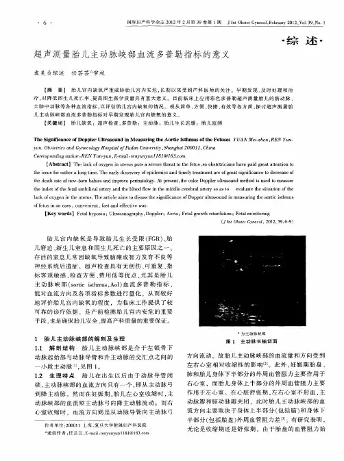
超声测量胎儿主动脉峡部血流多普勒指标的意义
袁美贞综述任芸芸 审校 ・综述・
【摘要】胎儿宫内缺氧严重威胁胎儿宫内安危,长期以来受到产科医师的关注。早期发现、及时处理和治 疗,对降低围生儿死亡率、提高同生医学质量具有重大意义。目前临床上应用彩色多普勒超声测量胎儿的脐动脉、 大脑中动脉等各种血流指标。以评估胎儿宫内缺氧的情况。现从简单、方便、快捷、有效等各方面,探讨超声测量胎 儿主动脉峡部血流多普勒指标对早期发现胎儿宫内缺氧的意义。 【关键词】life缺氧;超声检查,多普勒;主动脉;胎儿生长迟缓;胎儿监测
The Significance of Doppler Ultrasound in Measuring the Aortic Isthmus of the Fetuses YUA N Mei-zhen,REN Yun— yun.Obstetrics and GynecologyHospital ofFudan University,Shanghai 200011,China Correspondingauthor:REN Yun-yun,E—mail:renyunyun1181@163.com 【Abstract】The lack of oxygen in uterus puts a severe threat to the fetus,SO obstetricians have paid great attention to the issue for rather a long time.The early discovery of epidemics and timely treatment are of great significance to decrease of the death rate of new-born babies and improve perinatology.At present,the color Doppler ultrasound method is used to measure the index of the fetal umbilical artery and the blood flow in the middle cerebral artery SO as to evaluate the situation of the lack of oxygen in the uterus.The article aims to disuss the significance of Dopper ultrasound in measuring the aortic isthmus offetusin an easy,convenient,fast and effectiveway. 【Key words】Fetal hypoxia;Ultrasonography,Doppler;Aorta;Fetal growth retardation;Fetal monitoring (., £Obstet Gynecol,2012,39:6—9)
__________________________________________________
__________________________________________________ 常用胎儿超声测量数值
1、 妊娠龄: 妊娠龄(天)=妊娠囊平均内径(mm)+30(适用于孕7周内)
妊娠龄(天)=胚长(mm)+42(适用于孕7周—12周)
妊娠龄(周)=CRL(cm)+6.5(适用于孕7周—12周)
2、 孕囊增长速度:1.2—1.5mm/d
3、 3mm
4、 胎心率: 6周前:100—115次/min
至8周:144—159次/min
9周后:137—144次/min
5、 妊娠囊内各结构出现时间:
① 孕5周出现孕囊双环状。
② 孕5—6周出现卵黄囊。
③ 孕6—7周可见胚芽及胎心管搏动。
④ 孕7—8周可见胚胎轮廓。
⑤ 孕8—9周可辨头体及肢芽。 __________________________________________________
__________________________________________________ ⑥ 孕9—10周可见胎头及脑泡。
⑦ 孕10—11周可见四肢骨及指趾。
⑧ 孕12周以后可见四腔心及脊柱。
6、羊膜与绒毛膜融合时间:孕12—16周。个别晚妊期仍可见,无意义。
7、胎儿颈部透明层测量:
测量时间:10—14周(CRL约4.5—8.4cm )
异常值:NT≥3mm:胎儿染色体异常风险增加。
NT≥4mm:即使染色体正常的胎儿其妊娠结果亦较差。
8、 胎儿颈后皮肤皱褶测量:
测量时间:14—20周。
异常值:≥6mm。
9、 颅骨骨化时间:第10周开始,第11—12周骨化明显。
10、脊椎骨化时间:第10周开始,骶尾部到16—18周完成。 __________________________________________________
.
. .
..
.
.
.z 常用胎儿超声测量数值
1、 妊娠龄: 妊娠龄(天)=妊娠囊平均内径(mm)+30(适用于孕7周内)
妊娠龄(天)=胚长(mm)+42(适用于孕7周—12周)
妊娠龄(周)=CRL(cm)+6.5(适用于孕7周—12周)
2、 孕囊增长速度:1.2—1.5mm/d
3、 3mm
4、 胎心率: 6周前:100—115次/min
至8周:144—159次/min
9周后:137—144次/min
5、 妊娠囊内各结构出现时间:
① 孕5周出现孕囊双环状。
② 孕5—6周出现卵黄囊。
③ 孕6—7周可见胚芽及胎心管搏动。
④ 孕7—8周可见胚胎轮廓。
⑤ 孕8—9周可辨头体及肢芽。
⑥ 孕9—10周可见胎头及脑泡。
⑦ 孕10—11周可见四肢骨及指趾。
⑧ 孕12周以后可见四腔心及脊柱。
6、羊膜与绒毛膜融合时间:孕12—16周。个别晚妊期仍可见,无意义。 .

超声测量足月胎儿生长参数预测胎儿体重
目的:评价B超测量胎儿双顶径、头围、股骨、腹围预测胎儿体重的准确性,并与临床常用公式估计胎儿体重作比较。方法:选择临产前0~7 d、单胎、足月产妇231例,B超测量上述径线预测胎儿体重(方法一),同时测量孕妇宫高、腹围,根据公式计算胎儿体重(方法二),与出生后实际体重比较,并比较不同操作者诊断符合率间的差别。结果:231例产妇的足月胎儿进行预测,出生体重与两种方法的相关系数r值分别为0.914、0.336(均P<0.01),|r|越接近1,相关程度越大,超声体重与出生体重更加符合。方法一和方法二预测正常产、巨大儿及异常产的符合率分别为88.1%、81.0%、64.7%;71.6%、47.6%、23.5%。甲、乙2位操作者的诊断符合率分别为85.3%、80.9%,差异无统计学意义(P>0.05)。结论:超声测量胎儿双顶径、头围、股骨、腹围用于预测胎儿体重是一种简单、准确、易于掌握的方法。
[Abstract] Objective: To evaluate the accuracy of fetal weight estimated by
ultrasonography measurement of biparietal diameter (BPD), head circumferences
(HC), abdominal circumference (AC) and femur length (FL), and to compare the
estimated fetal weight (EFW) with EFW predicted from the commonly clinic-used
formula. Methods: 231 cases of pregnant women who would be labor in 0-7 days but
a single child and full-term, the fetal weight through the ultrasonography