cAMP信号通路
- 格式:docx
- 大小:13.23 KB
- 文档页数:1
细胞信号通路的基本组成受体介导细胞信号通路包括:a.CAMP信号通路:由CM上的五种组分组成——激活型激素受体,Rs;与GDP结合的活化型调蛋白,Gs;腺来自苷酸环化酶,c;与G演他攻看很委酒继钢亮DP结合的抑制型调节蛋白,Gi;抑制型激素受体,Ri。
激素配体+Rs→Rs构象改变暴露出与Gs结合位点→与Gs结合→Gs2变化排斥GDP结合GTP360问答而活化→使三聚体Gs解离出α和βγ→暴露出α与腺苷酸环化酶结合位点→与A 环化E结合并使之活化→将ATP→CAMP→激活靶酶和开启基因表达→GTP水解,α恢复构象与A环化酶解离→C的环化作用终止→α和βγ结合回复。
pip2信号通路:胞外signal+膜受体→PIP2IP3+DAG,IP3→内源钙→细胞溶质,胞内Ca2+浓度升高→启动Ca2+信号系统,DAGCM上活化蛋白激酶PKC→DG/PKC信号传递passwa。
细胞信号转导特点是了望析学省却析班:①高度亲和力,②高度特异性,③可饱和性1、受体:位于细胞膜上或细胞内,能特异性识别生物活性分子并与之结合,进而引起生物学效应的特殊蛋白质,膜受体多为镶嵌糖蛋白:胞内受体全部为DNA结合蛋白。
受体在细胞信息传递过程中起极为重要的作用。
2、G蛋白:即鸟苷酸结合蛋白,是一类位于细胞膜胞浆面、能与GDP或GTP结合的外周蛋白,由α、β、γ三个亚基组成。
以三聚体存在并与GDP结合者为非活化型。
当α亚基与GTP结合并导致βγ二聚体脱落时则变成活化型,可作用于面膜受体的不同激素,通过不同的G蛋白介导影响农场慢开你质膜上某些离子通道或酶的活性,继而影响细胞内第二信使浓度和后续的生物学效应。
第一个和第二个都是G蛋白偶连信号通路,第三个是与酶偶连的信号通路1、cAMP信号通路信号分子与受体结合后,通过与GTP结合的调节蛋白(G蛋白)的耦联,在细胞内产生第二信使,从而引起细胞的应答反应。
cAMP信号通路由质膜上的5种成分组成:①激活型激素受体(Rs);②抑制型激素受体(Ri);③与GDP结合的活化型调节蛋白(Gs);④与GDP 的抑制型调节蛋白(Gi);⑤腺苷酸环化酶( C )。


camp信号通路与能量代谢(中英文实用版)Title: camp Signal Pathway and Energy MetabolismTitle: cAMP信号通路与能量代谢Introduction:The cAMP signal pathway is a critical regulator of various cellular processes, including energy metabolism.cAMP, or cyclic adenosine monophosphate, serves as a second messenger that transmits extracellular signals into the cell.It activates protein kinase A (PKA), which in turn phosphorylates target proteins, leading to changes in energy metabolism.介绍:cAMP信号通路是调节包括能量代谢在内的多种细胞过程的关键调节器。
cAMP,即环状腺苷酸单磷酸,作为细胞外的第二信使,将细胞外的信号传递到细胞内。
它激活蛋白激酶A(PKA),进而使目标蛋白发生磷酸化,从而导致能量代谢的改变。
Significance:Understanding the cAMP signal pathway"s role in energy metabolism is crucial for unraveling the molecular mechanisms underlying various diseases, such as obesity, diabetes, and cancer.Disruptions in the cAMP signaling pathway have been linked to these diseases, highlighting its significance in maintaining energyhomeostasis.重要性:了解cAMP信号通路在能量代谢中的作用对于揭示包括肥胖、糖尿病和癌症在内的各种疾病的分子机制至关重要。
【文章标题】:camp和磷脂酰肌醇的信号传递通路的异同一、引言在生物体内,细胞之间的通信是通过信号传导通路来完成的,而camp和磷脂酰肌醇是两种重要的信号分子,在细胞内发挥着重要的作用。
本文将从不同的角度对它们的信号传递通路进行全面评估,并比较它们的异同。
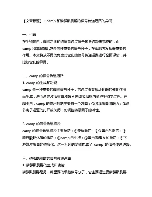
二、camp的信号传递通路1. camp的生成和功能camp是一种重要的细胞信号分子,它通过腺苷酸环化酶的催化作用而生成,进而通过激活蛋白激酶A来调节细胞内多种生物学过程。
在细胞内,camp的作用机制主要有三个方面:①激活蛋白激酶A;②调节离子通道的打开或关闭;③调控转录因子的活性。
2. camp的信号传递路径camp的信号传递路径主要包括:①受体激活;②G蛋白的激活;③腺苷酸环化酶的激活;④camp的生成;⑤蛋白激酶A的激活;⑥下游效应蛋白的磷酸化。
这一系列的步骤构成了camp的信号传递通路。
三、磷脂酰肌醇的信号传递通路1. 磷脂酰肌醇的生成和功能磷脂酰肌醇是另一种重要的细胞信号分子,它主要通过膜磷脂酰肌醇信号途径参与调节细胞内的生物学过程。
在细胞内,磷脂酰肌醇主要通过两种方式发挥作用:①参与细胞膜的组装和稳定;②作为二脂酰甘油的前体参与甘油分解。
2. 磷脂酰肌醇的信号传递路径磷脂酰肌醇的信号传递路径主要包括:①受体激活;②磷脂酰肌醇磷酸酶的激活;③二磷酸肌醇酶的激活;④磷脂酰肌醇的生成;⑤调节细胞内钙离子浓度的改变;⑥激活蛋白激酶C。
这一系列的步骤构成了磷脂酰肌醇的信号传递通路。
四、camp和磷脂酰肌醇的信号传递通路的异同1. 异同点的比较①生成方式:camp是通过腺苷酸环化酶的催化作用生成,而磷脂酰肌醇是通过磷酸肌醇途径生成。
②下游效应:camp主要通过激活蛋白激酶A来调节细胞内多种生物学过程,而磷脂酰肌醇则主要是通过激活蛋白激酶C。
③细胞内响应:camp主要影响蛋白的磷酸化,而磷脂酰肌醇则主要影响细胞内钙离子浓度的改变。
2. 个人观点和理解从我个人的观点来看,camp和磷脂酰肌醇作为两种重要的信号分子,在细胞内发挥着不可替代的作用。
cAMP信号通路介导全麻药遗忘和睡眠损害效应的
开题报告
一、研究背景
全麻药常用于手术操作过程中,通过抑制中枢神经系统的功能来达到麻醉效果。
然而,许多患者在手术后会出现遗忘和睡眠质量下降等不良反应。
近年来的研究表明,这些不良反应可能与全麻药对cAMP信号通路的影响有关。
cAMP信号通路是一种重要的第二信使系统,在细胞内调控重要生理过程。
实验证明,全麻药可以直接或间接地影响cAMP信号通路,从而引发一系列生理效应。
其中,cAMP信号通路的抑制似乎在全麻药导致遗忘和睡眠损害方面具有重要作用。
二、研究内容
本研究旨在探究cAMP信号通路对全麻药遗忘和睡眠损害效应的调控作用。
具体来说,我们将从以下两个方面进行研究:
1. 全麻药对cAMP信号通路的影响:通过用全麻药处理动物模型,检测其对cAMP信号通路的影响,包括cAMP水平、cAMP依赖性蛋白激酶(PKA)活性、cAMP响应元件结合蛋白(CREB)修饰等指标。
2. cAMP信号通路对全麻药遗忘和睡眠损害效应的调控:通过使用cAMP信号通路激动剂或抑制剂,调控cAMP信号通路的活性,观察其对全麻药遗忘和睡眠损害效应的影响。
三、研究意义
本研究的结果有助于深入理解全麻药对生理过程的调节机制,从而为深入研究全麻药的相关毒性和不良反应提供理论基础。
同时,该研究也有望为寻找并开发新型全麻药提供新思路和策略。

g蛋白偶联受体(G protein-coupled receptors,简称GPCRs)是一类重要的跨膜蛋白,能够感知细胞外的信号并将其转导到细胞内部。
而g蛋白偶联受体介导的cAMP信号通路被认为是细胞内信号传导中最为重要的一种通路,对于细胞的正常功能起着至关重要的作用。
在人体生理学中,g蛋白偶联受体介导的cAMP信号通路涉及多种重要的生理过程,包括细胞分化、增殖、代谢调节、离子平衡以及神经递质释放等。
以下将详细介绍g蛋白偶联受体介导的cAMP信号通路在这些生理过程中的作用和意义。
【1】细胞分化g蛋白偶联受体介导的cAMP信号通路在细胞分化过程中发挥重要作用。
研究表明,特定的GPCRs激活cAMP信号通路后,能够促进干细胞向特定细胞系的分化,从而在组织发育和再生过程中发挥关键作用。
这一发现为干细胞治疗和再生医学领域的研究提供了重要的理论基础。
【2】细胞增殖cAMP信号通路对细胞增殖也具有调节作用。
在细胞周期调控中,cAMP信号通路能够通过激活蛋白激酶A(PKA)和调节蛋白激酶(Epac)等效应分子,调控细胞周期蛋白激酶的活性,影响细胞的增殖能力。
cAMP信号通路在肿瘤发生和发展过程中扮演着重要的角色,也成为肿瘤治疗的重要靶点。
【3】代谢调节在细胞能量代谢过程中,cAMP信号通路亦扮演着重要的调节作用。
通过激活PKA和Epac等效应分子,cAMP信号通路能够促进葡萄糖代谢、脂质代谢以及蛋白质合成等代谢过程,对维持细胞能量平衡起着至关重要的作用,并参与调节胰岛素分泌等内分泌功能。
【4】离子平衡cAMP信号通路对离子通道和转运蛋白的调节,也对细胞内离子平衡起着重要作用。
通过激活离子通道蛋白或转运蛋白,cAMP信号通路能够调节细胞内Ca2+、Na+、K+等离子的浓度,影响细胞内环境的稳态,参与细胞对外界环境的适应和响应。
【5】神经递质释放在神经系统中,cAMP信号通路对神经递质的释放及神经元兴奋性起着重要调节作用。
《槲皮素通过ERβ-cAMP信号通路保护细胞损伤机制的探索》篇一槲皮素通过ERβ-cAMP信号通路保护细胞损伤机制的探索一、引言细胞损伤是许多疾病发生和发展的重要机制之一,如何有效保护细胞免受损伤,已成为当前生命科学研究的热点问题。
近年来,研究表明天然产物如槲皮素,对细胞具有显著的保护作用。
本篇论文将就槲皮素如何通过ERβ/cAMP信号通路保护细胞损伤的机制进行深入探讨。
二、槲皮素及其生物活性槲皮素,一种天然的黄酮类化合物,广泛存在于植物中。
其具有抗氧化、抗炎、抗肿瘤等多种生物活性,对多种细胞具有保护作用。
其作用机制复杂,涉及多个信号通路和靶点。
三、ERβ/cAMP信号通路简介雌激素受体β(ERβ)是细胞内重要的信号分子,与细胞生长、分化、凋亡等生理过程密切相关。
环磷酸腺苷(cAMP)是一种重要的第二信使,参与多种细胞内信号转导过程。
ERβ与cAMP之间的相互作用,在细胞损伤保护中具有重要作用。
四、槲皮素对ERβ/cAMP信号通路的影响研究表明,槲皮素能够激活ERβ,进而影响cAMP的合成和降解,从而调控ERβ/cAMP信号通路。
这一过程可能涉及多个酶和蛋白的相互作用,最终导致细胞损伤的减轻。
五、槲皮素保护细胞损伤的机制1. 抗氧化作用:槲皮素能够清除自由基,减轻氧化应激对细胞的损伤。
2. 抗炎作用:槲皮素能够抑制炎症反应,减轻炎症对细胞的损伤。
3. 激活ERβ/cAMP信号通路:槲皮素能够激活ERβ,进而影响cAMP的合成和降解,从而调节细胞内的信号转导过程,保护细胞免受损伤。
六、实验研究我们通过建立细胞损伤模型,观察槲皮素对细胞损伤的保护作用。
实验结果显示,槲皮素能够显著减轻细胞损伤,提高细胞存活率。
进一步的研究表明,槲皮素通过激活ERβ/cAMP信号通路,调节细胞内信号转导过程,从而发挥其保护作用。
七、结论本研究表明,槲皮素通过激活ERβ/cAMP信号通路,对细胞损伤具有显著的保护作用。
其机制涉及抗氧化、抗炎以及调节细胞内信号转导等多个方面。
camp通路相关基因
camp通路相关基因是指能够调控细胞内cAMP浓度的基因,cAMP 是一种重要的第二信使分子,参与了多种细胞信号传导路径的调节,包括紧张、分泌、分化、增殖等多种生物学过程。
camp通路相关基因主要包括cAMP合成酶、cAMP磷酸酶、蛋白激酶、离子通道等,这些基因在细胞内起到了重要的调节作用,对于维持正常的生理功能具有重要的意义。
与camp通路相关的基因突变或过度表达会导致多种疾病的发生,如癌症、免疫系统疾病、神经系统疾病等,因此camp 通路相关基因的研究对于疾病的预防和治疗具有重要的意义。
- 1 -。
胰高血糖素通过cAMP信号通路对胰岛β细胞胰岛素分泌的调节作用及机制的研究胰高血糖素通过 cAMP 信号通路对胰岛β 细胞胰岛素分泌的调节作用及机制的研究胰高血糖素(glucagon)是一种由胰岛的α 细胞分泌的多肽激素,主要起到升高血糖浓度的作用。
胰岛素则由胰岛的β 细胞分泌,用来调节血糖浓度,从而维持机体内部环境的稳定。
胰高血糖素通过激活细胞内的 cAMP(环磷酸腺苷)信号通路来调节胰岛素的分泌。
本文将就胰高血糖素通过 cAMP 信号通路对胰岛β 细胞胰岛素分泌的调节作用及机制进行研究。
胰高血糖素与胰岛素分泌之间存在着相互制约的关系。
当血糖浓度升高时,胰岛β 细胞释放胰岛素的反应受到抑制,而胰高血糖素的分泌则增加。
当血糖浓度降低时,胰岛β 细胞开始释放胰岛素,抑制胰高血糖素的分泌。
这种相互制约的机制使得胰岛素和胰高血糖素在血糖调节中保持平衡。
cAMP 信号通路是一个广泛存在于生物体中的重要信号传导途径。
cAMP 通过激活蛋白激酶 A (PKA)来介导与激素相关的信号转导,并通过调节细胞内多种靶蛋白的活性来影响细胞的功能。
在胰岛β 细胞中,胰高血糖素与其受体结合后,活化细胞膜上的 G 蛋白,进而使腺苷酸环化酶(AC)催化合成 cAMP。
cAMP 结合到 PKA 上,使得 PKA 激活并磷酸化特定的靶蛋白。
这些靶蛋白包括离子通道以及糖酵解途径中的多个关键酶,调节了胰岛β 细胞内的电位变化、胰岛素的分泌前体合成、胰岛素的分泌泡膜定位等过程。
在研究中,通过不同的实验手段发现了不同的 cAMP/PKA信号通路相关分子与胰岛素释放之间的关联。
一方面,胰高血糖素通过 cAMP 信号通路激活 PKA 会导致乙酰胆碱受体磷酸化,从而增强细胞膜去极化过程中的钙离子内流。
另一方面,活化的 PKA 还会通过磷酸化离子通道 Kv2.1 来调节细胞膜电位变化。
这些离子通道的激活和关闭对细胞膜去极化与复极化起着重要的调节作用,进而影响胰岛素分泌。
cAMP信号通路的名词解释在细胞生物学中,信号转导通路起着至关重要的作用,负责将细胞外的信号传导到细胞内,调控各种生物过程。
cAMP信号通路是一种重要的信号转导通路,具有广泛的生理和病理生理学意义。
本文将对cAMP信号通路的相关名词进行解释,并探讨其在细胞生物学研究中的应用。
1. cAMP简介cAMP,即环磷酸腺苷(cyclic adenosine monophosphate),是一种环状的核苷酸,由腺苷酸和磷酸分子组成。
它在细胞内广泛存在,是一种重要的细胞信号分子。
cAMP的合成和降解受到多种酶的调控,其中最主要的是腺苷酸环化酶和磷酸酶。
2. Adenylyl cyclase(腺苷酸环化酶)腺苷酸环化酶是合成cAMP的关键酶。
当细胞受到外界信号刺激时,腺苷酸环化酶被激活,将细胞内的腺苷酸转化为cAMP。
腺苷酸环化酶包含多个亚型,例如AC1、AC2和AC3,它们分布在细胞的不同亚细胞结构中,具有不同的调控方式和生物学功能。
3. cAMP依赖性蛋白激酶(cAMP-dependent protein kinase)cAMP依赖性蛋白激酶(PKA)是一种重要的激酶,它与cAMP密切相关。
在细胞中,当cAMP浓度上升时,cAMP结合到PKA的R亚基上,引发R亚基的解离和C亚基的激活。
激活的C亚基进一步磷酸化细胞内亚细胞器和蛋白质,进而调控细胞的生物学功能。
4. cAMP反应元件结合蛋白(CREB)cAMP反应元件结合蛋白(CREB)是一种转录因子,与cAMP信号通路密切相关。
在细胞受到激活剂刺激时,PKA激活后磷酸化CREB,并促使CREB与转录共激活因子形成复合物,进而激活下游基因的转录。
CREB调控的基因包括许多与细胞分化、增殖和存活相关的基因。
5. cAMP信号通路的生理功能cAMP信号通路具有广泛的生理功能。
它在神经系统中参与记忆和学习的调控,对心血管系统起着重要的调节作用,对胰岛素分泌和代谢调节具有关键作用。
cAMP (Cyclic Adenosine 3',5'-monophosphate) is the first identified second messenger, which has a fundamental role in the cellular response to many extracellular stimuli. The cAMP signaling pathway controls a diverse range of cellular processes. Indeed, not only did cAMP provide the paradigm for the second messenger concept, but also provided the paradigm for signaling compartmentalization. The different receptors, chiefly the GPCRs (G-Protein Coupled Receptors), Alpha andBeta-ADRs (Adrenergic Receptors), Growth Factor receptors, CRHR (Corticotropin Releasing Hormone Receptor), GcgR (Glucagon Receptor), DCC (Deleted in Colorectal Carcinoma), etc are responsible for cAMP accumulation in cells that cause different physiological outcomes, and changes in cAMP levels effects the selective activation of PKA (cAMP dependent Protein Kinase-A) isoforms. The chief source of cAMP is from ATP (Adenosine Triphosphate). In mammals, the conversion of ATP to cAMP is mediated by members of the Class-III AC (Adenylyl Cyclase)/ADCY (Adenylate Cyclase) family (E.C 4.6.1.1), which in humans comprises nine trans-membrane AC enzymes or tmACs and one soluble AC or sAC (Ref.1 & 2). The sAC predominantly occurs in mature spermatozoa. tmACs are regulated by heterotrimeric G-proteins in response to the stimulation of various types of GPCRs and therefore play a key role in the cellular response to extracellular signals. sAC, in contrast, is insensitive to G-proteins. Instead, sAC is directly activated by Ca2+ (Calcium Ions) and the metabolite HCO3- (Bicarbonate Ions), rendering the enzyme an intracellular metabolic sensor. Together, tmACs and sAC regulate a diverse set of essential biological processes, such as differentiation and gene transcription, and this makes cAMP signaling, an important mediator of intra- and extracellular signals in organisms from prokaryotes to higher eukaryotes (Ref.2 & 3).The extracellular stimuli like neurotransmitters, hormones, inflammatory stimuli, stress, epinephrine, norepinephrine, etc activate the G-proteins through receptors like GPCRs and ADR-Alpha/Beta. The major G-proteins that regulate activation of ACs are the GN-AlphaS (GN-AlphaS Complex Locus), GN-AlphaQ (Guanine Nucleotide-Binding Protein-Alpha-Q) andGN-AlphaI (Guanine Nucleotide Binding Protein-Alpha Inhibiting Activity Polypeptide). Upon activation these subunits are separated from GN-Beta (Guanine Nucleotide-Binding Protein-Beta) and GN-Gamma (Guanine Nucleotide-BindingProtein-Gamma) subunits and are converted to their GTP bound states that exhibit distinctive regulatory features on the nine tmACs in order to regulate intracellular cAMP levels (Ref.1 & 4). Other ligands like Gcg (Glucagon), Ucns (Urocortins) and Ntn1 (Netrin-1), etc either directly regulate activity of ACs or via G-protein activation through their respective receptors like GcgR, CRHR and DCC. GN-AlphaS and GN-AlphaQ activate ACs to increase intracellular cAMP levels, where as, GN-AlphaI decrease intracellular cAMP levels by inhibiting ACs. GN-Beta and GN-Gamma subunits act synergistically with GN-AlphaS and GN-AlphaQ only to activate ACII, IV and VII. However the Beta and Gamma-subunits along with GN-AlphaI inhibit the activity of ACI, V and VI. ACI, III, V, VI and IX isoforms play a vital role in spinal pain transmission and are up-regulated by chronic Opoids, for which they are often inhibited by Ca2+ and other proteins acting downstream to cAMP signaling. GN-AlphaI activity is counteracted by RGS (Regulator of G-Protein Signaling) proteins (Ref.5, 6 & 7). However, Beta-Arrestins desensitize many GPCRs and ADRs and this blocks interaction between receptors and their cellular effectors, thereby inhibiting GPCR activity. Beta-Arrestins contol intracellular cAMP levels by switching of signaling from GN-AlphaS to GN-AlphaI by recruiting PDE4D (cAMP Specific Phosphodiesterase-4D) isoforms (Ref.8). Growth factors and PI3K (Phosphatidylinositde-3 Kinase) crosstalk with cAMP signaling by activating Akt (v-Akt Murine Thymoma Viral Oncogene Homolog), which further activates PDE (Phosphodiesterase) to facilitate the conversion of cAMP to AMP through Akt Signaling. This modulates cardiac contractility and release of metabolic energy. G-proteins indirectly influence cAMP signaling by activating PI3K and PLC (Phospholipase-C). PLC cleaves PIP2 (Phosphatidylinositol 4,5-bisphosphate) to generate DAG (Diacylglycerol) and IP3 (Inositol 1,4,5-trisphosphate). DAG in turn activates PKC (Protein Kinase-C). IP3 modulates proteins upstream to cAMP signaling with the release of Ca2+ through IP3R (IP3 Receptor) facilitating activation of Src (v-Src Avian Sacroma(Schmidt-Ruppin A-2)Viral Oncogene) along with PYK2 (Proline-Rich Tyrosine Kinase-2). Activation of Src by PI3K and growth factors enhance activation of Raf1 (v-Raf1 Murine Leukemia Viral Oncogene Homolog-1) through Ras. Raf1 facilitate MEK1 (MAPK/ERK Kinase-1) and MEK2 activation, which in turn activates ERKs (Extracellular Signal-Regulated Kinases) and this ultimately leads to induction of transcription regulator Elk1 (ETS-domain protein Elk1) mediated gene expression. PKCmodulate cAMP signaling by activation of Raf1, PYK2 and ACs like ACI, II, III, V and VII, but inhibits ACVI (Ref.9).Once active, the tmACs and sAC produces the second messenger cAMP in response to a wide range of signal transduction pathways. Three main targets of cAMP are PKA, the GTP-exchange protein, EPACs (Exchange Protein Activated by cAMP) and the CNG (Cyclic-Nucleotide Gated Ion Channel). CNG activation by cAMP provides passage to Ca2+ influx. cAMP activate Rap1A (Ras-Related Protein-1A) and Rap1B (Ras-Related Protein Rap1B) through the PKA-independent and EPAC (Exchange Protein Activated by cAMP)-dependent pathway. cAMP activates cAMP-GEFI (cAMP-Regulated Guanine Nucleotide Exchange Factor-I)/EPAC1 and cAMP-GEFII (cAMP-Regulated Guanine Nucleotide Exchange Factor-II)/EPAC2 that in turn activate Rap1A and Rap1B, respectively. Rap1A and Rap1B then forms an active complex with BRaf (v-Raf Murine Sarcoma Viral Oncogene Homolog-B1) for MEK1/2 activation finally resulting in Elk1 activation. Rap1A and Rap1B further stimulate Rap1 and Rap2 pathways that are vital for cell survival (Ref.10 & 11). Apart from CNG, PKC, and EPACs, other direct targets of cAMP includes, PDE, mTOR (Mammalian Target of Rapamycin), p70S6K/RPS6KB1 (Ribosomal Protein-S6kinase-70kDa-Polypeptide-1), PLA2 (Phospholipase-A2). cAMP-activated mTOR and p70S6K promote cell growth via the mTOR and p70S6K signaling route, whereas, PLA2 facilitates release of stored energy by enhancing the Fatty acid metabolism processes. The Urocortin-cAMP mediated induction of PKC and p38 results in Apoptosis and Cytokine production (like that of IL-6 (Interleukin-6)), downstream to the Urocortin-cAMP pathway. Although cAMP directly regulates the activities of some molecules, PKA appears to be the major 'read-out' for cAMP and is the predominant cellular effector of cAMP (Ref.12). PKA is tethered to specific cellular locations by a growing class of proteins called AKAPs (A-Kinase Anchor Proteins). Targeting of PKA isozymes by AKAPs is important for an increasing number of physiological processes such as cAMP regulation of ion channels in the nervous system, regulation of the cell cycle which involves microtubule dynamics, chromatin condensation and decondensation, nuclear envelope disassembly and reassembly, Steroidogenesis, reproductive function, immune responses and numerous intracellular transport mechanisms (Ref.13).In the following sections, the role of localized pools of PKA in the context of some selected physiological processes where regulation by cAMP plays a major role has been analyzed. The ADR-Alpha/Beta stimulation of cAMP-PKA phosphorylates several proteins related to excitation-contraction coupling like activation of L-Type CaCn (Calcium Channels), KCn (Potassium Channel), SCn (Sodium Channels), ClCn (Chloride Channels), RyR (Ryanodine Receptor), Pln (Phospholamban), CRP(C-Reactive Protein) but inhibts TnnI (Troponin-I). PKA phosphorylates Pln that regulates the activity of SERCA2 (Sarcoplasmic Reticulum Ca2+-ATPase-2). It leads to increased reuptake of Ca2+, Cl- (Chloride Ions), K+ (Potassium Ions), Na+ (Sodium Ions) and this process is affected in failing hearts. Ca2+ uptake activates Caln (Calcineurin), which further facilitates NFAT (Nuclear Factor of Activated T-Cells) translocation to the nucleus, a process that is quite essential for axonal growth. cAMP plays a vital role in regulation of cardiovascular function by controlling the process of myocardial contraction. Increase in higher concentration of Ca2+ and PKA activation enhances eNOS (Endothelial NOS) enzyme activity by phosphorylation of Serine residue (Ser635) in order to stimulate eNOS signaling, which is essential to maintain cardiovascular homeostasis (Ref.13). In mammals, Ca2+ and HCO3- play a critical role in the regulation of sperm function, most likely by regulation of cAMP levels. sAC is active in human spermatozoa and is a sensor for both HCO3- and Ca2+. Ca2+ release by CaCn and CNG, activate Calm (Calmodulin) and CamKs (Calcium/Calmodulin-Dependent Protein Kinases). Calm further activate Caln, CamK2 (Calcium/Calmodulin-Dependent Protein Kinase-II) and CamK4. These in turn modulate cAMP production by regulating the activity of ACs and PDEs. The CamKs along with Caln inhibit PDE and ACIX, whereas CamK2 and CamK4 inhibit the function of ACIX and ACI, respectively (Ref.2). ACIX is also inhibited by PKC thus controlling cAMP signaling in the hippocampal neurons. cAMP activated PKA represses ERK activation by the formation of an inactive Rap1/Raf1 that interferes with ERK activation through seizure of MEK1,2 activation by sequestering Raf1 activity. 14-3-3 like the PKA also aids the process of Raf1 inactivation. Therefore, the effect of cAMP on ERK differs depending on the balance of the Raf1, BRaf and PKA isoforms occurring inside a cell (Ref.10). Long PDE4A (cAMP Specific Phosphodiesterase-4A) isoforms are activated bycAMP, PKA and Akt kinases. The PDE sequester cAMP activity by converting it back to AMP. This negative-feedback loop terminates the cAMP signal locally. cAMP further represses the activity PDE1 (Phosphodiesterase-1) to enhance the duration and intensity of cAMP signaling (Ref.14).cAMP follows a distinct route and activates a single PKA-AKAP complex close to the substrate to mediate a distinct biological effect. Accordingly, each substrate appears to have its own, private anchored pool of PKA and its own local gradient of cAMP. PKA inhibit the interaction of 14-3-3 proteins with BAD (through 14-3-3/BAD signaling) and NFAT to promote cell survival. PKA inhibits Adducin action by limiting its role during assembly of Spectrin-Actin network in erythrocytes, thereby reducing the chances of Erythroleukemia. It activates KDELR (KDEL (Lys-Asp-Glu-Leu) Endoplasmic Reticulum Protein Retention Receptor) to promote retrieval of proteins (protein retention) from golgi complex to endoplasmic reticulum thereby maintaining steady state of the cell. Increased cAMP levels promote survival of neuronal cells by inactivating GSK3Alpha (Glycogen Synthase Kinase-3-Alpha) and GSK3Beta (Glycogen Synthase Kinase-3-Beta) via a PKA dependent mechanism and thus prevents Oncogenesis and neurodegeneration (Ref.13 & 15). PKA interferes at different levels with other signaling pathways. Inactivation of PTP (Protein Tyrosine Phosphatase) results in dissociation from and consequent activation of ERKs. Inactivation of PCTK1 (PCTAIRE Protein Kinase-1) and APC (Anaphase-Promoting Complex) helps to maintain control cell proliferation and anaphase initiation and late mitotic events, respectively, thereby checking the degradation cell cycle regulators. PKA activation by cAMP enhances release of stored energy in cells by phosphorylation of HSL (Hormone-Sensitive Lipase) in white adipose tissue, which leads to the hydrolysis of triglycerides (vital intermediates of Tricaylglycerol Metabolism). Hydrolysis of triglycerides by HSL generates free fatty acids, the major gateway for the release of stored energy, and this process is termed as Lipolysis. Gcg binds to on the surface of liver cells and triggers an increase in cAMP production leading to an increased rate of Glycogenolysis by activating PHK (Phosphorylase Kinase) via the PKA-mediated cascade. PHK further activate PYG (Glycogen Phosphorylase), which converts Glycogen to Glucose-1-Phosphate. Phosphoglucomutase then transfers phosphate to C-6 of Glucose-1-Phosphate generating Glucose-1,6-phosphate as an intermediate. The phosphate on C-1 is then transferred to the enzyme regenerating it and Glucose-6-Phospahte is the released product that enters Glycolysis. This is the same response hepatocytes have to Epinephrine release through the ADR-Alpha/Beta. PKA further inhibits GYS (Glycogen Synthase) leading to seizure of energy consuming process like Glycogen Synthesis (Ref.13 & 16).Upon stimulation from hormones, cAMP increases phosphorylation of RhoA that inactivates Rho Kinase. Rho Kinase regulates Myosin-II and cell contraction by catalyzing phosphorylation of the regulatory subunit of Myosin phosphatase, PPtase1 (Protein Phosphatase-1), by inhibiting its catalytic activity, which results in an indirect increase in RLC (Regulatory Light Chain of Myosin) phosphorylation. Inactivation of Rho Kinase also directly increases RLC phosphorylation. Such increased intracellular cAMP and PKA activation on RLC phosphorylation decreases Thrombin-induced isometric tension development in endothelial cells and this decrease the development of Edema, a hallmark of acute and chronic inflammation (Ref.17). Interestingly, PKA controls phosphatase activity by phosphorylation of specific PPtase1 inhibitors, such as DARPP32 (Dopamine-andcAMP-Regulated Phosphoprotein). Neurotransmitters enhance DARPP32 interaction via GPCRs, which leads to suppression of PPtase1 activity, when DARPP32 is phosphorylated at Thr-34 (Threonine-34) position. Phosphorylation is a crucial event in transcriptional activation by CREB (cAMP Response Element-Binding Protein), CREM (cAMP Response Element Modulator) and ATF1 (Activating Transcription Factor-1), because it allows interaction with the transcriptional co-activators CBP(CREB-Binding Protein) and p300. CREM gene encodes many different isoforms, some of which have repressive functions. Particularly the repressor ICER (Inducible cAMP Early Repressor), participates in the downregulation of cAMP-induced transcription by competing with the binding of CREB and CREM activators to their DNA binding sites. PPtase1 checks the phosphorylation events in order to inactivate the formation of repressor isoforms like ICER so that CREB, CREM and ATF1 are able to interact with the co-activators like CBP and p300. Hence, under physiological conditions, ICER induction is a transient phenomenon that allows cAMP signaling to return to the basal state. In contrast, prolonged or inappropriate induction of ICERelicits pathological consequences (Ref.18). Similarly, phosphorylation of NK-KappaB (Nuclear Factor-KappaB) by PKA is necessary for transcriptional activation and interaction with CBP. PKA modulates the activity of transcription factors, such as nuclear receptors and HMG (High Mobility Group)-containing proteins, influencing their dimerization or DNA-binding properties.A peculiar example is the mechanism by which PKA regulates Gli3 (Gli-Kruppel Family Member-3) under the influence from Hedgehog signaling. Function of Gli3 is similar to that of Drosophila gene CI (Cubitus Interruptus) activity. In this case phosphorylation stimulates a specific cleavage of Gli3 which transforms the protein from an activator to a repressor. However, proteins like PKIs (Protein Kinase Inhibitors) and Mep1B (Meprin-A-Beta) down regulate PKA activity to prevent aberrant gene expression (Ref.13 & 16).Normally, the level of intracellular cAMP is regulated by the balance between the activities of two types of enzymes; ACs and the cyclic nucleotide PDEs chiefly in response to hormones and neurotransmitters. The cAMP signaling is involved in controlling exocytotic events in polarized epithelial cells with implication for Diabetes Insipidus, Hypertension, Gastric ulcers, Thyroid disease, Diabetes Mellitus, and Asthma. Heterologous sensitization of cAMP signaling contributes to fundamental physiological processes such as the timing of circadian rhythms, sexual behavior, and neurotransmitter crosstalk, and also to neurological disorders such as substance abuse and drug-induced Dyskinesias (Ref.19). This provide insight into the mechanisms of addiction and many other central nervous system disorders reflective of altered neuronal cell functioning. Besides it also represent very likely roles for ACs and open up avenues for therapeutics targeting ACs that may prove useful in the development of male contraceptives (Ref.1 & 20).References:1. Watts VJ,Neve KA.Sensitization of adenylate cyclase by Galpha i/o-coupled receptors.Pharmacol. Ther. 2005 Jun;106(3):405-21.2. Jaiswal BS,Conti M.Calcium regulation of the soluble adenylyl cyclase expressed in mammalian spermatozoa.Proc. Natl. Acad. Sci. U S A. 2003 Sep 16;100(19):10676-81.3. Baillie GS,Houslay MD.Arrestin times for compartmentalised cAMP signalling and phosphodiesterase-4 enzymes.Curr. Opin. Cell Biol. 2005 Apr;17(2):129-34.4. Pullar CE,Isseroff RR.Cyclic AMP mediates keratinocyte directional migration in an electric field.J. Cell Sci. 2005 May 1;118(Pt 9):2023-34.5. Bie B,Peng Y,Zhang Y,Pan ZZ.cAMP-mediated mechanisms for pain sensitization during opioid withdrawal.J. Neurosci. 2005 Apr 13;25(15):3824-32.6. Cumbay MG,Watts VJ.Galphaq potentiation of adenylate cyclase type 9 activity through a Ca2+/calmodulin-dependent pathway.Biochem. Pharmacol. 2005 Apr 15;69(8):1247-56.7. Yamada RX,Matsuki N,Ikegaya Y.cAMP differentially regulates axonal and dendritic development of dentate granule cells.J. Biol. Chem. 2005 Nov 11;280(45):38020-8.8. Dessauer CW,Nguyen BT.Relaxin stimulates multiple signaling pathways: activation of cAMP,PI3K,and PKCzeta in THP-1 cells.Ann. N Y. Acad. Sci. 2005 May;1041:272-9.9. Leblais V,Jo SH,Chakir K,Maltsev V,Zheng M,Crow MT,Wang W,Lakatta EG,Xiao RP.Phosphatidylinositol 3-kinase offsets cAMP-mediated positive inotropic effect via inhibiting Ca2+ influx incardiomyocytes.Circ. Res. 2004 Dec 10;95(12):1183-90.10. Kim SS,Choi JM,Kim JW,Ham DS,Ghil SH,Kim MK,Kim-Kwon Y,Hong SY,Ahn SC,Kim SU,Lee YD,Suh-Kim H.cAMP induces neuronal differentiation of mesenchymal stem cells via activation of extracellular signal-regulated kinase/MAPK.Neuroreport. 2005 Aug 22;16(12):1357-61.11. Kiermayer S,Biondi RM,Imig J,Plotz G,Haupenthal J,Zeuzem S,Piiper A.Epac Activation Converts cAMP from a Proliferative into a Differentiation Signal in PC12 Cells.Mol. Biol. Cell. 2005 Oct 5.12. Malbon CC,Tao J,Wang HY.AKAPs (A-kinase anchoring proteins) and molecules that compose their G-protein-coupled receptor signalling complexes.Biochem. J. 2004 Apr 1;379(Pt 1):1-9.13. Tasken K,Aandahl EM.Localized effects of cAMP mediated by distinct routes of protein kinase A.Physiol. Rev. 2004 Jan;84(1):137-67.14. Dumaz N,Marais R.Integrating signals between cAMP and the RAS/RAF/MEK/ERK signalling pathways.FEBS J. 2005 Jul;272(14):3491-504.15. Chin PC,Majdzadeh N,D'Mello SR.Inhibition of GSK3beta is a common event in neuroprotection by different survival factors.Brain. Res. Mol. Brain Res. 2005 Jun 13;137(1-2):193-201.16. Taylor SS,Kim C,Vigil D,Haste NM,Yang J,Wu J,Anand GS.Dynamics of signaling by PKA.Biochim. Biophys. Acta. 2005 Sep 22.17. Goeckeler ZM,Wysolmerski RB.Myosin phosphatase and cofilin mediate cAMP/cAMP-dependent protein kinase-induced decline in endothelial cell isometric tension and myosin II regulatory light chain phosphorylation.J. Biol. Chem. 2005 Sep 23;280(38):33083-95.18. Ding B,Abe J,Wei H,Xu H,Che W,Aizawa T,Liu W,Molina CA,Sadoshima J,Blaxall BC,Berk BC,Yan C.A positive feedback loop of phosphodiesterase 3 (PDE3) and inducible cAMP early repressor (ICER) leads tocardiomyocyte apoptosis.Proc. Natl. Acad. Sci. U S A. 2005 Oct 11;102(41):14771-6.19. Shankar DB,Cheng JC,Sakamoto KM.Role of cyclic AMP response element binding protein in human leukemias.Cancer. 2005 Nov 1;104(9):1819-24.20. Linder JU,Schultz JE.The class III adenylyl cyclases: multi-purpose signalling modules.Cell. Signal. 2003 Dec;15(12):1081-9.。
cAMP (Cyclic Adenosine 3',5'-monophosphate) is the first identified second messenger, which has a fundamental role in the cellular response to many extracellular stimuli. The cAMP signaling pathway controls a diverse range of cellular processes. Indeed, not only did cAMP provide the paradigm for the second messenger concept, but also provided the paradigm for signaling compartmentalization. The different receptors, chiefly the GPCRs (G-Protein Coupled Receptors), Alpha andBeta-ADRs (Adrenergic Receptors), Growth Factor receptors, CRHR (Corticotropin Releasing Hormone Receptor), GcgR (Glucagon Receptor), DCC (Deleted in Colorectal Carcinoma), etc are responsible for cAMP accumulation in cells that cause different physiological outcomes, and changes in cAMP levels effects the selective activation of PKA (cAMP dependent Protein Kinase-A) isoforms. The chief source of cAMP is from ATP (Adenosine Triphosphate). In mammals, the conversion of ATP to cAMP is mediated by members of the Class-III AC (Adenylyl Cyclase)/ADCY (Adenylate Cyclase) family (E.C 4.6.1.1), which in humans comprises nine trans-membrane AC enzymes or tmACs and one soluble AC or sAC (Ref.1 & 2). The sAC predominantly occurs in mature spermatozoa. tmACs are regulated by heterotrimeric G-proteins in response to the stimulation of various types of GPCRs and therefore play a key role in the cellular response to extracellular signals. sAC, in contrast, is insensitive to G-proteins. Instead, sAC is directly activated by Ca2+ (Calcium Ions) and the metabolite HCO3- (Bicarbonate Ions), rendering the enzyme an intracellular metabolic sensor. Together, tmACs and sAC regulate a diverse set of essential biological processes, such as differentiation and gene transcription, and this makes cAMP signaling, an important mediator of intra- and extracellular signals in organisms from prokaryotes to higher eukaryotes (Ref.2 & 3).The extracellular stimuli like neurotransmitters, hormones, inflammatory stimuli, stress, epinephrine, norepinephrine, etc activate the G-proteins through receptors like GPCRs and ADR-Alpha/Beta. The major G-proteins that regulate activation of ACs are the GN-AlphaS (GN-AlphaS Complex Locus), GN-AlphaQ (Guanine Nucleotide-Binding Protein-Alpha-Q) andGN-AlphaI (Guanine Nucleotide Binding Protein-Alpha Inhibiting Activity Polypeptide). Upon activation these subunits are separated from GN-Beta (Guanine Nucleotide-Binding Protein-Beta) and GN-Gamma (Guanine Nucleotide-BindingProtein-Gamma) subunits and are converted to their GTP bound states that exhibit distinctive regulatory features on the nine tmACs in order to regulate intracellular cAMP levels (Ref.1 & 4). Other ligands like Gcg (Glucagon), Ucns (Urocortins) and Ntn1 (Netrin-1), etc either directly regulate activity of ACs or via G-protein activation through their respective receptors like GcgR, CRHR and DCC. GN-AlphaS and GN-AlphaQ activate ACs to increase intracellular cAMP levels, where as, GN-AlphaI decrease intracellular cAMP levels by inhibiting ACs. GN-Beta and GN-Gamma subunits act synergistically with GN-AlphaS and GN-AlphaQ only to activate ACII, IV and VII. However the Beta and Gamma-subunits along with GN-AlphaI inhibit the activity of ACI, V and VI. ACI, III, V, VI and IX isoforms play a vital role in spinal pain transmission and are up-regulated by chronic Opoids, for which they are often inhibited by Ca2+ and other proteins acting downstream to cAMP signaling. GN-AlphaI activity is counteracted by RGS (Regulator of G-Protein Signaling) proteins (Ref.5, 6 & 7). However, Beta-Arrestins desensitize many GPCRs and ADRs and this blocks interaction between receptors and their cellular effectors, thereby inhibiting GPCR activity. Beta-Arrestins contol intracellular cAMP levels by switching of signaling from GN-AlphaS to GN-AlphaI by recruiting PDE4D (cAMP Specific Phosphodiesterase-4D) isoforms (Ref.8). Growth factors and PI3K (Phosphatidylinositde-3 Kinase) crosstalk with cAMP signaling by activating Akt (v-Akt Murine Thymoma Viral Oncogene Homolog), which further activates PDE (Phosphodiesterase) to facilitate the conversion of cAMP to AMP through Akt Signaling. This modulates cardiac contractility and release of metabolic energy. G-proteins indirectly influence cAMP signaling by activating PI3K and PLC (Phospholipase-C). PLC cleaves PIP2 (Phosphatidylinositol 4,5-bisphosphate) to generate DAG (Diacylglycerol) and IP3 (Inositol 1,4,5-trisphosphate). DAG in turn activates PKC (Protein Kinase-C). IP3 modulates proteins upstream to cAMP signaling with the release of Ca2+ through IP3R (IP3 Receptor) facilitating activation of Src (v-Src Avian Sacroma(Schmidt-Ruppin A-2)Viral Oncogene) along with PYK2 (Proline-Rich Tyrosine Kinase-2). Activation of Src by PI3K and growth factors enhance activation of Raf1 (v-Raf1 Murine Leukemia Viral Oncogene Homolog-1) through Ras. Raf1 facilitate MEK1 (MAPK/ERK Kinase-1) and MEK2 activation, which in turn activates ERKs (Extracellular Signal-Regulated Kinases) and this ultimately leads to induction of transcription regulator Elk1 (ETS-domain protein Elk1) mediated gene expression. PKCmodulate cAMP signaling by activation of Raf1, PYK2 and ACs like ACI, II, III, V and VII, but inhibits ACVI (Ref.9).Once active, the tmACs and sAC produces the second messenger cAMP in response to a wide range of signal transduction pathways. Three main targets of cAMP are PKA, the GTP-exchange protein, EPACs (Exchange Protein Activated by cAMP) and the CNG (Cyclic-Nucleotide Gated Ion Channel). CNG activation by cAMP provides passage to Ca2+ influx. cAMP activate Rap1A (Ras-Related Protein-1A) and Rap1B (Ras-Related Protein Rap1B) through the PKA-independent and EPAC (Exchange Protein Activated by cAMP)-dependent pathway. cAMP activates cAMP-GEFI (cAMP-Regulated Guanine Nucleotide Exchange Factor-I)/EPAC1 and cAMP-GEFII (cAMP-Regulated Guanine Nucleotide Exchange Factor-II)/EPAC2 that in turn activate Rap1A and Rap1B, respectively. Rap1A and Rap1B then forms an active complex with BRaf (v-Raf Murine Sarcoma Viral Oncogene Homolog-B1) for MEK1/2 activation finally resulting in Elk1 activation. Rap1A and Rap1B further stimulate Rap1 and Rap2 pathways that are vital for cell survival (Ref.10 & 11). Apart from CNG, PKC, and EPACs, other direct targets of cAMP includes, PDE, mTOR (Mammalian Target of Rapamycin), p70S6K/RPS6KB1 (Ribosomal Protein-S6kinase-70kDa-Polypeptide-1), PLA2 (Phospholipase-A2). cAMP-activated mTOR and p70S6K promote cell growth via the mTOR and p70S6K signaling route, whereas, PLA2 facilitates release of stored energy by enhancing the Fatty acid metabolism processes. The Urocortin-cAMP mediated induction of PKC and p38 results in Apoptosis and Cytokine production (like that of IL-6 (Interleukin-6)), downstream to the Urocortin-cAMP pathway. Although cAMP directly regulates the activities of some molecules, PKA appears to be the major 'read-out' for cAMP and is the predominant cellular effector of cAMP (Ref.12). PKA is tethered to specific cellular locations by a growing class of proteins called AKAPs (A-Kinase Anchor Proteins). Targeting of PKA isozymes by AKAPs is important for an increasing number of physiological processes such as cAMP regulation of ion channels in the nervous system, regulation of the cell cycle which involves microtubule dynamics, chromatin condensation and decondensation, nuclear envelope disassembly and reassembly, Steroidogenesis, reproductive function, immune responses and numerous intracellular transport mechanisms (Ref.13).In the following sections, the role of localized pools of PKA in the context of some selected physiological processes where regulation by cAMP plays a major role has been analyzed. The ADR-Alpha/Beta stimulation of cAMP-PKA phosphorylates several proteins related to excitation-contraction coupling like activation of L-Type CaCn (Calcium Channels), KCn (Potassium Channel), SCn (Sodium Channels), ClCn (Chloride Channels), RyR (Ryanodine Receptor), Pln (Phospholamban), CRP(C-Reactive Protein) but inhibts TnnI (Troponin-I). PKA phosphorylates Pln that regulates the activity of SERCA2 (Sarcoplasmic Reticulum Ca2+-ATPase-2). It leads to increased reuptake of Ca2+, Cl- (Chloride Ions), K+ (Potassium Ions), Na+ (Sodium Ions) and this process is affected in failing hearts. Ca2+ uptake activates Caln (Calcineurin), which further facilitates NFAT (Nuclear Factor of Activated T-Cells) translocation to the nucleus, a process that is quite essential for axonal growth. cAMP plays a vital role in regulation of cardiovascular function by controlling the process of myocardial contraction. Increase in higher concentration of Ca2+ and PKA activation enhances eNOS (Endothelial NOS) enzyme activity by phosphorylation of Serine residue (Ser635) in order to stimulate eNOS signaling, which is essential to maintain cardiovascular homeostasis (Ref.13). In mammals, Ca2+ and HCO3- play a critical role in the regulation of sperm function, most likely by regulation of cAMP levels. sAC is active in human spermatozoa and is a sensor for both HCO3- and Ca2+. Ca2+ release by CaCn and CNG, activate Calm (Calmodulin) and CamKs (Calcium/Calmodulin-Dependent Protein Kinases). Calm further activate Caln, CamK2 (Calcium/Calmodulin-Dependent Protein Kinase-II) and CamK4. These in turn modulate cAMP production by regulating the activity of ACs and PDEs. The CamKs along with Caln inhibit PDE and ACIX, whereas CamK2 and CamK4 inhibit the function of ACIX and ACI, respectively (Ref.2). ACIX is also inhibited by PKC thus controlling cAMP signaling in the hippocampal neurons. cAMP activated PKA represses ERK activation by the formation of an inactive Rap1/Raf1 that interferes with ERK activation through seizure of MEK1,2 activation by sequestering Raf1 activity. 14-3-3 like the PKA also aids the process of Raf1 inactivation. Therefore, the effect of cAMP on ERK differs depending on the balance of the Raf1, BRaf and PKA isoforms occurring inside a cell (Ref.10). Long PDE4A (cAMP Specific Phosphodiesterase-4A) isoforms are activated bycAMP, PKA and Akt kinases. The PDE sequester cAMP activity by converting it back to AMP. This negative-feedback loop terminates the cAMP signal locally. cAMP further represses the activity PDE1 (Phosphodiesterase-1) to enhance the duration and intensity of cAMP signaling (Ref.14).cAMP follows a distinct route and activates a single PKA-AKAP complex close to the substrate to mediate a distinct biological effect. Accordingly, each substrate appears to have its own, private anchored pool of PKA and its own local gradient of cAMP. PKA inhibit the interaction of 14-3-3 proteins with BAD (through 14-3-3/BAD signaling) and NFAT to promote cell survival. PKA inhibits Adducin action by limiting its role during assembly of Spectrin-Actin network in erythrocytes, thereby reducing the chances of Erythroleukemia. It activates KDELR (KDEL (Lys-Asp-Glu-Leu) Endoplasmic Reticulum Protein Retention Receptor) to promote retrieval of proteins (protein retention) from golgi complex to endoplasmic reticulum thereby maintaining steady state of the cell. Increased cAMP levels promote survival of neuronal cells by inactivating GSK3Alpha (Glycogen Synthase Kinase-3-Alpha) and GSK3Beta (Glycogen Synthase Kinase-3-Beta) via a PKA dependent mechanism and thus prevents Oncogenesis and neurodegeneration (Ref.13 & 15). PKA interferes at different levels with other signaling pathways. Inactivation of PTP (Protein Tyrosine Phosphatase) results in dissociation from and consequent activation of ERKs. Inactivation of PCTK1 (PCTAIRE Protein Kinase-1) and APC (Anaphase-Promoting Complex) helps to maintain control cell proliferation and anaphase initiation and late mitotic events, respectively, thereby checking the degradation cell cycle regulators. PKA activation by cAMP enhances release of stored energy in cells by phosphorylation of HSL (Hormone-Sensitive Lipase) in white adipose tissue, which leads to the hydrolysis of triglycerides (vital intermediates of Tricaylglycerol Metabolism). Hydrolysis of triglycerides by HSL generates free fatty acids, the major gateway for the release of stored energy, and this process is termed as Lipolysis. Gcg binds to on the surface of liver cells and triggers an increase in cAMP production leading to an increased rate of Glycogenolysis by activating PHK (Phosphorylase Kinase) via the PKA-mediated cascade. PHK further activate PYG (Glycogen Phosphorylase), which converts Glycogen to Glucose-1-Phosphate. Phosphoglucomutase then transfers phosphate to C-6 of Glucose-1-Phosphate generating Glucose-1,6-phosphate as an intermediate. The phosphate on C-1 is then transferred to the enzyme regenerating it and Glucose-6-Phospahte is the released product that enters Glycolysis. This is the same response hepatocytes have to Epinephrine release through the ADR-Alpha/Beta. PKA further inhibits GYS (Glycogen Synthase) leading to seizure of energy consuming process like Glycogen Synthesis (Ref.13 & 16).Upon stimulation from hormones, cAMP increases phosphorylation of RhoA that inactivates Rho Kinase. Rho Kinase regulates Myosin-II and cell contraction by catalyzing phosphorylation of the regulatory subunit of Myosin phosphatase, PPtase1 (Protein Phosphatase-1), by inhibiting its catalytic activity, which results in an indirect increase in RLC (Regulatory Light Chain of Myosin) phosphorylation. Inactivation of Rho Kinase also directly increases RLC phosphorylation. Such increased intracellular cAMP and PKA activation on RLC phosphorylation decreases Thrombin-induced isometric tension development in endothelial cells and this decrease the development of Edema, a hallmark of acute and chronic inflammation (Ref.17). Interestingly, PKA controls phosphatase activity by phosphorylation of specific PPtase1 inhibitors, such as DARPP32 (Dopamine-andcAMP-Regulated Phosphoprotein). Neurotransmitters enhance DARPP32 interaction via GPCRs, which leads to suppression of PPtase1 activity, when DARPP32 is phosphorylated at Thr-34 (Threonine-34) position. Phosphorylation is a crucial event in transcriptional activation by CREB (cAMP Response Element-Binding Protein), CREM (cAMP Response Element Modulator) and ATF1 (Activating Transcription Factor-1), because it allows interaction with the transcriptional co-activators CBP(CREB-Binding Protein) and p300. CREM gene encodes many different isoforms, some of which have repressive functions. Particularly the repressor ICER (Inducible cAMP Early Repressor), participates in the downregulation of cAMP-induced transcription by competing with the binding of CREB and CREM activators to their DNA binding sites. PPtase1 checks the phosphorylation events in order to inactivate the formation of repressor isoforms like ICER so that CREB, CREM and ATF1 are able to interact with the co-activators like CBP and p300. Hence, under physiological conditions, ICER induction is a transient phenomenon that allows cAMP signaling to return to the basal state. In contrast, prolonged or inappropriate induction of ICERelicits pathological consequences (Ref.18). Similarly, phosphorylation of NK-KappaB (Nuclear Factor-KappaB) by PKA is necessary for transcriptional activation and interaction with CBP. PKA modulates the activity of transcription factors, such as nuclear receptors and HMG (High Mobility Group)-containing proteins, influencing their dimerization or DNA-binding properties.A peculiar example is the mechanism by which PKA regulates Gli3 (Gli-Kruppel Family Member-3) under the influence from Hedgehog signaling. Function of Gli3 is similar to that of Drosophila gene CI (Cubitus Interruptus) activity. In this case phosphorylation stimulates a specific cleavage of Gli3 which transforms the protein from an activator to a repressor. However, proteins like PKIs (Protein Kinase Inhibitors) and Mep1B (Meprin-A-Beta) down regulate PKA activity to prevent aberrant gene expression (Ref.13 & 16).Normally, the level of intracellular cAMP is regulated by the balance between the activities of two types of enzymes; ACs and the cyclic nucleotide PDEs chiefly in response to hormones and neurotransmitters. The cAMP signaling is involved in controlling exocytotic events in polarized epithelial cells with implication for Diabetes Insipidus, Hypertension, Gastric ulcers, Thyroid disease, Diabetes Mellitus, and Asthma. Heterologous sensitization of cAMP signaling contributes to fundamental physiological processes such as the timing of circadian rhythms, sexual behavior, and neurotransmitter crosstalk, and also to neurological disorders such as substance abuse and drug-induced Dyskinesias (Ref.19). This provide insight into the mechanisms of addiction and many other central nervous system disorders reflective of altered neuronal cell functioning. Besides it also represent very likely roles for ACs and open up avenues for therapeutics targeting ACs that may prove useful in the development of male contraceptives (Ref.1 & 20).References:1. Watts VJ,Neve KA.Sensitization of adenylate cyclase by Galpha i/o-coupled receptors.Pharmacol. Ther. 2005 Jun;106(3):405-21.2. Jaiswal BS,Conti M.Calcium regulation of the soluble adenylyl cyclase expressed in mammalian spermatozoa.Proc. Natl. Acad. Sci. U S A. 2003 Sep 16;100(19):10676-81.3. Baillie GS,Houslay MD.Arrestin times for compartmentalised cAMP signalling and phosphodiesterase-4 enzymes.Curr. Opin. Cell Biol. 2005 Apr;17(2):129-34.4. Pullar CE,Isseroff RR.Cyclic AMP mediates keratinocyte directional migration in an electric field.J. Cell Sci. 2005 May 1;118(Pt 9):2023-34.5. Bie B,Peng Y,Zhang Y,Pan ZZ.cAMP-mediated mechanisms for pain sensitization during opioid withdrawal.J. Neurosci. 2005 Apr 13;25(15):3824-32.6. Cumbay MG,Watts VJ.Galphaq potentiation of adenylate cyclase type 9 activity through a Ca2+/calmodulin-dependent pathway.Biochem. Pharmacol. 2005 Apr 15;69(8):1247-56.7. Yamada RX,Matsuki N,Ikegaya Y.cAMP differentially regulates axonal and dendritic development of dentate granule cells.J. Biol. Chem. 2005 Nov 11;280(45):38020-8.8. Dessauer CW,Nguyen BT.Relaxin stimulates multiple signaling pathways: activation of cAMP,PI3K,and PKCzeta in THP-1 cells.Ann. N Y. Acad. Sci. 2005 May;1041:272-9.9. Leblais V,Jo SH,Chakir K,Maltsev V,Zheng M,Crow MT,Wang W,Lakatta EG,Xiao RP.Phosphatidylinositol 3-kinase offsets cAMP-mediated positive inotropic effect via inhibiting Ca2+ influx incardiomyocytes.Circ. Res. 2004 Dec 10;95(12):1183-90.10. Kim SS,Choi JM,Kim JW,Ham DS,Ghil SH,Kim MK,Kim-Kwon Y,Hong SY,Ahn SC,Kim SU,Lee YD,Suh-Kim H.cAMP induces neuronal differentiation of mesenchymal stem cells via activation of extracellular signal-regulated kinase/MAPK.Neuroreport. 2005 Aug 22;16(12):1357-61.11. Kiermayer S,Biondi RM,Imig J,Plotz G,Haupenthal J,Zeuzem S,Piiper A.Epac Activation Converts cAMP from a Proliferative into a Differentiation Signal in PC12 Cells.Mol. Biol. Cell. 2005 Oct 5.12. Malbon CC,Tao J,Wang HY.AKAPs (A-kinase anchoring proteins) and molecules that compose their G-protein-coupled receptor signalling complexes.Biochem. J. 2004 Apr 1;379(Pt 1):1-9.13. Tasken K,Aandahl EM.Localized effects of cAMP mediated by distinct routes of protein kinase A.Physiol. Rev. 2004 Jan;84(1):137-67.14. Dumaz N,Marais R.Integrating signals between cAMP and the RAS/RAF/MEK/ERK signalling pathways.FEBS J. 2005 Jul;272(14):3491-504.15. Chin PC,Majdzadeh N,D'Mello SR.Inhibition of GSK3beta is a common event in neuroprotection by different survival factors.Brain. Res. Mol. Brain Res. 2005 Jun 13;137(1-2):193-201.16. Taylor SS,Kim C,Vigil D,Haste NM,Yang J,Wu J,Anand GS.Dynamics of signaling by PKA.Biochim. Biophys. Acta. 2005 Sep 22.17. Goeckeler ZM,Wysolmerski RB.Myosin phosphatase and cofilin mediate cAMP/cAMP-dependent protein kinase-induced decline in endothelial cell isometric tension and myosin II regulatory light chain phosphorylation.J. Biol. Chem. 2005 Sep 23;280(38):33083-95.18. Ding B,Abe J,Wei H,Xu H,Che W,Aizawa T,Liu W,Molina CA,Sadoshima J,Blaxall BC,Berk BC,Yan C.A positive feedback loop of phosphodiesterase 3 (PDE3) and inducible cAMP early repressor (ICER) leads tocardiomyocyte apoptosis.Proc. Natl. Acad. Sci. U S A. 2005 Oct 11;102(41):14771-6.19. Shankar DB,Cheng JC,Sakamoto KM.Role of cyclic AMP response element binding protein in human leukemias.Cancer. 2005 Nov 1;104(9):1819-24.20. Linder JU,Schultz JE.The class III adenylyl cyclases: multi-purpose signalling modules.Cell. Signal. 2003 Dec;15(12):1081-9.。
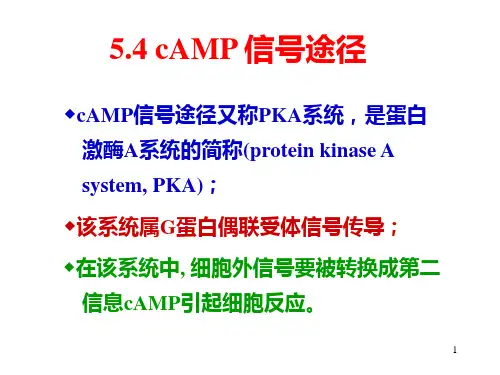
cAMP信号通路
信号分子:1.激素 2.局部介质3.神经递质
受体:G蛋白偶联受体
胞内应答过程:激素→G蛋白耦联受体→G蛋白→腺苷酸环化酶→cAMP→依赖cAMP的蛋白激酶A→基因调控蛋白→基因转录
举例:1.多发性骨髓瘤:通过调变细胞内环腺苷酸浓度可以诱导多种肿瘤细胞增殖阻滞和凋亡,成为肿瘤治疗新途径。
2.肝损伤:对乙酰氨基酚致药物性肝脏损伤可能与cAMP-PKA 信号通路有关。
3.研究人员已经确定了这其中的机制,现在,一种能抑制Epac的新的候选药物——称为ESI Epac特异性抑制剂,也已经被证明能够保护正常小鼠免受致命性立克次氏体感染。
目前,研究人员正在设计第二代ESI——更有效,即使在最高剂量也无毒。
也有来自预备试验的迹象表明,ESI能够保护动物抗击一些致命的病毒感染。
磷脂酰肌醇信号通路
信号分子:1.激素 2.局部介质3.神经递质
受体:酶耦联型受体
胞内应答过程:Ca2+活化各种Ca2+结合蛋白引起细胞反应,钙调素(calmodulin,CaM)由单一肽链构成,具有四个钙离子结合部位。
结合钙离子发生构象改变,可激活钙调素依赖性激酶(CaM-Kinase)。
细胞对Ca2+的反应取决于细胞内钙结合蛋白和钙调素依赖性激酶的种类。
IP3信号的终止是通过去磷酸化形成IP2,或被磷酸化形成IP4。
Ca2+由质膜上的Ca2+泵和Na+-Ca2+交换器将抽出细胞,或由内质网膜上的钙泵抽进内质网
DG通过两种途径终止其信使作用:一是被DG-激酶磷酸化成为磷脂酸,进入磷脂酰肌醇循环;二是被DG酯酶水解成单酯酰甘油。
由于DG代谢周期很短,不可能长期维持PKC活性,而细胞增殖或分化行为的变化又要求PKC长期活性所产生的效应。
现发现另一种DG生成途径,即由磷脂酶催化质膜上的磷脂酰胆碱断裂产生的DG,用来维持PKC的长期效应。
举例:1.肿瘤治疗:该通路调节肿瘤细胞的增殖和存活,其活性异常不仅能导致细胞恶性转化,而且与肿瘤细胞的迁移、黏附、肿瘤血管生成以及细胞外基质的降解等相关。
2.肝癌:PIK3R1在肝癌组织中表达上调,PIK3R1可能通过激活PI3K/AKT信号通路促进HepG2细胞的增殖.
生物技术15-1
曹文祥。